Intervertebral disc
Also called spinal disc, intervertebral disk, IVD
The pads of fibrocartilage between the bones of the spine. What a disc is made of, how it carries load and takes in nutrients, how it changes with age, and what studies found about how it heals and adapts.
- Region
- Spine
- Updated
- October 9, 2026
- Reviewed
- October 9, 2026 · how entries are checked
Function
A pad between each pair of vertebrae
An intervertebral disc is a pad of fibrocartilage that fills the gap between the bodies of two neighbouring vertebrae1. It is anchored to both1. The adult spine has 23 discs: 6 in the neck, 12 in the mid-back and 5 in the lower back2. The lowest sits between the last lumbar (lower back) vertebra and the sacrum, the triangle-shaped bone at the base of the spine2.
Discs are thin in the neck1. They are thickest in the lower back, the region that bears the most body weight1. A lumbar disc is about 7 to 10 mm thick and about 4 cm from front to back3.
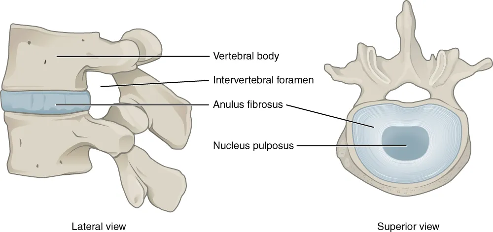
A gel core inside a wall of rings
Each disc has three main parts2:
- The nucleus pulposus is the inner part, a gel-like core that sits slightly behind the centre of the disc2.
- The annulus fibrosus is the outer part, built from 15 to 25 rings, one inside the next2,3.
- The cartilage endplates anchor the disc to the vertebrae above and below2. Each is a thin layer of hyaline cartilage (a smooth, glassy kind of cartilage), usually under 1 mm thick3.
Within each ring of the annulus the collagen fibres lie parallel, at about 60 degrees to the vertical3. Collagen is a strong, rope-like protein. The fibres lean the opposite way in the next ring3. This collagen network gives the disc its tensile strength, its strength against being pulled apart, and anchors it to the bone3.
The nucleus is about 80% water by wet weight, and the annulus about 70%3. The nucleus also holds more proteoglycan, a large protein-and-sugar molecule that holds water3. Aggrecan, the main proteoglycan of the disc, keeps the tissue hydrated through the osmotic pressure its sugar chains create3. Osmotic pressure is the pull that draws water toward dissolved substances.
What the disc does
The discs are the spinal column’s main joints3. They constantly pass on loads from body weight and muscle activity, and they let the spine bend and twist3. They spread compressive (squeezing) loads along the spine and keep neighbouring vertebral bodies from touching bone to bone2.
The gel-like nucleus lets the disc change shape as one vertebra rocks on the next1. Bending forward compresses the front of the disc and expands the back1.
How much pressure a disc carries
One set of figures comes from a single 45-year-old male volunteer with a pressure sensor in a healthy disc4. The sensor sat in his L4/L5 disc, between the fourth and fifth bones of the lower back4. MPa stands for megapascals.
| Position or task, one volunteer | Pressure in the L4/L5 disc |
|---|---|
| Lying face down | 0.1 MPa4 |
| Sitting unsupported | 0.46 MPa4 |
| Relaxed standing | 0.5 MPa4 |
| Sitting fully flexed (bent) | 0.83 MPa4 |
| Standing bent forward | 1.1 MPa4 |
| Lifting 20 kg held close to the body | 1.1 MPa4 |
| Lifting 20 kg with flexed knees | 1.7 MPa4 |
| Lifting 20 kg with a rounded, flexed back | 2.3 MPa4 |
How water and nutrients move
The changing loads on the spine over a day alter the water content and height of the discs9. In eight young adults, whole-body height varied by an average of 19.3 mm over the day10.
The disc has no blood supply of its own11. Its cells rely on diffusion, the slow spread of a substance through a tissue, from blood vessels at the edges of the disc11. Nutrients reach the nucleus from capillaries (tiny, thin-walled blood vessels) by diffusing through the cartilage endplate3. Nucleus cells can lie up to 8 mm from those capillaries3.
Breakdown
Discs change early, and in most people scanned in one study
The disc shows degenerative and ageing changes earlier than any other connective tissue in the body3. The Wakayama Spine Study scanned the whole spine of 975 people aged 21 to 97 with MRI12. It found degeneration somewhere in the spine in 71% of men and 77% of women under 5012. Over 50, the figure was more than 90% in both12.
The largest chemical change in degeneration is loss of proteoglycan, which lowers the osmotic pressure of the matrix and so its water content3. The matrix is the material around the cells. Degenerated discs hold less water than normal discs of the same age3. Under load they lose height and fluid faster, and they tend to bulge3.
What studies link to degeneration
Four sources looked at what goes with degeneration5,6,12,13:
- Inheritance. An MRI study scored disc degeneration in 172 identical and 154 non-identical twins not selected for back pain5. Heritability of the overall score was 74% in the lumbar spine and 73% in the neck, after adjusting for age, weight, smoking and other factors5. A narrative review (a summary of research written without a set, checkable search method) covered the Twin Spine Study13. It concluded that genetic influences appeared to determine degeneration in great part13.
- Physical loading. In the same review, twin siblings whose physical loading at work and leisure differed greatly through adulthood showed surprisingly little difference in disc degeneration13.
- Several causes together. A review of the literature named four underlying causes: genetic inheritance, age, poor transport of nutrients and wastes, and loading history6. It held that each can weaken a disc enough for it to fail structurally during everyday activity6.
- Age and obesity. The Wakayama Spine Study found age and obesity associated with degeneration in every region of the spine12.
How a disc herniates
A disc herniation is when the soft centre of a disc pushes out through its tough outer ring. The disc bulges or ruptures backward, or backward and to one side3. It then presses on nerve roots inside the spinal canal, the channel in the spine that holds the spinal cord3. A nerve root is the part of a nerve where it leaves the spinal cord. Herniation is most common at the L4/L5 and L5/S1 discs, the two lowest lumbar levels1,14.
In laboratory tests, a healthy disc herniates only under forces larger than those normally met3. In most such tests the vertebral body fails before the disc, and some degenerative change seems to be needed first3. Three studies looked at how discs fail:
- Cadaver (donated body) spines. Fifty-two lumbar segments were fatigue-loaded (loaded over and over) in compression and bending, and six discs herniated gradually15.
- Pig neck segments. In the laboratory, herniation at the back of the annulus occurred with modest compression but highly repetitive bending forward and back16.
- People having surgery. In 181 consecutive patients having surgery for a lumbar herniation at one level, 117 had failed where the disc joins the endplate17. That was more common than rupture of the annulus17.
Herniation of the L4/L5 or L5/S1 disc can cause sciatica, a widespread pain that spreads from the lower back into the leg1,18. The disc herniation page covers who is affected and what trials found, and the sciatica page covers the leg pain.
Recovery
A tissue that renews slowly
One review of the literature states that structural failure of a disc is irreversible, because adult discs have limited potential to heal6. In 21 adult sheep given a 5 mm cut in the outer annulus of lumbar discs, the outermost annulus showed the ability to heal19. The inner annulus failed progressively in all the sheep, in most discs within 4 to 12 months of the operation19.
A 2011 evidence review states that on repeated MRI scans the herniated part of a lumbar disc tends to shrink over time20. A 2024 meta-analysis (a study that pools earlier studies) covered 2,233 people who had non-surgical care for a lumbar disc herniation21. Its pooled overall incidence of disc resorption, the herniated material being absorbed, was 70.39%21.
Disc cells continually break the molecules of the matrix down and make new ones3. Disc collagen, though, turns over very slowly, and more slowly with age22. A laboratory study of collagen from normal discs estimated an average half-life of 95 years between ages 20 and 4022. A half-life is the time it takes for half of something to be replaced or broken down.
In seven healthy men, 21 days of head-down bed rest changed disc volume and height23. Those measures had not returned to their starting values 153 days later, although back muscle size had recovered23.
What studies of loading and running found
Studies in runners, twins and animals bear on how discs respond to loading24,25:
- Runners. A cross-sectional study, one that looks at a group once, compared adults aged 25 to 3524. Long-distance runners and joggers had lumbar disc T2 times, an MRI measure, 11.4% and 9.2% higher than people who did no sport24. The authors read this as better hydration, and the two running groups did not differ significantly24.
- Twins. The narrative review of the Twin Spine Study reported some indications that routine loading may have some benefits for the disc13.
- Animals. A brief narrative review reported that animal studies suggest a dose-response relationship between loading and the disc25. High loading at high volume and frequency might speed degeneration or injure the disc25. High loading at low volume and low frequency appears to switch on potentially regenerative mechanisms25.
Connections
Each disc joins the bodies of the vertebrae above and below it, and the discs are the main joints of the spine1,3. Five discs sit in the lower back, the region of the lumbar vertebrae2. Neighbouring vertebrae also meet at the facet joints, the small joints that guide and limit movement2. The disc joints between the vertebral bodies carry most of the weight the spine bears2.
A spinal nerve leaves through the intervertebral foramen, the opening formed between neighbouring vertebrae1. A nucleus bulging backward can press on the nerve there1,3. Herniation of the L4/L5 or L5/S1 disc can cause sciatica, and the disc herniation page covers that condition1,18. In the Wakayama Spine Study, low back pain was associated with degeneration in the lumbar region12.
Related exercise pages: good morning, child’s pose, knee-to-chest and running. Four male nationally ranked weightlifters did good mornings with a mean of 67.5 kg on the bar26. A model estimated the mean peak compressive force at L5/S1 at 6712.7 newtons26.
One published description of child’s pose is a forward bend with the spine rounded27. In one trial in people with chronic low back pain, the flexion-based (forward-bending) lumbar exercise programme included knee-to-chest28. Bending forward compresses the front of a disc and expands the back1.
Intervertebral disc forms part of
Intervertebral disc joins
Intervertebral disc is injured in
Intervertebral disc is related to
Intervertebral disc has been studied in people with
Intervertebral disc is loaded by
Intervertebral disc is bent forward in
Intervertebral disc is bent forward in flexion programmes that include
Intervertebral disc has studies of runners in
Sources
- Betts JG, Young KA, Wise JA, Johnson E, Poe B, Kruse DH, Korol O, Johnson JE, Womble M, DeSaix P (2022). Anatomy and Physiology 2e. OpenStax. Link
- Waxenbaum JA, Reddy V, Futterman B (2026). Anatomy, Back, Intervertebral Discs. StatPearls. PMID 29262063
- Urban JP, Roberts S (2003). Degeneration of the intervertebral disc.. Arthritis research & therapy. PMID 12723977
- Wilke HJ, Neef P, Caimi M, Hoogland T, Claes LE (1999). New in vivo measurements of pressures in the intervertebral disc in daily life.. Spine. doi:10.1097/00007632-199904150-00005 PMID 10222525
- Sambrook PN, MacGregor AJ, Spector TD (1999). Genetic influences on cervical and lumbar disc degeneration: a magnetic resonance imaging study in twins.. Arthritis and rheumatism. doi:10.1002/1529-0131(199902)42:2<366::AID-ANR20>3.0.CO;2-6 PMID 10025932
- Adams MA, Roughley PJ (2006). What is intervertebral disc degeneration, and what causes it?. Spine. doi:10.1097/01.brs.0000231761.73859.2c PMID 16915105
- Sato K, Kikuchi S, Yonezawa T (1999). In vivo intradiscal pressure measurement in healthy individuals and in patients with ongoing back problems.. Spine. doi:10.1097/00007632-199912010-00008 PMID 10626309
- Nachemson AL (1981). Disc pressure measurements.. Spine. doi:10.1097/00007632-198101000-00020 PMID 7209680
- Adams MA, Dolan P, Hutton WC, Porter RW (1990). Diurnal changes in spinal mechanics and their clinical significance.. The Journal of bone and joint surgery. British volume. doi:10.1302/0301-620X.72B2.2138156 PMID 2138156
- Tyrrell AR, Reilly T, Troup JD (1985). Circadian variation in stature and the effects of spinal loading.. Spine. doi:10.1097/00007632-198503000-00011 PMID 4002039
- Urban JP, Smith S, Fairbank JC (2004). Nutrition of the intervertebral disc.. Spine. doi:10.1097/01.brs.0000146499.97948.52 PMID 15564919
- Teraguchi M, Yoshimura N, Hashizume H, Muraki S, Yamada H, Minamide A, Oka H, Ishimoto Y, Nagata K, Kagotani R, Takiguchi N, Akune T, Kawaguchi H, Nakamura K, Yoshida M (2014). Prevalence and distribution of intervertebral disc degeneration over the entire spine in a population-based cohort: the Wakayama Spine Study.. Osteoarthritis and cartilage. doi:10.1016/j.joca.2013.10.019 PMID 24239943
- Battié MC, Videman T, Kaprio J, Gibbons LE, Gill K, Manninen H, Saarela J, Peltonen L (2009). The Twin Spine Study: contributions to a changing view of disc degeneration.. The spine journal. doi:10.1016/j.spinee.2008.11.011 PMID 19111259
- Hincapié CA, Kroismayr D, Hofstetter L, Kurmann A, Cancelliere C, Raja Rampersaud Y, Boyle E, Tomlinson GA, Jadad AR, Hartvigsen J, Côté P, Cassidy JD (2025). Incidence of and risk factors for lumbar disc herniation with radiculopathy in adults: a systematic review.. European spine journal. doi:10.1007/s00586-024-08528-8 PMID 39453541
- Adams MA, Hutton WC (1985). Gradual disc prolapse.. Spine. doi:10.1097/00007632-198507000-00006 PMID 4081867
- Callaghan JP, McGill SM (2001). Intervertebral disc herniation: studies on a porcine model exposed to highly repetitive flexion/extension motion with compressive force.. Clinical biomechanics (Bristol, Avon). doi:10.1016/s0268-0033(00)00063-2 PMID 11114441
- Rajasekaran S, Bajaj N, Tubaki V, Kanna RM, Shetty AP (2013). ISSLS Prize winner: The anatomy of failure in lumbar disc herniation: an in vivo, multimodal, prospective study of 181 subjects.. Spine. PMID 23680832
- Stafford MA, Peng P, Hill DA (2007). Sciatica: a review of history, epidemiology, pathogenesis, and the role of epidural steroid injection in management.. British journal of anaesthesia. doi:10.1093/bja/aem238 PMID 17704089
- Osti OL, Vernon-Roberts B, Fraser RD (1990). 1990 Volvo Award in experimental studies. Anulus tears and intervertebral disc degeneration. Spine. doi:10.1097/00007632-199008010-00005 PMID 2237626
- Jordan J, Konstantinou K, O'Dowd J (2011). Herniated lumbar disc.. BMJ clinical evidence. PMID 21711958
- Zou T, Liu XY, Wang PC, Chen H, Wu PG, Feng XM, Sun HH (2024). Incidence of Spontaneous Resorption of Lumbar Disc Herniation: A Meta-analysis.. Clinical spine surgery. doi:10.1097/BSD.0000000000001490 PMID 37559207
- Sivan SS, Wachtel E, Tsitron E, Sakkee N, van der Ham F, Degroot J, Roberts S, Maroudas A (2008). Collagen turnover in normal and degenerate human intervertebral discs as determined by the racemization of aspartic acid.. The Journal of biological chemistry. doi:10.1074/jbc.M709885200 PMID 18250164
- Belavý DL, Bansmann PM, Böhme G, Frings-Meuthen P, Heer M, Rittweger J, Zange J, Felsenberg D (2011). Changes in intervertebral disc morphology persist 5 mo after 21-day bed rest.. Journal of applied physiology (Bethesda, Md. : 1985). doi:10.1152/japplphysiol.00695.2011 PMID 21799122
- Belavý DL, Quittner MJ, Ridgers N, Ling Y, Connell D, Rantalainen T (2017). Running exercise strengthens the intervertebral disc.. Scientific reports. doi:10.1038/srep45975 PMID 28422125
- Steele J, Bruce-Low S, Smith D, Osborne N, Thorkeldsen A (2015). Can specific loading through exercise impart healing or regeneration of the intervertebral disc?. The spine journal. doi:10.1016/j.spinee.2014.08.446 PMID 26409630
- Burnett A, Beard A, Netto K (2002). Back stress and assistance exercises in weightlifting. ISBS - Conference Proceedings Archive (20th International Symposium on Biomechanics in Sports, Cáceres, Spain), pp. 421-424. Link
- Zuhair M, Seitler S, Kaza N, Lim PB, Keene D, Khiani R (2025). Yoga in the emergency room: termination of SVT with the Child's pose.. Oxford medical case reports. doi:10.1093/omcr/omaf049 PMID 40443873
- Park CH, Beom J, Chung CK, Kim CH, Lee MY, Park MW, Kim K, Chung SG (2024). Long-term effects of lumbar flexion versus extension exercises for chronic axial low back pain: a randomized controlled trial.. Scientific reports. doi:10.1038/s41598-024-51769-2 PMID 38302483
Changes
- First published.