Spine
Also called vertebral column, spinal column
The column of 24 vertebrae, with the discs and small joints between them. How each region moves, the loads it carries, how its joints wear with age, what studies found about adaptation, and what connects to it.
- Region
- Spine
- Muscles
- erector spinae, multifidus, quadratus lumborum
- Updated
- October 9, 2026
- Reviewed
- October 9, 2026 · how entries are checked
Function
Twenty-four vertebrae in three regions
The vertebral column, or spine, bends, holds up the head, neck and body, lets them move, and guards the spinal cord1. The adult column has 24 vertebrae, plus the sacrum (the triangle-shaped bone at the base of the spine) and the coccyx (tailbone)1. Seven cervical vertebrae form the neck, twelve thoracic (chest) vertebrae sit below them, and five lumbar vertebrae form the lower back1.
A typical vertebra is made of a body, an arch and seven processes (bony projections)1. The openings between the arch and body of each vertebra line up to form the vertebral canal1. This is the channel in the spine that holds the spinal cord1. The lumbar vertebrae carry more body weight than any other vertebrae, and have large, thick bodies as a result1.
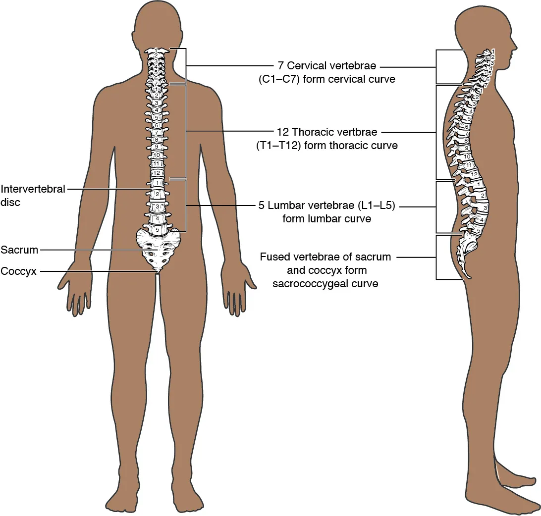
The adult column has four curves along its length, which add to its strength, flexibility and ability to absorb shock1.
Three joints at each level
Between two vertebral bodies sits an intervertebral disc, a thick pad of fibrocartilage (a tough, fibrous kind of cartilage)1. It unites the vertebrae strongly while allowing small movements between them1.
An articular process is a part of a vertebra that sticks up or down to form a joint with the next vertebra. Each vertebra has an upward-facing and a downward-facing articular process on each side, and these join the neighbouring vertebrae1. The joints they form are the facet joints1. These are plane synovial joints, which have nearly flat surfaces and a fluid-filled space, and they allow limited movement1.
A narrative review describes each spinal level below the first two neck vertebrae as a motion segment: one disc and two facet joints2. The spine is then a chain of motion segments, and its total motion is the sum of the motion in each2.
Ligaments (tough bands that hold bones together at a joint) run along the column1:
- The anterior longitudinal ligament runs down the front of the whole column and resists excess backward bending1.
- The posterior longitudinal ligament lies in front of the spinal cord and the elastic ligamentum flavum behind it1. Both support the column in forward bending1.
How each region moves
The direction the articular processes face differs between regions and sets which movements each region allows1. Ranges of motion are greatest in the neck and the lower back1.
- Cervical. The processes are flattened and face mostly up or down, which gives extensive bending in each direction and extensive rotation1.
- Thoracic. The overlapping spinous processes (the bony points at the back of the vertebrae) and the attached rib cage greatly limit bending forward, backward and sideways1.
- Lumbar. The region allows considerable bending forward, backward and sideways, but the way its processes face largely blocks rotation1.
One study had 60 adults without symptoms, aged 20 to 75, perform 15 simulated daily tasks3. The median share of full active lumbar motion used was 11% for bending forward and back, 17% for side bending and 13% for rotation3.
The loads it carries
The discs are the spinal column’s main joints, and they constantly pass on loads from body weight and muscle activity6. A narrative review states that the disc is usually the main load-bearing structure of a motion segment2. In models of healthy segments described in that review, the facets carried up to 33% of the load, more when the spine was arched back2.
One laboratory study used lumbar joints from cadavers (bodies donated for study)7. The facet joints resisted about 16% of the squeezing force in simulated upright standing7. That was after sustained loading had reduced disc height7.
One study placed a pressure sensor in a lower-back disc of a male volunteer, between the fourth and fifth lumbar vertebrae4:
| Position or task | Disc pressure, megapascals |
|---|---|
| Lying face down | 0.14 |
| Sitting unsupported | 0.464 |
| Relaxed standing | 0.54 |
| Standing bent forward | 1.14 |
| Lifting 20 kg, weight held close to the body | 1.14 |
| Lifting 20 kg with flexed knees | 1.74 |
| Lifting 20 kg with a rounded, flexed back | 2.34 |
What keeps it steady
Six fresh cadaver lumbar spines, stripped to bone, discs and ligaments, buckled sideways under an average squeezing load of 88 newtons9. One model, by Panjabi, divides the system that steadies the spine into three parts10. They are the vertebrae, discs and ligaments, the muscles and tendons around the column, and the nerves that direct the muscles10.
Breakdown
Facet joint wear on scans, and pain
Facet joint osteoarthritis involves the whole joint, including bone, cartilage, ligaments, capsule (the sleeve of tough tissue that encloses a joint) and the surrounding muscles2. A narrative review summarised CT findings in community-based adults in the Framingham Heart Study2:
| Age group | Share with moderate or severe lumbar facet osteoarthritis on CT |
|---|---|
| Under 45 | 36%2 |
| 45 to 64 | 67%2 |
| 65 and older | 89%2 |
In CT scans of 188 Framingham participants, facet osteoarthritis at any lumbar level showed no association with low back pain in the previous 12 months5. The narrative review states that studies of this link have given conflicting results2. Several found no relationship between facet osteoarthritis on CT or MRI (a scan that shows soft tissue in detail) and low back or neck pain2. The low back pain and neck pain pages cover those conditions.
How wear in one joint reaches the others
The same review states that the three joints of a motion segment depend on one another2. In most people, it says, degenerative (wear-type) change starts in the disc, and facet joint changes come after2. It adds that disc degeneration and loss of disc height markedly raise the force passing through the facet joints2. They can bear up to 70% of a load along the spine when the disc space is severely narrowed2. The review also states that bony spurs and joint enlargement can narrow the spinal canal, with potential pressure on the spinal nerve roots2.
Discs and the bony arch
In a herniated disc, the disc bulges or ruptures backward, or backward and to one side, and presses on nerve roots inside the spinal canal6. A nerve root is the part of a nerve where it leaves the spinal cord. The disc herniation and sciatica pages cover what studies found.
The pars interarticularis is a small segment of bone between the upper and lower articular processes of a vertebra11. One review describes stress fractures there as the result of repetitive loading, especially in activities needing spinal hyperextension and rotation11. Hyperextension is straightening a joint past its normal limit. Spondylolysis is a crack in the pars interarticularis of a vertebra. A critical review concluded that the kind called isthmic spondylolysis is considered a fatigue fracture of the pars, which the spondylolysis page covers12.
What reviews of lifting found
A 1999 systematic review (a study that searches for all the research on a question in a set way) compared stoop lifting with squat lifting13. Stoop lifting is lifting with the back bent forward and the knees nearly straight. Squat lifting is lifting with the knees bent and the back more upright.
Across biomechanical studies, it found no significant difference in spinal compression between them13. Squat lifting appeared to put lower shear (sliding) forces and bending moments on the spine13. A bending moment is the turning effect of a force that tends to bend a structure. The same review found that compression during lifting reaches magnitudes that can probably cause injury, while shear forces stayed below the injury threshold13.
Another systematic review, with a meta-analysis (pooled results), looked at lumbar flexion (forward bending) during lifting14. It found low-quality evidence that greater flexion was not a risk factor for the onset or persistence of low back pain14.
Curves that deepen or bend sideways
Kyphosis is an excessive backward curve of the thoracic region, and scoliosis is an abnormal sideways curve with twisting of the column1. A narrative review reports that the kyphosis angle begins to increase after age 40, faster in women than men15.
The review defines hyperkyphosis as an angle above 40 degrees15. Among older adults, it puts both the prevalence and the incidence at roughly 20% to 40%15. Prevalence is the share of people in a group who have a condition at a given time. Incidence is how many new cases appear in a group over a set time.
Recovery
Daily height loss, and disc changes after bed rest
In eight young adults, height varied by an average of 19.3 mm over the day16. About 70% of the daytime loss was regained in the first half of the night16. The changing loads on the spine over a day alter the water content and height of the discs17.
In another study, seven healthy men spent 21 days in head-down bed rest18. The changes in disc volume and height had not returned to their starting values 153 days later, although back muscle size had recovered18. A review of the literature states that adult discs have limited potential to heal19.
Bone in people who lift
In the eight power lifters, bone mineral content of the third lumbar vertebra was extremely high8. Bone mineral content is the amount of mineral in a bone. It correlated closely with the weight lifted in training over the last year8.
Exercise studies in adults with hyperkyphosis
A systematic review and meta-analysis included 24 exercise studies in adults aged 45 or older with hyperkyphosis20. Exercise or physiotherapy improved kyphosis outcomes, on moderate-certainty evidence20. Strength and endurance of the back extensors (the muscles that bend the spine backward) improved, on very-low and low-certainty evidence20. The included studies reported no serious adverse events (unwanted effects)20.
Connections
Where it meets the pelvis
The lower articular processes of the fifth lumbar vertebra join the upper articular processes of the sacrum1. The sacroiliac joint is where the sacrum meets the ilium, the wide upper part of the hip bone21. It forms the transition from the spine to the pelvis21. An anatomy reference gives its main roles as providing stability and passing the trunk’s load on to the lower limbs21. The SI joint pain page covers pain in that region.
The muscles that move it
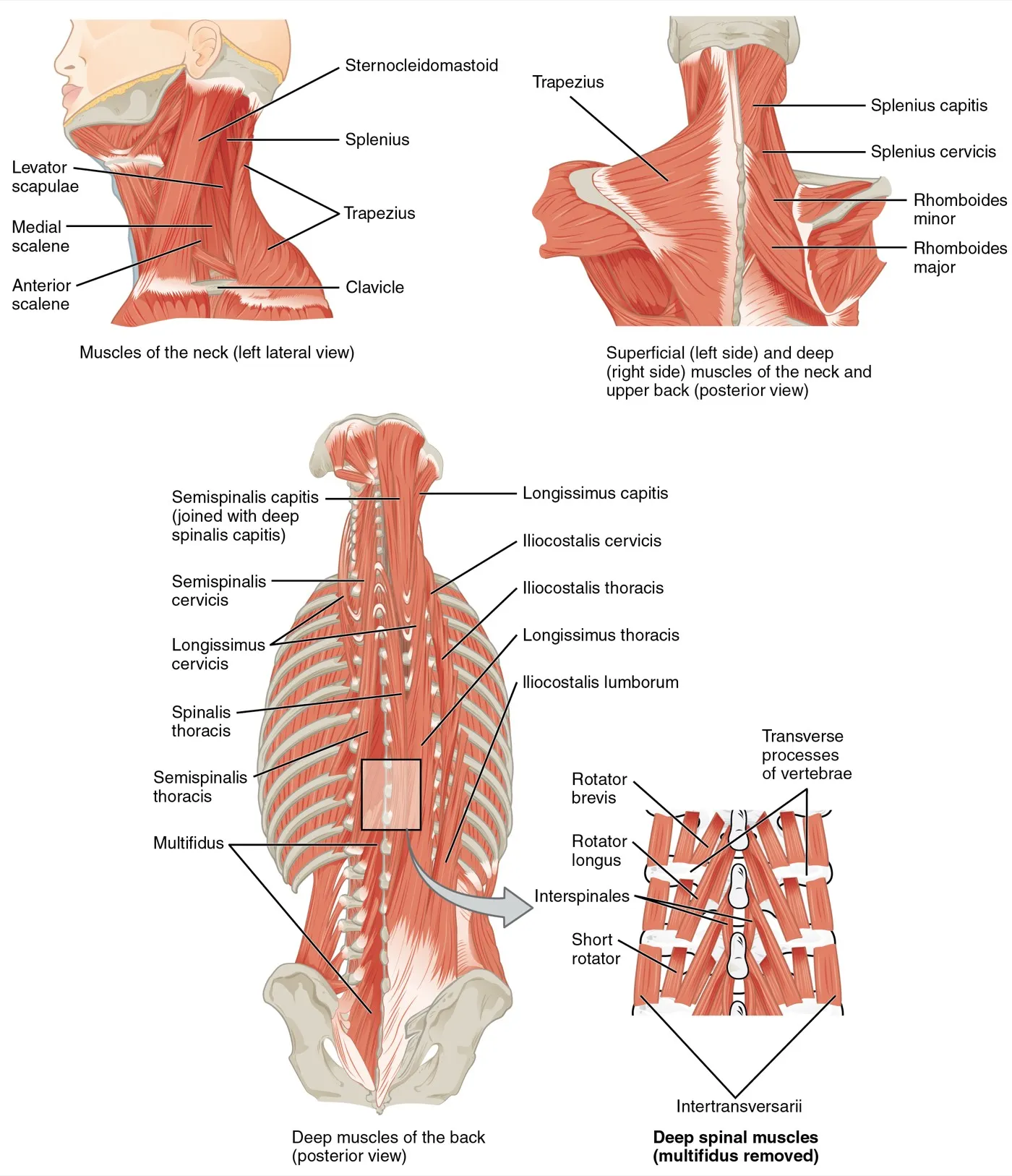
The erector spinae group is the column’s primary extensor and accounts for most of the back’s muscle mass1. The multifidus of the lumbar region helps extend the spine and bend it to the side1. One anatomy textbook lists the quadratus lumborum as the muscle for bending the column to the side1.
Exercises and stretches that involve it
Related exercise pages: good morning, back extension, bird dog, farmer’s carry and back squat22,23,24,25. A study of 15 trained men doing the good morning concluded that the spine did not stay neutral and moved through flexion and extension22. In 13 young men doing four back exercises, a model estimated the highest lower-back joint loads in those with active trunk extension23. Extending one leg with the spine held still reduced spine load23. A 2024 biomechanical review states that the squat needs high trunk-muscle recruitment to steady the spine and torso25.
One published description of child’s pose is a kneeling forward bend with the spine rounded26. In one randomised trial in people with chronic low back pain, the flexion programme included the knee-to-chest stretch27.
Spine is formed by
Spine meets the pelvis at
Spine is extended by
Spine is bent sideways by
Spine is moved in
Spine is loaded by
Spine is stabilised during
Spine is rounded in
Spine is related to
Sources
- Betts JG, Young KA, Wise JA, Johnson E, Poe B, Kruse DH, Korol O, Johnson JE, Womble M, DeSaix P (2022). Anatomy and Physiology 2e. OpenStax. Link
- Gellhorn AC, Katz JN, Suri P (2013). Osteoarthritis of the spine: the facet joints.. Nature reviews. Rheumatology. doi:10.1038/nrrheum.2012.199 PMID 23147891
- Bible JE, Biswas D, Miller CP, Whang PG, Grauer JN (2010). Normal functional range of motion of the lumbar spine during 15 activities of daily living.. Journal of spinal disorders & techniques. doi:10.1097/BSD.0b013e3181981823 PMID 20065869
- Wilke HJ, Neef P, Caimi M, Hoogland T, Claes LE (1999). New in vivo measurements of pressures in the intervertebral disc in daily life.. Spine. doi:10.1097/00007632-199904150-00005 PMID 10222525
- Kalichman L, Li L, Kim DH, Guermazi A, Berkin V, O'Donnell CJ, Hoffmann U, Cole R, Hunter DJ (2008). Facet joint osteoarthritis and low back pain in the community-based population.. Spine. doi:10.1097/BRS.0b013e318184ef95 PMID 18923337
- Urban JP, Roberts S (2003). Degeneration of the intervertebral disc.. Arthritis research & therapy. PMID 12723977
- Adams MA, Hutton WC (1980). The effect of posture on the role of the apophysial joints in resisting intervertebral compressive forces.. The Journal of bone and joint surgery. British volume. doi:10.1302/0301-620X.62B3.6447702 PMID 6447702
- Granhed H, Jonson R, Hansson T (1987). The loads on the lumbar spine during extreme weight lifting.. Spine. doi:10.1097/00007632-198703000-00010 PMID 3589805
- Crisco JJ, Panjabi MM, Yamamoto I, Oxland TR (1992). Euler stability of the human ligamentous lumbar spine. Part II: Experiment.. Clinical biomechanics (Bristol, Avon). doi:10.1016/0268-0033(92)90004-N PMID 23915613
- Panjabi MM (1992). The stabilizing system of the spine. Part I. Function, dysfunction, adaptation, and enhancement.. Journal of spinal disorders. doi:10.1097/00002517-199212000-00001 PMID 1490034
- Madden V, Ayoub A, Thomas J, Thomas I (2026). Spondylolysis: A Narrative Review of Etiology, Diagnosis, and Management.. International journal of environmental research and public health. doi:10.3390/ijerph23020153 PMID 41752236
- Standaert CJ, Herring SA (2000). Spondylolysis: a critical review.. British journal of sports medicine. doi:10.1136/bjsm.34.6.415 PMID 11131228
- van Dieën JH, Hoozemans MJ, Toussaint HM (1999). Stoop or squat: a review of biomechanical studies on lifting technique.. Clinical biomechanics (Bristol, Avon). doi:10.1016/s0268-0033(99)00031-5 PMID 10545622
- Saraceni N, Kent P, Ng L, Campbell A, Straker L, O'Sullivan P (2020). To Flex or Not to Flex? Is There a Relationship Between Lumbar Spine Flexion During Lifting and Low Back Pain? A Systematic Review With Meta-analysis.. The Journal of orthopaedic and sports physical therapy. doi:10.2519/jospt.2020.9218 PMID 31775556
- Katzman WB, Wanek L, Shepherd JA, Sellmeyer DE (2010). Age-related hyperkyphosis: its causes, consequences, and management.. The Journal of orthopaedic and sports physical therapy. doi:10.2519/jospt.2010.3099 PMID 20511692
- Tyrrell AR, Reilly T, Troup JD (1985). Circadian variation in stature and the effects of spinal loading.. Spine. doi:10.1097/00007632-198503000-00011 PMID 4002039
- Adams MA, Dolan P, Hutton WC, Porter RW (1990). Diurnal changes in spinal mechanics and their clinical significance.. The Journal of bone and joint surgery. British volume. doi:10.1302/0301-620X.72B2.2138156 PMID 2138156
- Belavý DL, Bansmann PM, Böhme G, Frings-Meuthen P, Heer M, Rittweger J, Zange J, Felsenberg D (2011). Changes in intervertebral disc morphology persist 5 mo after 21-day bed rest.. Journal of applied physiology (Bethesda, Md. : 1985). doi:10.1152/japplphysiol.00695.2011 PMID 21799122
- Adams MA, Roughley PJ (2006). What is intervertebral disc degeneration, and what causes it?. Spine. doi:10.1097/01.brs.0000231761.73859.2c PMID 16915105
- Ponzano M, Tibert N, Bansal S, Katzman W, Giangregorio L (2021). Exercise for improving age-related hyperkyphosis: a systematic review and meta-analysis with GRADE assessment.. Archives of osteoporosis. doi:10.1007/s11657-021-00998-3 PMID 34546447
- Wong M, Sinkler MA, Kiel J (2023). Anatomy, Abdomen and Pelvis, Sacroiliac Joint. StatPearls. PMID 29939578
- Vigotsky AD, Harper EN, Ryan DR, Contreras B (2015). Effects of load on good morning kinematics and EMG activity.. PeerJ. doi:10.7717/peerj.708 PMID 25653899
- Callaghan JP, Gunning JL, McGill SM (1998). The relationship between lumbar spine load and muscle activity during extensor exercises.. Physical Therapy. doi:10.1093/ptj/78.1.8 PMID 9442191
- Hindle BR, Lorimer A, Winwood P, Keogh JWL (2019). The Biomechanics and Applications of Strongman Exercises: a Systematic Review.. Sports Medicine - Open. doi:10.1186/s40798-019-0222-z PMID 31820223
- Straub RK, Powers CM (2024). A Biomechanical Review of the Squat Exercise: Implications for Clinical Practice.. International Journal of Sports Physical Therapy. doi:10.26603/001c.94600 PMID 38576836
- Zuhair M, Seitler S, Kaza N, Lim PB, Keene D, Khiani R (2025). Yoga in the emergency room: termination of SVT with the Child's pose.. Oxford medical case reports. doi:10.1093/omcr/omaf049 PMID 40443873
- Park CH, Beom J, Chung CK, Kim CH, Lee MY, Park MW, Kim K, Chung SG (2024). Long-term effects of lumbar flexion versus extension exercises for chronic axial low back pain: a randomized controlled trial.. Scientific reports. doi:10.1038/s41598-024-51769-2 PMID 38302483
Changes
- First published.