Articular cartilage
Also called joint cartilage
The thin, smooth layer that covers the ends of bones in a joint. What it is made of, how it carries load, how it gets damaged, why it heals slowly, and how it responds to use.
- Region
- Knee
- Updated
- September 30, 2026
- Reviewed
- September 30, 2026 · how entries are checked
Function
What it is made of
Articular cartilage is hyaline cartilage, between 2 and 4 mm thick1. Hyaline cartilage, the most common kind in the body, is smooth, strong and flexible2. It covers the ends of bones where they meet in movable joints2.
The tissue is a dense framework, or matrix, made mostly of water, collagen (a strong, rope-like protein) and proteoglycans (large protein-and-sugar molecules)1. Cartilage cells, called chondrocytes, are scattered thinly through it1. Each sits in a small space in the matrix called a lacuna2. The cells make up about 2% of the tissue’s volume1.
Water makes up 65% to 80% of its weight1. Collagen is about 60% of the dry weight, and 90% to 95% of that collagen is one type, type II1. The largest and most common proteoglycan, aggrecan, gives the tissue properties that are key to resisting squeezing1.
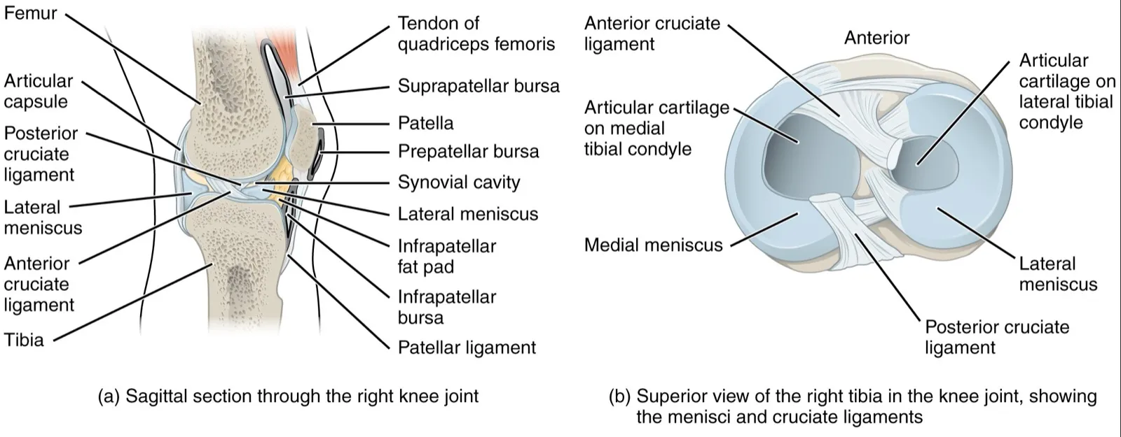
Articular cartilage has no blood vessels, lymph vessels or nerves1. In adults, the subchondral plate (the bone plate just beneath the cartilage) separates it from the blood spaces in that bone1. Instead, it is fed by substances seeping in from the synovial fluid (the slippery fluid inside a joint that oils its moving surfaces)1.
Four layers, from the surface down
The tissue is built in four zones, or layers, from the surface down1:
| Layer | Share of the tissue | Collagen fibres run | Main job |
|---|---|---|---|
| Superficial | About 10% to 20% of thickness1 | Parallel to the surface, tightly packed1 | Shields deeper layers from forces that push surfaces to slide across each other1 |
| Middle | 40% to 60% of volume1 | At a slant1 | First line of resistance to squeezing1 |
| Deep | About 30% of volume1 | Upright, at right angles to the surface1 | Gives the most resistance to squeezing1 |
| Calcified | — | Deep-zone fibres are anchored here1 | Fixes the cartilage to the bone beneath1 |
How it carries load
Its main job is to give a joint a smooth, slippery surface and pass loads across it with low friction1. It passes load to the bone beneath and can bear high, repeated loads with little or no sign of damage or wear1.
When a joint takes load, pressure in the cartilage’s fluid rises and fluid flows out of the matrix1. When the load comes off, the fluid flows back in1. The tissue lets fluid through slowly, which stops it being squeezed out quickly1. This pressurised water carries a significant share of the load and eases the stress on the solid matrix1.
Together, that resistance to flow and the water pressure let the cartilage bear loads often several times body weight1. Under a steady squeeze, the tissue keeps slowly deforming until it settles, a behaviour called creep1. Water moving through and across the cartilage also helps carry nutrients to the cells and oil the joint1.
Why thickness differs
In 11 sets of ankle, knee and hip joints from cadavers (bodies donated for study), knee cartilage was on average the thickest5. Thick cartilage was found in joints whose surfaces fit together less closely, like the knee5.
In 372 adults aged 26 to 61, men had 33% to 42% more knee cartilage volume than women6. Allowing for body and bone size cut the gap to 8% to 18%, and the gap was larger over age 506.
Breakdown
Three kinds of injury
Impact and twisting loads on a joint can injure articular cartilage7. A review sorts these injuries into three classes7:
- damage inside the tissue while the surface stays whole7
- a break that stays within the cartilage7
- a break through both the cartilage and the bone beneath7
Injuries to the anterior cruciate ligament (ACL, a ligament inside the knee) are linked with damage to the joint cartilage8. A meta-analysis (a study that pools the results of several earlier studies) covered 37,423 ACL reconstructions9. Cartilage damage was over three times as common at a repeat reconstruction as at the first one9.
How often damage is found
In 25,124 knee arthroscopies (keyhole surgery inside a joint, using a small camera), cartilage damage was found in 60% of patients10. Of the damage recorded, 67% was local patches in the cartilage, or in cartilage and bone10. Another 29% was osteoarthritis (a long-term joint disease in which joint cartilage wears away)10. In the same study, 70% of the damage came alongside other knee injuries10. The most common were a tear of the inner meniscus (a pad of cartilage in the knee) (37%) or an ACL injury (36%)10.
A systematic review of 11 studies covered 931 athletes3. It put full-thickness patches of damage in athletes’ knees at 36% overall, from 2.4% to 75% between studies3. Its authors concluded this damage is more common in athletes than in the general population3. Of the athletes, 14% had no symptoms when the damage was found3. More than half of the athletes without symptoms had full-thickness damage3.
The two studies recorded where the damage sat3,10:
| Where in the knee | 25,124 arthroscopies | Athletes’ knees |
|---|---|---|
| Kneecap (back of it, or its joint) | 36%10 | 37%3 |
| Condyles (rounded knobs) of the femur (thigh bone) | 34%, inner condyle10 | 35%, both condyles3 |
| Top of the tibia (shinbone) | — | 25%3 |
Osteochondritis dissecans
Osteochondritis dissecans is a disorder of the cartilage and the bone just beneath it11. In a US health system of over 1 million patients, it occurred in the knee at 9.5 per 100,000 in 6- to 19-year-olds11. The rate was 15.4 per 100,000 in males and 3.3 in females11. In that group, 12- to 19-year-olds had 3.3 times the risk of 6- to 11-year-olds11.
In adults aged 20 to 45 in one US health system, the knee rate was 1.21 per 100,000 person-years (a count of people multiplied by the years each was followed)12. Men had 3.6 times the risk of women12.
Wear and ageing
One review notes that osteoarthritis comes with sharp changes in how cartilage is built and broken down1. These changes happen when the cells’ breakdown and building of matrix fall out of balance1. It adds that with age, cartilage holds less water and grows stiffer under squeezing1. The number of cells stays about the same1.
Recovery
Why it heals slowly
Articular cartilage has a limited capacity to heal and repair itself1. Each cell is trapped in its own matrix and cannot move to other parts of the cartilage1. The cells also have a limited ability to divide, which adds to the tissue’s poor healing after injury1.
One review gives the half-life (the time for half of it to be replaced) of its collagen as several decades to up to 400 years1. One study calculated it at 117 years from age-related changes in human tissue, against 15 years for skin collagen13.
How damage changes over time
In 325 adults with an average age of 45, MRI (a scan that shows soft tissue in detail) was done two years apart14. Over about 2.3 years, knee cartilage damage got worse in 33% and got better in 37%14. Worsening was linked with female sex, older age, higher body mass index (a measure of weight for height) and bony spurs at the start14.
What surgery studies found
A 2009 systematic review of 28 studies covered 3,122 patients who had microfracture (a cartilage repair technique)15. Knee function improved in the first 24 months, but reports on whether that improvement lasted were conflicting15. The repair tissue held little hyaline cartilage and varied in amount, and function might decline15. At the time, there was too little data on long-term results15.
Squashing and bouncing back
A systematic review and meta-analysis of 40 studies covered 653 adults16. In healthy adults, knee cartilage got thinner or smaller on MRI right after each loading activity studied, though the evidence was of very low certainty16. Kneecap cartilage fell by 5.0% after body-weight knee bends16. The review suggested knee cartilage regains its shape within 30 minutes of walking and 90 minutes of 100 knee bends16.
In an MRI study of 12 volunteers, kneecap cartilage volume fell after each activity17:
| Activity | Fall in kneecap cartilage volume, % |
|---|---|
| Knee bends | 5.917 |
| Running | 5.017 |
| Squatting | 4.717 |
| Cycling | 4.517 |
| Normal walking | 2.817 |
Does training make it thicker?
One review notes that joint motion and load matter for keeping cartilage normal, and joint inactivity has been shown to break it down1. In one study, knee cartilage thickness did not differ significantly between 18 triathletes training over 10 hours a week and 18 people never regularly active4. The authors suggested that joint size can be shaped during growth, but that cartilage thickness does not adapt to load, unlike muscle and bone4.
One systematic review looked at randomised controlled trials (studies that assign people to groups by chance, then compare the groups) of exercise18. In people with knee osteoarthritis, six study comparisons reported no effect of exercise on cartilage thickness, volume or damage on MRI18. Kneecap cartilage squashing after knee bends did not differ significantly between 14 athletes (seven weight lifters and seven sprinters) and 14 untrained volunteers17.
In 74 children aged 9 to 18, measured about 1.6 years apart, knee cartilage volume rose at all sites19. Boys gained cartilage faster than girls19. Those above the median (middle value) for sport intensity gained about twice as much at the tibia as those below, though not at the kneecap19.
What happens without load
In 20 people with ankle fractures who bore partial weight for 7 weeks, knee cartilage thinned20. The loss ranged from 2.9% at the kneecap to 6.6% at the inner tibia20. The other knee showed no significant change20. In people with a complete spinal cord injury, knee cartilage was thinner than in healthy volunteers and kept thinning21. By 12 months after injury, it was 21% to 24% thinner at the kneecap and inner tibia, and 23% to 25% thinner by 24 months21.
Connections
The joints and bones it covers
In the knee, each bone surface in the joint has a thin coat of hyaline cartilage that lets the bones move smoothly2. The hip and knee, at each end of the femur, are covered by cartilage that cuts friction22. The back of the patella (kneecap) was a common damage site in the arthroscopy study10. In the athletes’ review, the kneecap joint and the top of the tibia were3. In 10 healthy adults, single-leg hopping thinned ankle cartilage on the tibia and talus (the ankle bone under the shinbone) by about 3% and 2%23.
The structures injured with it
The two menisci are C-shaped pads of fibrocartilage (a tough, fibrous kind of cartilage) between the femur and tibia2. In knee arthroscopies, cartilage damage often came with a meniscus tear or ACL injury, the subjects of the meniscus tear and ACL tear pages10. In one review of athletes’ knees, 37% of cartilage damage was in the kneecap joint3. Patellofemoral pain (pain around or behind the kneecap) has its own page.
Exercises that load it
In 20 healthy people, the modelled peak force at the kneecap joint in a single-leg squat was 6.9 times body weight24. In 16 experienced men doing drop jumps, force on the inner half of the knee rose with drop height25. Related exercise pages: back squat, single-leg squat, drop landing.
Articular cartilage lines
Articular cartilage covers the ends of
Articular cartilage covers the top of
Articular cartilage covers the back of
Articular cartilage works with
Articular cartilage is often damaged along with
Articular cartilage lines the joint involved in
Articular cartilage is loaded by
Sources
- Sophia Fox AJ, Bedi A, Rodeo SA (2009). The basic science of articular cartilage: structure, composition, and function.. Sports health. doi:10.1177/1941738109350438 PMID 23015907
- Betts JG, Young KA, Wise JA, Johnson E, Poe B, Kruse DH, Korol O, Johnson JE, Womble M, DeSaix P (2022). Anatomy and Physiology 2e. OpenStax. Link
- Flanigan DC, Harris JD, Trinh TQ, Siston RA, Brophy RH (2010). Prevalence of chondral defects in athletes' knees. Medicine and science in sports and exercise. doi:10.1249/MSS.0b013e3181d9eea0 PMID 20216470
- Eckstein F, Faber S, Mühlbauer R, Hohe J, Englmeier KH, Reiser M, Putz R (2002). Functional adaptation of human joints to mechanical stimuli.. Osteoarthritis and cartilage. doi:10.1053/joca.2001.0480 PMID 11795982
- Shepherd DE, Seedhom BB (1999). Thickness of human articular cartilage in joints of the lower limb.. Annals of the rheumatic diseases. doi:10.1136/ard.58.1.27 PMID 10343537
- Ding C, Cicuttini F, Scott F, Glisson M, Jones G (2003). Sex differences in knee cartilage volume in adults: role of body and bone size, age and physical activity.. Rheumatology (Oxford, England). doi:10.1093/rheumatology/keg374 PMID 12810930
- Buckwalter JA (1998). Articular cartilage: injuries and potential for healing.. The Journal of orthopaedic and sports physical therapy. doi:10.2519/jospt.1998.28.4.192 PMID 9785255
- Kiapour AM, Murray MM (2014). Basic science of anterior cruciate ligament injury and repair.. Bone & joint research. doi:10.1302/2046-3758.32.2000241 PMID 24497504
- Vega TF, Thamrongskulsiri N, Brinkman JC, Pérez Lloveras GO, Casanova F, Cerezal Á, San Roman AV, Borace AF, Hazime D, Moews LD, Chahla J (2026). Concomitant Intra-articular Injuries in 37,423 Primary and Revision ACL Reconstructions: A Systematic Review and Meta-analysis of Comparative Studies.. The American journal of sports medicine. doi:10.1177/03635465261439947 PMID 42116540
- Widuchowski W, Widuchowski J, Trzaska T (2007). Articular cartilage defects: study of 25,124 knee arthroscopies.. The Knee. doi:10.1016/j.knee.2007.02.001 PMID 17428666
- Kessler JI, Nikizad H, Shea KG, Jacobs JC Jr, Bebchuk JD, Weiss JM (2014). The demographics and epidemiology of osteochondritis dissecans of the knee in children and adolescents.. The American journal of sports medicine. doi:10.1177/0363546513510390 PMID 24272456
- Weiss JM, Shea KG, Jacobs JC Jr, Cannamela PC, Becker I, Portman M, Kessler JI (2018). Incidence of Osteochondritis Dissecans in Adults.. The American journal of sports medicine. doi:10.1177/0363546518764676 PMID 29613834
- Verzijl N, DeGroot J, Thorpe SR, Bank RA, Shaw JN, Lyons TJ, Bijlsma JW, Lafeber FP, Baynes JW, TeKoppele JM (2000). Effect of collagen turnover on the accumulation of advanced glycation end products.. The Journal of biological chemistry. doi:10.1074/jbc.M006700200 PMID 10976109
- Ding C, Cicuttini F, Scott F, Cooley H, Boon C, Jones G (2006). Natural history of knee cartilage defects and factors affecting change.. Archives of internal medicine. doi:10.1001/archinte.166.6.651 PMID 16567605
- Mithoefer K, McAdams T, Williams RJ, Kreuz PC, Mandelbaum BR (2009). Clinical efficacy of the microfracture technique for articular cartilage repair in the knee. The American journal of sports medicine. doi:10.1177/0363546508328414 PMID 19251676
- Coburn SL, Crossley KM, Kemp JL, Warden SJ, West TJ, Bruder AM, Mentiplay BF, Culvenor AG (2023). Immediate and Delayed Effects of Joint Loading Activities on Knee and Hip Cartilage: A Systematic Review and Meta-analysis.. Sports medicine - open. doi:10.1186/s40798-023-00602-7 PMID 37450202
- Eckstein F, Lemberger B, Gratzke C, Hudelmaier M, Glaser C, Englmeier KH, Reiser M (2005). In vivo cartilage deformation after different types of activity and its dependence on physical training status.. Annals of the Rheumatic Diseases. doi:10.1136/ard.2004.022400 PMID 15647438
- Bricca A, Juhl CB, Steultjens M, Wirth W, Roos EM (2019). Impact of exercise on articular cartilage in people at risk of, or with established, knee osteoarthritis: a systematic review of randomised controlled trials.. British journal of sports medicine. doi:10.1136/bjsports-2017-098661 PMID 29934429
- Jones G, Ding C, Glisson M, Hynes K, Ma D, Cicuttini F (2003). Knee articular cartilage development in children: a longitudinal study of the effect of sex, growth, body composition, and physical activity.. Pediatric research. doi:10.1203/01.PDR.0000072781.93856.E6 PMID 12736391
- Hinterwimmer S, Krammer M, Krötz M, Glaser C, Baumgart R, Reiser M, Eckstein F (2004). Cartilage atrophy in the knees of patients after seven weeks of partial load bearing.. Arthritis and rheumatism. doi:10.1002/art.20378 PMID 15334465
- Vanwanseele B, Eckstein F, Knecht H, Stüssi E, Spaepen A (2002). Knee cartilage of spinal cord-injured patients displays progressive thinning in the absence of normal joint loading and movement.. Arthritis and rheumatism. doi:10.1002/art.10462 PMID 12209511
- Chang A, Breeland G, Black AC, Hubbard JB (2023). Anatomy, Bony Pelvis and Lower Limb: Femur.. StatPearls. PMID 30422577
- Cher WL, Utturkar GM, Spritzer CE, Nunley JA, DeFrate LE, Collins AT (2016). An analysis of changes in in vivo cartilage thickness of the healthy ankle following dynamic activity.. Journal of Biomechanics. doi:10.1016/j.jbiomech.2016.05.030 PMID 27289415
- Song K, Scattone Silva R, Hullfish TJ, Silbernagel KG, Baxter JR (2023). Patellofemoral Joint Loading Progression Across 35 Weightbearing Rehabilitation Exercises and Activities of Daily Living.. The American journal of sports medicine. doi:10.1177/03635465231175160 PMID 37272685
- Bi G, Hua L, Sun J, Xu Q, Li G (2024). Impact of different landing heights on the contact force in the medial tibiofemoral compartment and the surrounding muscle force characteristics in drop jumps.. PloS one. doi:10.1371/journal.pone.0307538 PMID 39028745
Changes
- A supporting claim was narrowed to match its source; the page text is unchanged.