Knee joint
Also called knee
The joint where the thigh bone, shin bone and kneecap meet. How it bends and locks, what holds it together, how it gets injured or wears, what studies found about recovery, and what crosses it.
- Region
- Knee
- Muscles
- quadriceps, hamstrings, gastrocnemius, popliteus, sartorius, gracilis
- Updated
- October 15, 2026
- Reviewed
- October 15, 2026 · how entries are checked
Revised October 15, 2026: Corrects a statement that the gastrocnemius is the only lower-leg muscle crossing both the knee and the ankle.
Function
Three joints in one
The knee is the largest joint in the body1. It has three parts, all inside one joint capsule1:
- the patellofemoral joint, between the patella (kneecap) and the end of the femur (thigh bone)1
- the inner (medial) tibiofemoral joint, between the femur and the tibia (shinbone)1
- the outer (lateral) tibiofemoral joint, also between the femur and tibia1
Condyles are rounded knobs at the end of a bone, where it meets another bone. At the two tibiofemoral joints, the rounded condyles of the femur meet the flatter condyles on top of the tibia1. At the patellofemoral joint, the kneecap slides up and down in a groove at the lower end of the femur1. The kneecap forms a joint with the femur only, not with the tibia1.
Each bone surface in the joint has a thin coat of cartilage (a firm, rubbery body tissue, softer than bone)1. It lets the bones move smoothly against each other1. The lining of the capsule makes synovial fluid, which oils the joint1. The fluid also feeds the cartilage, which has no blood vessels of its own1.
How it bends, locks and unlocks
The knee works mainly as a hinge, bending and straightening the leg1. The femur both rolls and glides on the tibia to do this1. Rolling makes the bend, while gliding keeps the rounded femur centred over the flat tibia1. That gives the most bony support for weight in every knee position1.
Near full straightening, the femur turns slightly inward on the tibia1. This happens because the outer condyle is a little smaller and finishes rolling first1. The small turn locks the knee in its most stable, fully straight position1. To bend again, the popliteus, a muscle at the back of the leg, turns the femur slightly outward and unlocks it1. The leg can rotate a little at the knee when it is bent, but not when it is straight1.
What holds it together
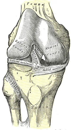
Ligaments (tough bands that hold bones together at a joint) cross inside the knee and run down its sides1:
| Ligament | Where it runs | What it does |
|---|---|---|
| Anterior cruciate (ACL) | Inside the joint, from the tibia up to a femoral condyle1 | Tightens as the knee straightens, resisting overstraightening1 |
| Posterior cruciate (PCL) | Inside the joint, crossing the ACL in an X1 | Stops the femur sliding forward off the tibia when the knee is bent and bearing weight1 |
| Tibial (medial) collateral | Outside the capsule, from the femur to the inner tibia1 | Taut when the knee is straight, steadying it against sideways and twisting movement1 |
| Fibular (lateral) collateral | Outside the capsule, from the femur to the top of the fibula (the thin outer bone of the lower leg)1 | Taut when the knee is straight, steadying it against sideways and twisting movement1 |
At the back of the knee, extra ligaments thicken the capsule and help resist overstraightening1. The tibial collateral ligament is firmly joined on its deep side to the capsule and the inner meniscus1.
The menisci
Two menisci sit between the femur and tibia, one inner and one outer1. Each is a C-shaped pad of fibrocartilage (a tough, fibrous kind of cartilage), thin at its inner edge and thick at its outer edge1. They cushion the space between the bones and help fill the gap between the rounded femur and flatter tibia1. They attach to the tibia but not the femur, and both can shift as the knee moves1. The inner one moves less, because its outer edge is anchored to the capsule and the tibial collateral ligament1.
A review of meniscus research reports that about 70% of the load on the knee’s outer side passes through the meniscus5. On the inner side, the share is about 50%5. The same review reports that removing the inner meniscus cuts the contact area on the femur by 50% to 70%5. It also doubles the contact stress5.
The loads it carries
In one study of 5 people with a knee implant that measured force, average peak forces were as follows2:
| Activity | Peak knee force, times body weight |
|---|---|
| Walking on level ground | 2.62 |
| Climbing stairs | 3.22 |
| Going down stairs | 3.52 |
Breakdown
What video shows about ACL injuries
A systematic review (a study that searches for all the research on a question in a set, checkable way) studied ACL injuries on video6. It counted 1,551 of them: 745 with no contact, 533 with indirect contact and 273 with direct contact6. In the no-contact injuries, the most common action was cutting (sharp changes of direction, 53.8%)6. Next came pressing or tackling an opponent (50.2%), slowing down (38.9%) and landing (30.1%)6. Most of them, 70.7%, happened while moving fast across the ground6.
Who has ACL injuries most
A meta-analysis found that female athletes were more likely than male athletes to injure the ACL7. The authors noted it is unknown whether this holds in every sport or level7. A systematic review and meta-analysis compared injury rates by sport type8. In contact sports, it put the female rate at about 3 times the male rate8.
In collision, limited-contact and no-contact sports, that review found rates were similar between the sexes8. Sports with hard, twisting landings on fixed objects, such as gymnastics, had the highest rates for both sexes in that review8.
Meniscus tears are often silent
In 991 adults aged 50 to 90 from Framingham, Massachusetts, MRI scans looked for meniscus damage4. The share with a torn or worn-away meniscus in the right knee varied by age and sex group4. It ranged from 19% of women aged 50 to 59 to 56% of men aged 70 to 904. In the same sample, 61% of those with a tear had had no knee pain, aching or stiffness in the previous month4.
Osteoarthritis
An anatomy textbook calls osteoarthritis the most common type of arthritis, linked with ageing and wear of the joint cartilage1. The same textbook describes the cartilage slowly thinning, the joint sometimes swelling with extra fluid, and the bone beneath thickening and roughening1. A meta-analysis of 88 population studies estimated knee osteoarthritis at 16.0% of people aged 15 and over worldwide3. In those aged 40 and over, the estimate was 22.9%3.
A systematic review of cohort studies (studies that follow a group over time) looked at people aged 50 and over9. It linked several factors to new knee pain, in a review aimed at knee pain and osteoarthritis9. It reported them as odds ratios, how many times higher the odds were in one group than another9:
| Factor | Odds ratio |
|---|---|
| Previous knee injury | 2.839 |
| Obesity | 2.669 |
| Being overweight | 1.989 |
| Female sex | 1.689 |
A meta-analysis of 53 studies compared injured knees with uninjured ones10. The odds of knee osteoarthritis were about 4 times higher after an ACL injury10. They were about 6 times higher after a meniscus injury, or an ACL and meniscus injury together10.
Recovery
Blood supply and healing
Only the outer rim of each meniscus is well supplied with blood, which a review of meniscus research links to how tears heal5. That rim is about the outer 10% to 30% of the inner meniscus and 10% to 25% of the outer one5.
What trials of ACL tears found
A randomised trial (a study that assigns people to groups by chance) studied 121 young, active adults with a fresh ACL tear11. Both groups had a return-to-training programme11. One group also had early reconstruction surgery (surgery to rebuild the ligament)11. The other group could choose surgery later11. At five years, early surgery gave no better self-reported or X-ray results11.
A secondary analysis of a randomised trial looked at adults treated with a return-to-training programme alone, without surgery, after an ACL tear12. In 16 of 30, MRI showed signs of the ligament healing at two years12. Those 16 reported better results than those whose ligament had not healed12.
A systematic review with meta-analysis took in 18 studies of 33,540 people with ACL injuries, followed for 12.3 years on average13. Across 15 of those studies, X-ray signs of osteoarthritis did not differ significantly between ACL reconstruction and no surgery13. Reconstruction was linked with fewer total knee replacements13.
How cartilage responds to load
A systematic review of 9 trials concluded that knee-loading exercise seems not to harm cartilage on MRI14. It studied people with, or at higher risk of, knee osteoarthritis, and the evidence quality was low14. Among that review’s trials in people with knee osteoarthritis, six study comparisons found no effect of exercise on cartilage thickness, volume or defects on MRI14.
Connections
The bones that form it
The two condyles at the lower end of the femur meet the tibia to form the knee1. The top of the tibia widens into two condyles with flat tops that meet the femur1.
The muscles and tendons that cross it
The quadriceps, the four muscles on the front of the thigh, straightens the knee, working through the patella and patellar ligament (the patellar tendon)1. It also acts like a “dynamic ligament” that supports and steadies the knee1. Its four muscles share the quadriceps tendon, which fixes into the kneecap and carries on below it as the patellar tendon1. The kneecap protects the quadriceps tendon from rubbing against the end of the femur15.
The hamstrings, three long muscles at the back of the thigh, bend the knee, so they and the quadriceps oppose each other there1. Other muscles also cross it1,16,17:
- the gracilis, a long thin muscle of the inner thigh, which bends the knee1
- the sartorius, which runs from the front of the pelvis to the inner upper tibia and bends the knee1
- the gastrocnemius, the large calf muscle nearest the skin, which crosses both the knee and the ankle16,17
- the popliteus at the back of the leg, which unlocks the straight knee1
Exercises that load it
A 2001 review of the back squat reports that the cruciate ligaments hold back sliding forces at the knee during the lift18. In 24 adults doing the Copenhagen adductor plank with the support below the knee, the exercise produced turning forces at the knee19. A narrative review found few studies linking the deadlift to specific injuries, and knee meniscus injuries were among those reported20.
Knee joint is formed by
Knee joint is straightened by
Knee joint is bent by
Knee joint is crossed by
Knee joint is loaded by
Sources
- Betts JG, Young KA, Wise JA, Johnson E, Poe B, Kruse DH, Korol O, Johnson JE, Womble M, DeSaix P (2022). Anatomy and Physiology 2e. OpenStax. Link
- Kutzner I, Heinlein B, Graichen F, Bender A, Rohlmann A, Halder A, et al (2010). Loading of the knee joint during activities of daily living measured in vivo in five subjects.. Journal of biomechanics. doi:10.1016/j.jbiomech.2010.03.046 PMID 20537336
- Cui A, Li H, Wang D, Zhong J, Chen Y, Lu H (2020). Global, regional prevalence, incidence and risk factors of knee osteoarthritis in population-based studies.. EClinicalMedicine. doi:10.1016/j.eclinm.2020.100587 PMID 34505846
- Englund M, Guermazi A, Gale D, Hunter DJ, Aliabadi P, Clancy M, et al (2008). Incidental meniscal findings on knee MRI in middle-aged and elderly persons.. The New England journal of medicine. doi:10.1056/NEJMoa0800777 PMID 18784100
- Fox AJ, Bedi A, Rodeo SA (2012). The Basic Science of Human Knee Menisci. Sports health. doi:10.1177/1941738111429419 PMID 23016106
- Mai P, Willwacher S, Rahlf L, Hoenig T, Braun L, von Diecken C, et al (2026). Situations Leading to Anterior Cruciate Ligament Injury in Sports: A Systematic Review with Meta-analysis.. Sports medicine (Auckland, N.Z.). doi:10.1007/s40279-026-02436-1 PMID 42000943
- Norman D, Terrian L, Novosel J, Guzman A, Montalvo AM, Myer GD, et al (2026). Anterior Cruciate Ligament Injury Incidence Across Sex, Sport, and Competition Level: A Systematic Review and Meta-Analysis.. Journal of athletic training. doi:10.4085/1062-6050-0559.24 PMID 41816313
- Montalvo AM, Schneider DK, Webster KE, Yut L, Galloway MT, Heidt RS Jr, et al (2019). Anterior Cruciate Ligament Injury Risk in Sport: A Systematic Review and Meta-Analysis of Injury Incidence by Sex and Sport Classification.. Journal of athletic training. doi:10.4085/1062-6050-407-16 PMID 31009238
- Silverwood V, Blagojevic-Bucknall M, Jinks C, Jordan JL, Protheroe J, Jordan KP (2015). Current evidence on risk factors for knee osteoarthritis in older adults: a systematic review and meta-analysis.. Osteoarthritis and cartilage. doi:10.1016/j.joca.2014.11.019 PMID 25447976
- Poulsen E, Goncalves GH, Bricca A, Roos EM, Thorlund JB, Juhl CB (2019). Knee osteoarthritis risk is increased 4-6 fold after knee injury - a systematic review and meta-analysis.. British journal of sports medicine. doi:10.1136/bjsports-2018-100022 PMID 31072840
- Frobell RB, Roos HP, Roos EM, Roemer FW, Ranstam J, Lohmander LS (2013). Treatment for acute anterior cruciate ligament tear: five year outcome of randomised trial.. BMJ (Clinical research ed.). doi:10.1136/bmj.f232 PMID 23349407
- Filbay SR, Roemer FW, Lohmander LS, Turkiewicz A, Roos EM, Frobell R, et al (2023). Evidence of ACL healing on MRI following ACL rupture treated with rehabilitation alone may be associated with better patient-reported outcomes: a secondary analysis from the KANON trial.. British journal of sports medicine. doi:10.1136/bjsports-2022-105473 PMID 36328403
- Blackman B, Juric M, Ybema S, Bouchard MD, Macciacchera M, Matache BA, et al (2026). Reconstruction After an Anterior Cruciate Ligament Injury Does Not Reduce the Osteoarthritis Risk But Lowers the Total Knee Arthroplasty Rate: A Systematic Review and Meta-analysis.. Orthopaedic journal of sports medicine. doi:10.1177/23259671261451241 PMID 42539917
- Bricca A, Juhl CB, Steultjens M, Wirth W, Roos EM (2019). Impact of exercise on articular cartilage in people at risk of, or with established, knee osteoarthritis: a systematic review of randomised controlled trials.. British journal of sports medicine. doi:10.1136/bjsports-2017-098661 PMID 29934429
- Cox CF, Sinkler MA, Black AC, Launico MV, Hubbard JB (2023). Anatomy, Bony Pelvis and Lower Limb, Knee Patella. StatPearls. PMID 30137819
- Pedret C, Palermi S, Mechó S, Frutos EJ, Rovira J, Vicente O, Balius R (2026). Calf Injuries: An Anatomy-Based Approach to Diagnosis, Management and Return to Sport.. Current reviews in musculoskeletal medicine. doi:10.1007/s12178-026-10059-2 PMID 42799963
- Bryan Dixon J (2009). Gastrocnemius vs. soleus strain: how to differentiate and deal with calf muscle injuries.. Current reviews in musculoskeletal medicine. doi:10.1007/s12178-009-9045-8 PMID 19468870
- Escamilla RF (2001). Knee biomechanics of the dynamic squat exercise.. Medicine and Science in Sports and Exercise. doi:10.1097/00005768-200101000-00020 PMID 11194098
- Dæhlin TE, Dmytriieva D, Rutherford E, Chiu LZF (2026). Three-dimensional hip and knee loading during the Copenhagen adductor exercise.. Journal of biomechanics. doi:10.1016/j.jbiomech.2025.113116 PMID 41370869
- Bengtsson V, Berglund L, Aasa U (2018). Narrative review of injuries in powerlifting with special reference to their association to the squat, bench press and deadlift.. BMJ Open Sport & Exercise Medicine. doi:10.1136/bmjsem-2018-000382 PMID 30057777
Changes
- Revised. Corrects a statement that the gastrocnemius is the only lower-leg muscle crossing both the knee and the ankle.
- A supporting claim was narrowed to match its source; the page text is unchanged.
- First published.