Femur
Also called thigh bone
The thigh bone, the longest and strongest bone in the body. How it forms the hip and knee, how much load it carries, how it breaks, how it heals and responds to load, and what attaches to it.
- Region
- Thigh
- Muscles
- gluteus maximus, gluteus medius, gluteus minimus, hip adductors, hamstrings, gastrocnemius, Hip flexors, Deep hip rotators
- Updated
- September 30, 2026
- Reviewed
- September 30, 2026 · how entries are checked
Function
The body’s longest bone
The femur, also called the thigh bone, is the single bone in the thigh1. It is the longest and strongest bone in the body1,2. It makes up about a quarter of a person’s height1,2.
The hips are wider than the knees, so the femur runs at a slant down the thigh1. This slant is the Q-angle (the angle between the line of the thigh and the line of the shin, seen from the front)1. It is normally 10 to 15 degrees, and it is typically larger in females1.
The top end: head, neck and trochanters
The rounded head at the top of the femur fits into the acetabulum (hip socket) of the hip bone1. Together they form the hip joint1.
Below the head sits the femoral neck, a narrowed region1. In the average adult, the neck meets the shaft at about 128 degrees2. This angle gets smaller with age2.
The greater trochanter (the bony bump at the outer hip) anchors several muscles that act across the hip1. Because it sticks out from the bone, it gives those muscles extra leverage1. The lesser trochanter is a small bump on the inner side, just below the neck1. A single powerful muscle attaches there1.
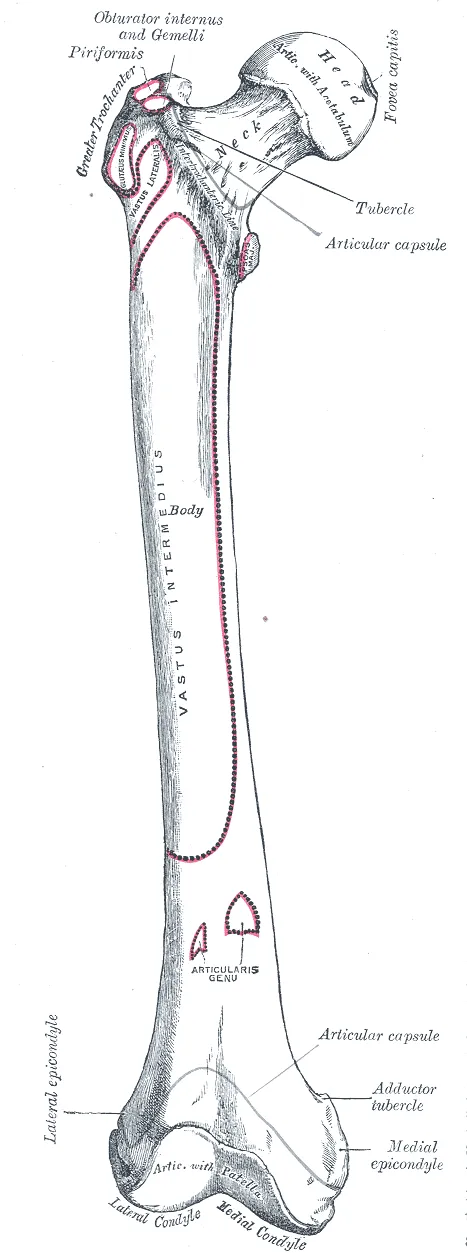
Down the shaft to the knee
The linea aspera is a rough ridge running down the back of the shaft1. Many hip and thigh muscles attach along it in long, narrow strips1. Another feature is the adductor tubercle2. The back half of the adductor magnus (a large muscle on the inner side of the thigh) attaches there2.
At the lower end are two condyles (rounded knobs where a bone meets another bone at a joint), one inner and one outer1. They meet the tibia to form the knee joint1. Beside them sit the epicondyles (bumps on the sides of the condyles)1. They anchor muscles and the ligaments (tough bands that hold bones together at a joint) that support the knee1.
At the front of the lower end is a smooth groove, the patellar surface1. The patella (kneecap) sits in this groove and glides on it1. This keeps the quadriceps tendon from rubbing on the lower femur1.
The hip and knee sit at the two ends of the femur2. Both are synovial joints (joints with a fluid-filled space between the bones, which lets them move freely)2. Their surfaces are covered with cartilage (a firm, rubbery tissue, softer than bone), which reduces friction2.
How much load passes through it
A review describes the upper femur as built to resist force in the direction of weight-bearing6. The same review describes it as less able to resist force from a sideways fall6. One study measured the force at the hip in four patients with instrumented hip implants (artificial hips that record the force through them)3.
| Activity (four patients with instrumented hip implants) | Hip contact force (% of body weight) |
|---|---|
| Walking at about 4 km/h (average)3 | 2383 |
| Going upstairs3 | 2513 |
| Going downstairs3 | 2603 |
Breakdown
Hip fractures in older adults
A systematic review (a study that searches the research on a question in a checkable way) covered hip fracture rates in 63 countries7. It found more than a 10-fold difference in risk between countries7. A worldwide systematic review compared age-standardised rates (adjusted so groups with different age mixes can be compared fairly)7. Within countries, the rate in men was about half the rate in women7.
In one study of 206 consecutive hip fracture patients, 98% of the fractures came from a fall4. In that study, 76% said they fell straight to the side4. A fresh bruise over the greater trochanter was found in 56% of them, against 6% of fallers without a fracture4. A review describes sideways falls as raising neck fracture risk by loading the upper outer part of the neck6. It describes that part as relatively weak6.
After a hip fracture, studies report these outcomes8,9:
- A meta-analysis of cohorts aged 50 and over found a 5- to 8-fold higher risk of death from any cause in the first 3 months8.
- The same meta-analysis found that extra yearly deaths persisted over time in older women and men, and were higher in men at any given age8.
- A review of cohort studies (which follow people over time) found that 40 to 60% of people regained their pre-fracture mobility9.
- That review found that most recovery of walking and daily activities took place within 6 months9.
- It also reports that in Western nations, 10 to 20% of patients move into institutional care (long-term living in a care home) after the fracture9.
Stress fractures of the femoral neck
An international consensus on bone stress injuries in athletes (a statement agreed by a panel of experts) describes them as overuse injuries to bone10. These include stress reactions (early bone stress injuries, before a crack forms) and stress fractures (small cracks from repeated load)10. A review describes femoral neck stress fractures as relatively rare11. It says they most often affect military recruits and endurance athletes11.
One systematic review pooled case reports (reports on single patients)12. It found a sizable share in people with low activity and abnormal bone metabolism (the building and breakdown of bone)12.
A review reports that a stress fracture here that is left untreated can progress to a complete, displaced break (bone ends moved out of line)11. Such a break can be associated with nonunion (a break that fails to join) and long-term disability11. It can also be associated with osteonecrosis (death of bone tissue) in the femoral head (the ball at the top of the thigh bone)11.
A systematic review of bone stress injuries in athletes placed the femoral neck among the sites with the highest complication rates13. In the review of case reports, 71% of the 53 patients followed for at least a year returned to full pre-injury activity12.
Breaks of the shaft
A population-based prospective study (a study that follows people forward in time) covered a region of 850,000 people14. It found femoral shaft fractures in 21 per 100,000 people a year14.
Recovery
How a broken bone mends
A review of fracture healing describes bone repair in general, not the femur alone15. It describes repair as rebuilding bone, repeating many of the steps by which the skeleton first forms before birth15. It reports that repair usually restores a bone’s structure and function15. It also reports that about 10% of fractures do not heal normally15.
How the femur responds to load
A review of bone mechanics reports that bone adapts to the loads it usually carries16. It adjusts its structure to the strain (how far something stretches as a share of its resting length) on it16. The review names the strain’s size, rate, frequency and spread16.
One modelling study (a study that estimates forces inside the body with a model rather than measuring them directly) looked at 15 activities17. In it, neck strain closely tracked the force at the hip joint17. Where the peak load fell depended on the activity17.
The study estimated that 8 of the 15 weight-bearing and resistance activities were likely to trigger bone formation in the neck17. Three activities produced the largest estimated increase in strain energy17. They were isokinetic hip extension (on a machine that holds the limb to a fixed speed), the one-legged long jump and isokinetic knee flexion17.
A review reports that the neck adapts to activity mainly in its lower inner, weight-bearing region6. It found little or no adaptation in the upper outer region6.
Studies in people report these findings5,18:
- In a study of 24-year-old men, soccer players had 19.5% higher femoral neck bone density than non-athletic men of the same age18.
- In the same study, men who did nonspecific resistance exercise showed no higher bone density, and no better bone geometry (shape), than non-athletic men18.
- A meta-analysis of six studies in premenopausal women (before menopause) found that jumping exercise raised bone density at the femoral neck and trochanter19.
- A meta-analysis of trials lasting at least 6 months in postmenopausal women found a significant, low-to-moderate effect of dynamic resistance exercise on femoral neck density5.
- A meta-analysis of 10 randomised trials (studies that assign people to groups by chance) tested supervised exercise lasting 6 months or more20. It found gains in lower-limb bone strength at loaded sites for children, but not for adults20.
One study followed 14 crew members (13 men and 1 woman, aged 40 to 55) on 4- to 6-month space station missions21. Their hip bone density fell by 1.4 to 1.5% a month21.
Connections
Most muscles that attach to the femur and move it start on the pelvic girdle1. At the hip, the femoral head fits into the acetabulum of the pelvis1. Muscles attached to the femur include these:
- The gluteus maximus: its deeper fibres attach to the femur22.
- The gluteus medius: its tendon, one of the gluteal tendons, attaches to the greater trochanter at two sites23,24.
- The gluteus minimus: its tendon attaches to the front of the greater trochanter23,25.
- The hip adductors: they generally start on the pelvis, attach to the femur and adduct it (move it toward the body’s midline) at the hip1,26.
- The hamstrings: the short head of the biceps femoris (a hamstring muscle on the outer back of the thigh) starts from the linea aspera27. It crosses only the knee27.
- The gastrocnemius: it arises from the two condyles of the femur28.
The knee is the body’s largest joint, and all three of its joints involve the femur1. One lies between the kneecap and the femur, and two lie between the condyles of the femur and the tibia1. The patella forms a joint only with the femur, not with the tibia1.
Femur forms the hip joint with
Femur forms the knee joint with
Femur forms a joint with
Femur forms part of
Femur is attached to by
Sources
- Betts JG, Young KA, Wise JA, Johnson E, Poe B, Kruse DH, Korol O, Johnson JE, Womble M, DeSaix P (2022). Anatomy and Physiology 2e. OpenStax. Link
- Chang A, Breeland G, Black AC, Hubbard JB (2023). Anatomy, Bony Pelvis and Lower Limb: Femur.. StatPearls. PMID 30422577
- Bergmann G, Deuretzbacher G, Heller M, Graichen F, Rohlmann A, Strauss J, Duda GN (2001). Hip contact forces and gait patterns from routine activities.. Journal of Biomechanics. doi:10.1016/s0021-9290(01)00040-9 PMID 11410170
- Parkkari J, Kannus P, Palvanen M, Natri A, Vainio J, Aho H, Vuori I, Järvinen M (1999). Majority of hip fractures occur as a result of a fall and impact on the greater trochanter of the femur: a prospective controlled hip fracture study with 206 consecutive patients.. Calcified Tissue International. doi:10.1007/s002239900679 PMID 10441647
- Shojaa M, von Stengel S, Kohl M, Schoene D, Kemmler W (2020). Effects of dynamic resistance exercise on bone mineral density in postmenopausal women: a systematic review and meta-analysis with special emphasis on exercise parameters.. Osteoporosis International. doi:10.1007/s00198-020-05441-w PMID 32399891
- Fuchs RK, Kersh ME, Carballido-Gamio J, Thompson WR, Keyak JH, Warden SJ (2017). Physical Activity for Strengthening Fracture Prone Regions of the Proximal Femur.. Current Osteoporosis Reports. doi:10.1007/s11914-017-0343-6 PMID 28133707
- Kanis JA, Odén A, McCloskey EV, Johansson H, Wahl DA, Cooper C, IOF Working Group on Epidemiology and Quality of Life (2012). A systematic review of hip fracture incidence and probability of fracture worldwide.. Osteoporosis International. doi:10.1007/s00198-012-1964-3 PMID 22419370
- Haentjens P, Magaziner J, Colón-Emeric CS, Vanderschueren D, Milisen K, Velkeniers B, Boonen S (2010). Meta-analysis: excess mortality after hip fracture among older women and men.. Annals of Internal Medicine. doi:10.7326/0003-4819-152-6-201003160-00008 PMID 20231569
- Dyer SM, Crotty M, Fairhall N, Magaziner J, Beaupre LA, Cameron ID, Sherrington C, Fragility Fracture Network (FFN) Rehabilitation Research Special Interest Group (2016). A critical review of the long-term disability outcomes following hip fracture.. BMC Geriatrics. doi:10.1186/s12877-016-0332-0 PMID 27590604
- Hoenig T, Hollander K, Popp KL, Fredericson M, Kraus EA, Warden SJ, Tenforde AS, Bone Stress Injury Authorship Group (2025). International Delphi consensus on bone stress injuries in athletes. British journal of sports medicine. doi:10.1136/bjsports-2024-108616 PMID 39638438
- Bernstein EM, Kelsey TJ, Cochran GK, Deafenbaugh BK, Kuhn KM (2022). Femoral Neck Stress Fractures: An Updated Review.. Journal of the American Academy of Orthopaedic Surgeons. doi:10.5435/JAAOS-D-21-00398 PMID 35077440
- Yang K, Sambandam S, Yan MJ, Huo M (2023). Femoral neck stress fracture return to activity and the effect of metabolic dysfunction on recovery: A systematic review.. Journal of Orthopaedics. doi:10.1016/j.jor.2023.07.023 PMID 37545869
- Hoenig T, Eissele J, Strahl A, Popp KL, Stürznickel J, Ackerman KE, Hollander K, Warden SJ, Frosch KH, Tenforde AS, Rolvien T (2023). Return to sport following low-risk and high-risk bone stress injuries: a systematic review and meta-analysis. British journal of sports medicine. doi:10.1136/bjsports-2022-106328 PMID 36720584
- Enninghorst N, McDougall D, Evans JA, Sisak K, Balogh ZJ (2013). Population-based epidemiology of femur shaft fractures.. Journal of Trauma and Acute Care Surgery. doi:10.1097/TA.0b013e31828c3dc9 PMID 23694881
- Einhorn TA, Gerstenfeld LC (2015). Fracture healing: mechanisms and interventions.. Nature reviews. Rheumatology. doi:10.1038/nrrheum.2014.164 PMID 25266456
- Hart NH, Nimphius S, Rantalainen T, Ireland A, Siafarikas A, Newton RU (2017). Mechanical basis of bone strength: influence of bone material, bone structure and muscle action.. Journal of musculoskeletal & neuronal interactions. PMID 28860414
- Martelli S, Kersh ME, Schache AG, Pandy MG (2014). Strain energy in the femoral neck during exercise.. Journal of Biomechanics. doi:10.1016/j.jbiomech.2014.03.036 PMID 24746018
- Nilsson M, Ohlsson C, Mellström D, Lorentzon M (2013). Sport-specific association between exercise loading and the density, geometry, and microstructure of weight-bearing bone in young adult men.. Osteoporosis International. doi:10.1007/s00198-012-2142-3 PMID 23011682
- Zhao R, Zhao M, Zhang L (2014). Efficiency of jumping exercise in improving bone mineral density among premenopausal women: a meta-analysis.. Sports medicine (Auckland, N.Z.). doi:10.1007/s40279-014-0220-8 PMID 24981245
- Nikander R, Sievänen H, Heinonen A, Daly RM, Uusi-Rasi K, Kannus P (2010). Targeted exercise against osteoporosis: A systematic review and meta-analysis for optimising bone strength throughout life.. BMC Medicine. doi:10.1186/1741-7015-8-47 PMID 20663158
- Lang T, LeBlanc A, Evans H, Lu Y, Genant H, Yu A (2004). Cortical and trabecular bone mineral loss from the spine and hip in long-duration spaceflight.. Journal of Bone and Mineral Research. doi:10.1359/JBMR.040307 PMID 15125798
- Krause Neto W, Krause TLV, Gama EF (2025). The impact of resistance training on gluteus maximus hypertrophy: a systematic review and meta-analysis.. Frontiers in Physiology. doi:10.3389/fphys.2025.1542334 PMID 40276368
- Pfirrmann CW, Chung CB, Theumann NH, Trudell DJ, Resnick D (2001). Greater trochanter of the hip: attachment of the abductor mechanism and a complex of three bursae--MR imaging and MR bursography in cadavers and MR imaging in asymptomatic volunteers.. Radiology. doi:10.1148/radiol.2211001634 PMID 11687692
- Robertson WJ, Gardner MJ, Barker JU, Boraiah S, Lorich DG, Kelly BT (2008). Anatomy and dimensions of the gluteus medius tendon insertion.. Arthroscopy. doi:10.1016/j.arthro.2007.11.015 PMID 18237695
- Philippon MJ, Michalski MP, Campbell KJ, Goldsmith MT, Devitt BM, Wijdicks CA, LaPrade RF (2014). Surgically Relevant Bony and Soft Tissue Anatomy of the Proximal Femur.. Orthopaedic Journal of Sports Medicine. doi:10.1177/2325967114535188 PMID 26535335
- Jeno SH, Launico MV, Schindler GS (2023). Anatomy, Bony Pelvis and Lower Limb: Thigh Adductor Magnus Muscle.. StatPearls. PMID 30521263
- Koolmees DS, Klott JD, Poppe TR, Bernholt DL (2025). Distal Hamstring Injuries and Disorders.. Journal of the American Academy of Orthopaedic Surgeons: Global Research & Reviews. doi:10.5435/JAAOSGlobal-D-25-00248 PMID 40972536
- Bordoni B, Varacallo MA (2026). Anatomy, Bony Pelvis and Lower Limb: Gastrocnemius Muscle.. StatPearls. PMID 30422541
Changes
- First published.