Gluteus minimus
The smallest and deepest buttock muscle. How its two parts steady the hip, how it shrinks and fills with fat, what happens to its tendon, and how it responds to training.
- Region
- Hip
- Starts on
- the outer surface of the ilium between two gluteal lines, and at the sciatic notch (Pelvis)1,2
- Ends on
- the front (anterior) facet of the greater trochanter of the thigh bone, and the hip joint capsule (Gluteal tendons)1,2,3,7
- Tendons
- Gluteal tendons
- Updated
- September 30, 2026
- Reviewed
- September 30, 2026 · how entries are checked
Function
Where it sits and attaches
The gluteus minimus is the smallest of the three gluteal muscles, the muscles of the buttock1. It lies deepest, under the gluteus medius, which in turn lies under the gluteus maximus1. In 16 cadaver hips (bodies donated for study), it started on the outer surface of the ilium (the wide upper part of the hip bone)2. Some of it also started at the sciatic notch, on the inside of the pelvis2.
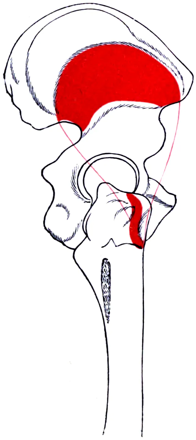
Its tendon (a tough cord of tissue that connects a muscle to a bone) attaches to the front of the greater trochanter3,7. In 14 cadaver half-pelvises, the attachment was crescent-shaped in 11 and bowtie-shaped in 33. Its fibres met and slightly overlapped those of the gluteus medius at one corner of the trochanter3.
On its way to the trochanter, the muscle also joins the capsule of the hip joint2,3. Hips of 17 adults and 3 full-term fetuses were dissected8. In every case, the deep side of the tendon was fixed to the capsule8.
Two parts in one muscle
In 18 adult cadavers, the gluteus minimus was made of two parts, each with its own nerve branch9. Studies of its activity call them the front and back segments10. In recordings from 15 healthy volunteers, the two segments worked at different levels in three of five all-out efforts10. The authors concluded that the two segments work independently10.
What it does at the hip
The gluteus minimus works mainly to steady the hip and to move the thigh out to the side12. With the gluteus medius, it is one of the hip abductors, the muscles that move the thigh out to the side, important for normal walking13. In one study of cadavers and a plastic bone model, its action changed with the thigh bone’s position and with which part was working2. It could bend the hip, lift the thigh sideways, or turn it in or out2.
Three dissection studies propose roles tied to the hip joint2,8,9:
- Steadying the femoral head (the ball of the hip joint) by tightening the capsule and pressing on the ball2.
- Pulling the capsule clear during movement so it is not pinched in the joint, as suggested from dissections8.
- From fibre directions in 18 cadavers, its front part appears suited to starting the sideways lift, and its back part to steadying the ball9.
A 1989 study described it as a main steadier of the hip in the middle and late part of each step14. In a study of 38 people after hip replacement, the authors concluded its main job is keeping the ball centred during walking15. They ranked that above lifting the thigh sideways and steadying the pelvis15.
How it works in walking
In 15 healthy volunteers walking, the back segment was more active in the first fifth of each stride10. The front segment peaked later, while the foot was still on the ground10. The authors suggested each segment may steady the hip at a different point in the step10.
Stance is the part of each step when the foot is on the ground. In 40 people, half with hip osteoarthritis and half matched controls, the front segment instead peaked early in stance16. That pattern differed from earlier reports in young adults16. A computer model of one healthy man walking estimated its force at peak loading at about 158 newtons (a unit of force)17. For the gluteus medius, it was about 897 newtons17.
Its nerve and blood supply
It shares its nerve and blood supply with the gluteus medius: the superior gluteal nerve and artery12. That nerve also carries feeling from the hip joint capsule, especially its upper part18. In 16 cadaver hips, the part starting at the sciatic notch shielded the nerve and artery2. A meta-analysis pools the results of earlier studies. One that pooled 41 anatomy studies found that the nerve’s branch to this muscle runs closest to the greater trochanter18.
Breakdown
Fat in the front part
MRI is a detailed scan of soft tissue. MRI scans of 83 patients over 65 were studied19. Fatty infiltration (fat building up inside a muscle) was uneven, mostly in the front part19. It began earlier than in the gluteus medius19. In 38 hips from 20 older cadavers, the fat was confined to deep within the front part20.
The cadaver study’s authors linked that fat to disuse and to faulty nerve loss and regrowth20. In another study, all 12 healthy controls had some fat in the front segment4. Its authors judged a little fat there a normal part of ageing4.
Size and fat in hip osteoarthritis
Studies of people with hip osteoarthritis do not all agree4,21:
- In 39 people with end-stage disease, the gluteus minimus was smaller than in 12 matched controls, while the gluteus medius was not4.
- The same people had more fat in its front segment than the controls4.
- In people with one affected hip, the muscle was smaller on that side, more so with worse disease5. It also held more fat on that side5.
- In 20 people with one affected hip, a difference between legs showed up with mild as well as worse symptoms22.
- In a small MRI study of 12 people with hip joint wear and 12 controls, it showed only a trend towards a difference between legs21. The trend seemed linked to the dominant leg21.
One meta-analysis found low to very low quality evidence that the muscle is smaller in people with hip osteoarthritis on one side23. Another, covering the three hip abductors, found no pooled difference in size and called the data limited24. That review, which included 22 studies, did find hip abductor strength lower in osteoarthritis, both against controls and against the unaffected leg24.
In 91 women awaiting hip replacement for one affected hip, front-segment muscle density was studied25. On the affected side, it was the only factor in their model that predicted walking speed25. In 20 people with one affected hip, the back segment fired in a larger burst early in stance than in 20 controls16.
Tendon wear and tears
Tendinopathy is local pain in a tendon that comes with activities that load it. Gluteal tendinopathy is now seen mainly as a tendon problem, without inflammation, where the gluteus medius or minimus attaches26. One review covered 194 hip MRI exams from 178 people with gluteus minimus tendon changes27:
| Finding on MRI | Exams |
|---|---|
| Tendinopathy at the attachment | 7227 |
| Partial tear | 8127 |
| Complete tear | 4027 |
Of the 178 people, 148 were women, and their average age was 6127. In 38 of the 40 complete tears, the tendon had not pulled back27. It stayed joined to the top of the vastus lateralis, the quadriceps muscle on the outer thigh27. Tears were associated with fluid by the trochanter, rough bone there, and fatty wasting of the muscle27.
In MRI scans of 185 people aged 50 or over, gluteus medius and minimus tendon changes went together with muscle wasting28. Both rose with age, from tendinosis (tendon wear seen on a scan) to low-grade and then high-grade tears28.
Changed activity with hip pain
- Greater trochanteric pain syndrome is pain and tenderness at the outer hip. In 8 postmenopausal women with it, the front segment fired in a reversed pattern while walking29. Its average activity was higher than in 10 controls29.
- Ultrasound scans of 35 people with long-standing front-of-hip pain were compared with 35 controls30. In the pain group, the muscle began moving 103 milliseconds earlier during a step-down30.
After hip replacement
In 38 people followed by MRI after hip replacement, nearly half had tendon defects and fatty wasting of the gluteus minimus15. That damage was not related to hip scores, pain, satisfaction or sideways strength15. A CT scan builds detailed pictures of the inside of the body from X-rays. In 20 people followed for two years, the gluteus medius and minimus, measured together, stayed lower in CT density than on the healthy side31.
Recovery
How it shrinks with disuse
In head-down-tilt bed rest, people stay in bed with the head tilted slightly below the feet. In 24 healthy people kept this way for 60 days, all three gluteal muscles shrank32. The gluteus minimus lost the most volume32:
| Muscle | Volume lost by day 59 |
|---|---|
| Gluteus minimus | 10.5%32 |
| Gluteus maximus | 9.2%32 |
| Gluteus medius | 8.0%32 |
Its fat-to-water ratio did not rise, unlike in the other two gluteal muscles32.
How it responds to training
In one randomised trial, 27 people with hip osteoarthritis did 12 weeks of targeted gluteal exercise or a sham (dummy) program6. The gluteus minimus grew in both legs in the exercise group and did not change with the sham6. The other hip muscles measured did not change significantly6. Hip strength improved to a similar degree in both groups6.
In 22 people with mild to moderate hip osteoarthritis in a randomised trial, the back segment’s walking activity shifted after 12 weeks of targeted exercise33. It was lower late in stance and peaked earlier, closer to patterns seen in healthy young people33.
Connections
The other hip abductors
A 1989 study described the tensor fasciae latae (a small muscle at the upper outer thigh), gluteus medius and gluteus minimus as one abductor system14. In 12 cadavers, the gluteus medius measured about 27.6 cm³ by volume, against 14.1 cm³ for the gluteus minimus34. The two share a nerve and an artery12. Their tendon attachments meet on the greater trochanter, where their fibres interlace3.
Exercises that load it
Studies using electromyography (EMG, measuring a muscle’s electrical signals) and models compared exercises35,36. Related exercise pages: side-lying hip abduction, side plank and single-leg Romanian deadlift36,37.
| Exercise | What one study or review found |
|---|---|
| Side-lying hip abduction | One study in a review of healthy people: 38% of maximum in the front segment and 44% in the back37 |
| Side-lying hip abduction | In 12 healthy men, the back part peaked at 55% of maximum with the hip extended and turned in, late in the lift38 |
| Clam | Low activity when two studies of healthy people were pooled: about 5% of maximum in the front segment and 12% in the back37 |
| Resisted abduction with extension | In 10 healthy young adults, high front-segment activity, at 51% of maximum35 |
| Single-leg bridge and single-leg squat | In the same 10 adults, the back segment reached 40% to 49% of maximum35 |
| Standing hip hitch | In 10 healthy postmenopausal women, ranked first or second of seven for every segment39 |
| Body-weight side plank and loaded single-leg Romanian deadlift | In a model of 14 female footballers, top tier for estimated gluteus minimus force36 |
Gluteus minimus works with as a hip abductor
Gluteus minimus lies deep to
Gluteus minimus attaches through
Gluteus minimus arises from
Gluteus minimus attaches to
Gluteus minimus is loaded by
Sources
- Betts JG, Young KA, Wise JA, Johnson E, Poe B, Kruse DH, Korol O, Johnson JE, Womble M, DeSaix P (2022). Anatomy and Physiology 2e. OpenStax. Link
- Beck M, Sledge JB, Gautier E, Dora CF, Ganz R (2000). The anatomy and function of the gluteus minimus muscle.. The Journal of Bone and Joint Surgery. British Volume. doi:10.1302/0301-620x.82b3.10356 PMID 10813169
- Philippon MJ, Michalski MP, Campbell KJ, Goldsmith MT, Devitt BM, Wijdicks CA, LaPrade RF (2014). Surgically Relevant Bony and Soft Tissue Anatomy of the Proximal Femur.. Orthopaedic Journal of Sports Medicine. doi:10.1177/2325967114535188 PMID 26535335
- Kivle K, Lindland ES, Mjaaland KE, Svenningsen S, Nordsletten L (2021). Gluteal atrophy and fatty infiltration in end-stage osteoarthritis of the hip. Bone & Joint Open. doi:10.1302/2633-1462.21.BJO-2020-0179.R1 PMID 33537675
- Zacharias A, Pizzari T, English DJ, Kapakoulakis T, Green RA (2016). Hip abductor muscle volume in hip osteoarthritis and matched controls.. Osteoarthritis and Cartilage. doi:10.1016/j.joca.2016.05.002 PMID 27163446
- Rostron ZPJ, Zacharias A, Semciw AI, Kingsley M, Pizzari T, Woodley SJ, Green R (2022). Effects of a targeted resistance intervention compared to a sham intervention on gluteal muscle hypertrophy, fatty infiltration and strength in people with hip osteoarthritis: analysis of secondary outcomes from a randomised clinical trial.. BMC Musculoskeletal Disorders. doi:10.1186/s12891-022-05907-4 PMID 36309690
- Pfirrmann CW, Chung CB, Theumann NH, Trudell DJ, Resnick D (2001). Greater trochanter of the hip: attachment of the abductor mechanism and a complex of three bursae--MR imaging and MR bursography in cadavers and MR imaging in asymptomatic volunteers.. Radiology. doi:10.1148/radiol.2211001634 PMID 11687692
- Walters J, Solomons M, Davies J (2001). Gluteus minimus: observations on its insertion.. Journal of Anatomy. doi:10.1046/j.1469-7580.2001.19820239.x PMID 11273048
- Al-Hayani A (2009). The functional anatomy of hip abductors.. Folia Morphologica. PMID 19449297
- Semciw AI, Green RA, Murley GS, Pizzari T (2014). Gluteus minimus: an intramuscular EMG investigation of anterior and posterior segments during gait.. Gait & Posture. doi:10.1016/j.gaitpost.2013.11.008 PMID 24314814
- Flack NA, Nicholson HD, Woodley SJ (2012). A review of the anatomy of the hip abductor muscles, gluteus medius, gluteus minimus, and tensor fascia lata.. Clinical Anatomy. doi:10.1002/ca.22004 PMID 22109658
- Greco AJ, Vilella RC (2023). Anatomy, Bony Pelvis and Lower Limb, Gluteus Minimus Muscle.. StatPearls. PMID 32310604
- Shah A, Bordoni B (2023). Anatomy, Bony Pelvis and Lower Limb, Gluteus Medius Muscle.. StatPearls. PMID 32491441
- Gottschalk F, Kourosh S, Leveau B (1989). The functional anatomy of tensor fasciae latae and gluteus medius and minimus.. Journal of Anatomy. PMID 2621137
- Müller M, Tohtz S, Winkler T, Dewey M, Springer I, Perka C (2010). MRI findings of gluteus minimus muscle damage in primary total hip arthroplasty and the influence on clinical outcome.. Archives of Orthopaedic and Trauma Surgery. doi:10.1007/s00402-010-1085-4 PMID 20221834
- Zacharias A, Pizzari T, Semciw AI, English DJ, Kapakoulakis T, Green RA (2019). Comparison of gluteus medius and minimus activity during gait in people with hip osteoarthritis and matched controls.. Scandinavian Journal of Medicine & Science in Sports. doi:10.1111/sms.13379 PMID 30615237
- Wang S, Serbin R, Bonvillain K, Jolissaint J, Habet N, Weeks D (2025). Influence of hip morphology on gluteal muscle biomechanics: a computational modeling study.. BMC Musculoskeletal Disorders. doi:10.1186/s12891-024-08136-z PMID 39789570
- Tempski J, Kotlarek A, Pękala J, Fibiger G, Krager E, Łazarz D, Rosa M, Glądys K, Walocha JA, Pękala P (2025). Variability and clinical anatomy of the superior gluteal nerve-A systematic review and meta-analysis.. Journal of Anatomy. doi:10.1111/joa.14174 PMID 39626199
- Kiyoshige Y, Watanabe E (2015). Fatty degeneration of gluteus minimus muscle as a predictor of falls.. Archives of Gerontology and Geriatrics. doi:10.1016/j.archger.2014.07.013 PMID 25440137
- Takano Y, Kobayashi H, Yuri T, Yoshida S, Naito A, Kiyoshige Y (2018). Fat infiltration in the gluteus minimus muscle in older adults.. Clinical Interventions in Aging. doi:10.2147/CIA.S157402 PMID 29872279
- Grimaldi A, Richardson C, Stanton W, Durbridge G, Donnelly W, Hides J (2009). The association between degenerative hip joint pathology and size of the gluteus medius, gluteus minimus and piriformis muscles.. Manual Therapy. doi:10.1016/j.math.2009.07.004 PMID 19695944
- Zacharias A, Green RA, Semciw A, English DJ, Kapakoulakis T, Pizzari T (2018). Atrophy of hip abductor muscles is related to clinical severity in a hip osteoarthritis population.. Clinical Anatomy. doi:10.1002/ca.23064 PMID 29446121
- Lawrenson PR, Crossley KM, Vicenzino BT, Hodges PW, James G, Croft KJ, King MG, Semciw AI (2019). Muscle size and composition in people with articular hip pathology: a systematic review with meta-analysis.. Osteoarthritis and Cartilage. doi:10.1016/j.joca.2018.10.008 PMID 30389399
- Marshall AR, Noronha M, Zacharias A, Kapakoulakis T, Green R (2016). Structure and function of the abductors in patients with hip osteoarthritis: Systematic review and meta-analysis.. Journal of Back and Musculoskeletal Rehabilitation. doi:10.3233/BMR-150614 PMID 26406195
- Yuri T, Nankaku M, Kawano T, Murao M, Hamada R, Goto K, Kuroda Y, Kawai T, Ikeguchi R, Matsuda S (2023). Evaluating the contribution of fat infiltration in anterior gluteus minimus muscle to walking ability in female with unilateral hip osteoarthritis and candidates for total hip arthroplasty.. Clinical Biomechanics. doi:10.1016/j.clinbiomech.2023.105909 PMID 36878079
- Grimaldi A, Mellor R, Nasser A, Vicenzino B, Hunter DJ (2024). Current and future advances in practice: tendinopathies of the hip.. Rheumatology Advances in Practice. doi:10.1093/rap/rkae022 PMID 38601140
- White LM, Oar DA, Naraghi AM, Griffin A, Safir OA (2021). Gluteus minimus tendon: MR imaging features and patterns of tendon tearing.. Skeletal Radiology. doi:10.1007/s00256-021-03745-4 PMID 33779785
- Chi AS, Long SS, Zoga AC, Read PJ, Deely DM, Parker L, Morrison WB (2015). Prevalence and pattern of gluteus medius and minimus tendon pathology and muscle atrophy in older individuals using MRI.. Skeletal Radiology. doi:10.1007/s00256-015-2220-7 PMID 26260535
- Ganderton C, Pizzari T, Harle T, Cook J, Semciw A (2017). A comparison of gluteus medius, gluteus minimus and tensor facia latae muscle activation during gait in post-menopausal women with and without greater trochanteric pain syndrome.. Journal of Electromyography and Kinesiology. doi:10.1016/j.jelekin.2017.01.004 PMID 28171786
- Dieterich AV, Deshon L, Strauss GR, McKay J, Pickard CM (2016). M-Mode Ultrasound Reveals Earlier Gluteus Minimus Activity in Individuals With Chronic Hip Pain During a Step-down Task.. Journal of Orthopaedic & Sports Physical Therapy. doi:10.2519/jospt.2016.6132 PMID 26954272
- Rasch A, Byström AH, Dalén N, Martinez-Carranza N, Berg HE (2009). Persisting muscle atrophy two years after replacement of the hip.. Journal of Bone and Joint Surgery (British Volume). doi:10.1302/0301-620X.91B5.21477 PMID 19407289
- Tran V, De Martino E, Hides J, Cable G, Elliott JM, Hoggarth M, Zange J, Lindsay K, Debuse D, Winnard A, Beard D, Cook JA, Salomoni SE, Weber T, Scott J, Hodges PW, Caplan N (2021). Gluteal Muscle Atrophy and Increased Intramuscular Lipid Concentration Are Not Mitigated by Daily Artificial Gravity Following 60-Day Head-Down Tilt Bed Rest.. Frontiers in Physiology. doi:10.3389/fphys.2021.745811 PMID 34867450
- Rostron ZPJ, Zacharias A, Semciw AI, Kingsley M, Pizzari T, Woodley SJ, Green R (2023). Comparison between a targeted exercise program and a sham intervention on gluteal muscle activity in people with hip osteoarthritis: Analysis of secondary outcomes from a randomised clinical trial.. Gait & Posture. doi:10.1016/j.gaitpost.2022.11.016 PMID 36469965
- Flack NA, Nicholson HD, Woodley SJ (2014). The anatomy of the hip abductor muscles.. Clinical Anatomy. doi:10.1002/ca.22248 PMID 23625344
- Moore D, Semciw AI, McClelland J, Wajswelner H, Pizzari T (2019). Rehabilitation Exercises for the Gluteus Minimus Muscle Segments: An Electromyography Study.. Journal of Sport Rehabilitation. doi:10.1123/jsr.2017-0262 PMID 29584533
- Collings TJ, Bourne MN, Barrett RS, Meinders E, Gonçalves BAM, Shield AJ, Diamond LE (2023). Gluteal Muscle Forces during Hip-Focused Injury Prevention and Rehabilitation Exercises.. Medicine and Science in Sports and Exercise. doi:10.1249/MSS.0000000000003091 PMID 36918403
- Moore D, Semciw AI, Pizzari T (2020). A SYSTEMATIC REVIEW AND META-ANALYSIS OF COMMON THERAPEUTIC EXERCISES THAT GENERATE HIGHEST MUSCLE ACTIVITY IN THE GLUTEUS MEDIUS AND GLUTEUS MINIMUS SEGMENTS.. International Journal of Sports Physical Therapy. doi:10.26603/ijspt20200856 PMID 33344003
- Uebayashi K, Okubo Y, Akasaka K, Adachi G, Oshikawa T, Matsunaga N, Kaneoka K (2026). Muscle activity of deep hip muscles across joint positions and elevation phases during side-lying hip abduction.. PeerJ. doi:10.7717/peerj.21536 PMID 42433387
- Ganderton C, Pizzari T, Cook J, Semciw A (2017). Gluteus Minimus and Gluteus Medius Muscle Activity During Common Rehabilitation Exercises in Healthy Postmenopausal Women.. Journal of Orthopaedic & Sports Physical Therapy. doi:10.2519/jospt.2017.7229 PMID 29034801
Changes
- First published.