Gluteus medius
The fan-shaped buttock muscle that moves the thigh out to the side and holds the pelvis level in walking. Where it sits, how it weakens or shrinks, how it responds to training, and what it works with.
- Region
- Hip
- Starts on
- the gluteal fossa of the ilium, the gluteal aponeurosis, and the back lower edge of the iliac crest (Pelvis)2,6
- Ends on
- the greater trochanter, the bony bump at the top of the thigh bone, on its outer and upper-back facets (Gluteal tendons)2,6,7,8
- Tendons
- Gluteal tendons
- Updated
- September 30, 2026
- Reviewed
- September 30, 2026 · how entries are checked
Function
Where it sits and attaches
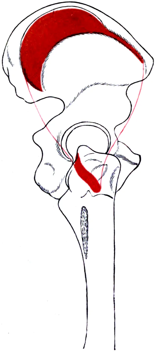
The gluteus medius lies beneath the gluteus maximus, the large muscle of the buttock, and over the gluteus minimus6. In 12 cadavers, the medius started from three sites2. One was the ilium, the wide upper part of the hip bone2. Another was the gluteal aponeurosis, a broad, flat sheet of tendon2. The third was the back lower edge of the iliac crest, the hip bone’s curved top rim2.
At its lower end, its tendon had an outer part and a back part2. Cadaver studies found the tendon attaching to the greater trochanter, the bony bump at the outer hip, at two sites7,8. These sit on the outer facet and the upper back facet, two of the bump’s four small flat areas7,8. The gluteal tendons page covers these attachments in detail.
By volume, the medius was the largest of the three hip abductors2:
| Muscle | Average volume in 12 cadavers (cm³) |
|---|---|
| Gluteus medius | 27.62 |
| Gluteus minimus | 14.12 |
| Tensor fasciae latae (a small muscle at the upper outer thigh that pulls on the iliotibial band) | 1.82 |
Its nerve and blood supply
The superior gluteal nerve serves three muscles: the medius, the minimus and the tensor fasciae latae9. It comes from the L4, L5 and S1 nerve roots, where nerves leave the spinal cord9. The medius and minimus also share one artery, the superior gluteal artery10.
In 20 muscles from 10 cadavers, the nerve left the pelvis through an opening called the greater sciatic foramen1. The pelvis is the ring of bone at the base of the spine. It passed above the piriformis, a small muscle deep in the buttock, then branched into the medius1.
Three parts, or four
Studies divide the muscle in different ways2,11. In 18 cadavers, the medius was made of three distinct parts, each with its own nerve branch11. In 15 healthy people, fine-wire electrodes (very thin wire sensors placed into a muscle) recorded its front, middle and back segments12. The segments’ share of a hardest effort differed in four of five test positions, which pointed to three segments working independently12.
Another dissection of 12 cadavers found three or four main nerve branches, each reaching a different region2. From its build and nerve supply, those authors judged it to have four compartments2. A systematic review, which searches for all the research on a question and sums it up, found little data on fibre direction and nerve branching13. So whether these muscles are divided into compartments was hard to judge13.
What it moves
The gluteus medius is the main abductor of the hip, moving the thigh away from the midline of the body1. Its front fibres also help with internal rotation, turning the thigh inward1. The hip abductors can also help steady the pelvis during walking13.
A study of fibre direction in 18 cadavers suggests the parts may split the work11. The front and middle parts seem to start the lift, which the tensor fasciae latae then completes11. The back part runs parallel to the femoral neck, the angled part of the thigh bone just below the ball of the hip11. It seems suited to holding the ball of the hip in its socket during walking11.
In walking and running
In computer simulations of eight people walking at four speeds, the medius was among the main muscles holding the body up and moving it forward14. Unlike the other main muscles, its share did not generally rise with walking speed14. In 15 healthy people walking, all three segments fired in two bursts during stance, while the foot is on the ground12. The front segment peaked later in both bursts12.
In ten active people without symptoms, every medius and minimus segment was more active in running than in walking15. All the gluteal segments peaked during stance in running15. The authors suggested that the medius segments’ greater stance activity may help steady the pelvis15.
Breakdown
When the muscle is weak
Weakness of the gluteus medius and minimus lets the pelvis drop toward the other side during walking3. This pattern is called Trendelenburg gait3. Damage to the superior gluteal nerve paralyses the medius and produces the same pattern9. The test for this gait was first reported by the German surgeon Friedrich Trendelenburg in 18953. It is used to reveal weakness or other problems in the hip abductors3.
Tendon tears and tendon pain
Tears of the medius or minimus tendons are the more severe end of gluteal tendon problems5. In 176 people having a hip replacement, 34 (20%) had tears from gradual wear in the hip abductors16.
Gluteal tendinopathy, local pain in a tendon that comes with activities that load it, mainly involves the medius and minimus17. It is now understood as a problem where these tendons attach, without inflammation17. In an MRI study of 185 people aged 50 or over, tendon changes and muscle wasting in the medius and minimus went together18. Both increased with age18.
Hip osteoarthritis
Osteoarthritis is a long-term joint disease in which the cartilage lining the joint wears away. A meta-analysis, which pools the results of several studies, covered 22 studies of hip osteoarthritis19. Hip abductor strength was lower in people with the disease than in controls, and lower than in their unaffected leg19.
Studies of the muscle’s size disagree19,20:
| Study | What it found |
|---|---|
| MRI study of people with osteoarthritis in one hip | Medius volume was lower on the affected side than on the other side and than in controls, and the gap tracked disease severity20 |
| Meta-analysis | Low to very low quality evidence that the medius is smaller on the painful side when one hip is affected21 |
| Systematic review and meta-analysis | Pooled data showed no difference in hip abductor size between patients and controls, or between legs; data were limited19 |
| 39 people with end-stage disease and 12 matched healthy controls | The gluteus minimus was smaller, but the medius was not22 |
In 20 people with osteoarthritis in one hip and 20 matched controls, fine-wire recordings found no differences in medius activity during walking23.
Weaker hip abduction alongside other conditions
Reviews and studies have compared hip abduction strength in people with and without several other conditions24,25:
- Low back pain. A review of 24 case-control studies found the medius tended to be weaker, with more trigger points (tender knots), in adults with the pain26. Results for its activity, timing, fatigue and size were unclear26.
- Knee osteoarthritis. A meta-analysis of five studies found moderate-quality evidence of hip abduction strength 7% to 24% lower in people with symptoms24.
- Pain around the kneecap. A review of five studies found lower hip abduction strength on the affected side in females with this pain25. A later review found lower hip strength in adults with the pain27.
- Distance running. In a review of 11 studies in distance runners, weak hip abduction may be linked with iliotibial band syndrome28. The syndrome is named for the long band of tough tissue running down the outer thigh. That link was suggested by several studies done at one point in time28. Its role in four other running injuries was unclear28.
- Falls. In 60 people over 65, the most hip abductor force they could make told apart those with and without past falls with good accuracy29.
For pain around the kneecap, the order of events is in question. A meta-analysis of 18 studies that followed 4,818 people forward found moderate evidence that weak hips did not raise the risk of later pain30. In teenagers, greater hip abduction strength was a risk factor30. The later review found no link between hip strength and developing the pain later27. It suggests the weakness may result from the pain rather than cause it27.
Recovery
Shrinking with disuse
Head-down-tilt bed rest is a research set-up in which people stay in bed with the head tilted slightly below the feet. In 24 healthy people, 59 days of it cut gluteus medius volume by 8.0%4. The fat level inside the muscle rose by 0.5% over the same period4.
Response to training
Two trials found little or no growth in the medius31,32. In untrained college-aged people, 9 weeks of hip thrusts or back squats gave the medius and minimus together little to no growth on MRI31. In a trial, 27 people with hip osteoarthritis were assigned by chance to 12 weeks of targeted gluteal exercise or a sham (dummy) program32. Medius volume did not change significantly, and hip strength improved similarly in both groups32.
A meta-analysis pooled randomised controlled trials in people with knee osteoarthritis33. These studies assign people to groups by chance, then compare the groups. Hip abductor strengthening lowered knee pain and raised function scores compared with control groups33.
Strength after hip replacement
A meta-analysis of 19 studies, covering 875 people, tracked hip abductor strength around hip replacement surgery34:
| Time | Change in hip abductor strength |
|---|---|
| Before surgery, against controls | About 19% lower34 |
| 4 to 6 months after | Rose by about 20%34 |
| 9 to 12 months after | Rose by about 30%34 |
The review concluded that strength may improve slowly over 24 months, possibly without full recovery34. It rated the evidence as low to moderate in quality34.
Connections
The other hip abductors
The medius and gluteus minimus act as a pair to move the thigh out to the side, and both matter for normal walking35. The tensor fasciae latae works with the medius to bend the hip and move the thigh out to the side6. A 1989 study described the three as one abductor system, with the medius steadying the hip early in each step36.
Compared with other leg muscles
In a dissection of 27 muscles from 21 legs, the soleus, a wide, flat calf muscle, was the strongest by one measure of size37. It came ahead of the vastus lateralis, the outer thigh muscle, and the gluteus medius37. Judging by force capacity, the authors rank the soleus as likely the key muscle at the ankle37. They give the vastus lateralis that role at the knee, and the medius at the hip37.
Exercises that load it
Studies of the muscle’s electrical activity show how exercises use it. Activity is given as a share of maximum voluntary contraction, a person’s hardest effort with the muscle38:
| Exercise | What studies found |
|---|---|
| Side-lying hip abduction | Best of 12 exercises for medius activity in 21 active people39. About 40% in the middle segment on average across 8 studies of healthy people in one review38 |
| Side plank | With hip abduction, the highest medius activity of 18 exercises in 24 healthy people, at 103%40 |
| Single-leg Romanian deadlift | In 15 trained men, about 78% against 59% in the two-leg deadlift41 |
| Farmer’s carry | In 16 trained men, about 26% in those with strong hip abductors. It was 42% to 47% where those muscles were weak next to the hamstrings or quadriceps42 |
| Wall sit | Holding a one-leg wall squat, 13% to 35% across the three segments in 15 pain-free people43 |
| Bird dog | In the moving version done standing on one leg, a peak of about 45%44 |
| Back squat | One review reports that stance width does not appear to change medius activity45 |
Related exercise pages: side-lying hip abduction, side plank, single-leg Romanian deadlift, farmer’s carry, wall sit, bird dog and back squat39,40,41,42,43,44,45.
Gluteus medius ends in
Gluteus medius lies beneath
Gluteus medius works with to move the thigh out to the side
Gluteus medius compared by force with
Gluteus medius starts on
Gluteus medius attaches to
Gluteus medius is loaded by
Sources
- Pinheiro RP, Macaya DAM, Itezerote AM, Saleh SO, Hojaij F, Andrade M, et al. (2026). Anatomical Investigation of the Gluteus Medius Muscle Innervation and Its Topographical Correspondence With Myofascial Trigger Points.. BioMed Research International. doi:10.1155/bmri/7544057 PMID 41561444
- Flack NA, Nicholson HD, Woodley SJ (2014). The anatomy of the hip abductor muscles.. Clinical Anatomy. doi:10.1002/ca.22248 PMID 23625344
- Gandbhir VN, Lam JC, Lui F, Rayi A (2024). Trendelenburg Gait.. StatPearls. PMID 31082138
- Tran V, De Martino E, Hides J, Cable G, Elliott JM, Hoggarth M, Zange J, Lindsay K, Debuse D, Winnard A, Beard D, Cook JA, Salomoni SE, Weber T, Scott J, Hodges PW, Caplan N (2021). Gluteal Muscle Atrophy and Increased Intramuscular Lipid Concentration Are Not Mitigated by Daily Artificial Gravity Following 60-Day Head-Down Tilt Bed Rest.. Frontiers in Physiology. doi:10.3389/fphys.2021.745811 PMID 34867450
- Spencer T, Nasser A, Brown NAT, Grimaldi A, Vicenzino B, Smith PN, Fearon A (2026). Low quality evidence supports surgery for gluteal tendon tears, no non-surgical evidence was identified: a systematic review.. BMC Musculoskeletal Disorders. doi:10.1186/s12891-026-09519-0 PMID 41634705
- Betts JG, Young KA, Wise JA, Johnson E, Poe B, Kruse DH, Korol O, Johnson JE, Womble M, DeSaix P (2022). Anatomy and Physiology 2e. OpenStax. Link
- Pfirrmann CW, Chung CB, Theumann NH, Trudell DJ, Resnick D (2001). Greater trochanter of the hip: attachment of the abductor mechanism and a complex of three bursae--MR imaging and MR bursography in cadavers and MR imaging in asymptomatic volunteers.. Radiology. doi:10.1148/radiol.2211001634 PMID 11687692
- Robertson WJ, Gardner MJ, Barker JU, Boraiah S, Lorich DG, Kelly BT (2008). Anatomy and dimensions of the gluteus medius tendon insertion.. Arthroscopy. doi:10.1016/j.arthro.2007.11.015 PMID 18237695
- Lung K, Lui F (2023). Anatomy, Abdomen and Pelvis: Superior Gluteal Nerve.. StatPearls. PMID 30571029
- Greco AJ, Vilella RC (2023). Anatomy, Bony Pelvis and Lower Limb, Gluteus Minimus Muscle.. StatPearls. PMID 32310604
- Al-Hayani A (2009). The functional anatomy of hip abductors.. Folia Morphologica. PMID 19449297
- Semciw AI, Pizzari T, Murley GS, Green RA (2013). Gluteus medius: an intramuscular EMG investigation of anterior, middle and posterior segments during gait.. Journal of Electromyography and Kinesiology. doi:10.1016/j.jelekin.2013.03.007 PMID 23587766
- Flack NA, Nicholson HD, Woodley SJ (2012). A review of the anatomy of the hip abductor muscles, gluteus medius, gluteus minimus, and tensor fascia lata.. Clinical Anatomy. doi:10.1002/ca.22004 PMID 22109658
- Liu MQ, Anderson FC, Schwartz MH, Delp SL (2008). Muscle contributions to support and progression over a range of walking speeds.. Journal of Biomechanics. doi:10.1016/j.jbiomech.2008.07.031 PMID 18822415
- Nunes GS, Pizzari T, Neate R, Barton CJ, Semciw A (2020). Gluteal muscle activity during running in asymptomatic people.. Gait & Posture. doi:10.1016/j.gaitpost.2020.06.008 PMID 32563058
- Howell GE, Biggs RE, Bourne RB (2001). Prevalence of abductor mechanism tears of the hips in patients with osteoarthritis.. The Journal of Arthroplasty. doi:10.1054/arth.2001.19158 PMID 11172282
- Grimaldi A, Mellor R, Nasser A, Vicenzino B, Hunter DJ (2024). Current and future advances in practice: tendinopathies of the hip.. Rheumatology Advances in Practice. doi:10.1093/rap/rkae022 PMID 38601140
- Chi AS, Long SS, Zoga AC, Read PJ, Deely DM, Parker L, Morrison WB (2015). Prevalence and pattern of gluteus medius and minimus tendon pathology and muscle atrophy in older individuals using MRI.. Skeletal Radiology. doi:10.1007/s00256-015-2220-7 PMID 26260535
- Marshall AR, Noronha M, Zacharias A, Kapakoulakis T, Green R (2016). Structure and function of the abductors in patients with hip osteoarthritis: Systematic review and meta-analysis.. Journal of Back and Musculoskeletal Rehabilitation. doi:10.3233/BMR-150614 PMID 26406195
- Zacharias A, Pizzari T, English DJ, Kapakoulakis T, Green RA (2016). Hip abductor muscle volume in hip osteoarthritis and matched controls.. Osteoarthritis and Cartilage. doi:10.1016/j.joca.2016.05.002 PMID 27163446
- Lawrenson PR, Crossley KM, Vicenzino BT, Hodges PW, James G, Croft KJ, King MG, Semciw AI (2019). Muscle size and composition in people with articular hip pathology: a systematic review with meta-analysis.. Osteoarthritis and Cartilage. doi:10.1016/j.joca.2018.10.008 PMID 30389399
- Kivle K, Lindland ES, Mjaaland KE, Svenningsen S, Nordsletten L (2021). Gluteal atrophy and fatty infiltration in end-stage osteoarthritis of the hip. Bone & Joint Open. doi:10.1302/2633-1462.21.BJO-2020-0179.R1 PMID 33537675
- Zacharias A, Pizzari T, Semciw AI, English DJ, Kapakoulakis T, Green RA (2019). Comparison of gluteus medius and minimus activity during gait in people with hip osteoarthritis and matched controls.. Scandinavian Journal of Medicine & Science in Sports. doi:10.1111/sms.13379 PMID 30615237
- Deasy M, Leahy E, Semciw AI (2016). Hip Strength Deficits in People With Symptomatic Knee Osteoarthritis: A Systematic Review With Meta-analysis.. Journal of Orthopaedic and Sports Physical Therapy. doi:10.2519/jospt.2016.6618 PMID 27374011
- Prins MR, van der Wurff P (2009). Females with patellofemoral pain syndrome have weak hip muscles: a systematic review.. Australian Journal of Physiotherapy. doi:10.1016/s0004-9514(09)70055-8 PMID 19226237
- Sadler S, Cassidy S, Peterson B, Spink M, Chuter V (2019). Gluteus medius muscle function in people with and without low back pain: a systematic review.. BMC Musculoskeletal Disorders. doi:10.1186/s12891-019-2833-4 PMID 31638962
- Rathleff MS, Rathleff CR, Crossley KM, Barton CJ (2014). Is hip strength a risk factor for patellofemoral pain? A systematic review and meta-analysis.. British Journal of Sports Medicine. doi:10.1136/bjsports-2013-093305 PMID 24687010
- Mucha MD, Caldwell W, Schlueter EL, Walters C, Hassen A (2017). Hip abductor strength and lower extremity running related injury in distance runners: A systematic review.. Journal of Science and Medicine in Sport. doi:10.1016/j.jsams.2016.09.002 PMID 27693442
- Gafner SC, Bastiaenen CHG, Ferrari S, Gold G, Trombetti A, Terrier P, et al. (2020). The Role of Hip Abductor Strength in Identifying Older Persons at Risk of Falls: A Diagnostic Accuracy Study.. Clinical Interventions in Aging. doi:10.2147/CIA.S246998 PMID 32440107
- Neal BS, Lack SD, Lankhorst NE, Raye A, Morrissey D, van Middelkoop M (2019). Risk factors for patellofemoral pain: a systematic review and meta-analysis.. British Journal of Sports Medicine. doi:10.1136/bjsports-2017-098890 PMID 30242107
- Plotkin DL, Rodas MA, Vigotsky AD, McIntosh MC, Breeze E, Ubrik R, Robitzsch C, Agyin-Birikorang A, Mattingly ML, Michel JM, Kontos NJ, Lennon S, Frugé AD, Wilburn CM, Weimar WH, Bashir A, Beyers RJ, Henselmans M, Contreras BM, Roberts MD (2023). Hip thrust and back squat training elicit similar gluteus muscle hypertrophy and transfer similarly to the deadlift.. Frontiers in Physiology. doi:10.3389/fphys.2023.1279170 PMID 37877099
- Rostron ZPJ, Zacharias A, Semciw AI, Kingsley M, Pizzari T, Woodley SJ, Green R (2022). Effects of a targeted resistance intervention compared to a sham intervention on gluteal muscle hypertrophy, fatty infiltration and strength in people with hip osteoarthritis: analysis of secondary outcomes from a randomised clinical trial.. BMC Musculoskeletal Disorders. doi:10.1186/s12891-022-05907-4 PMID 36309690
- Thomas DT, R S, Prabhakar AJ, Dineshbhai PV, Eapen C (2022). Hip abductor strengthening in patients diagnosed with knee osteoarthritis - a systematic review and meta-analysis.. BMC Musculoskeletal Disorders. doi:10.1186/s12891-022-05557-6 PMID 35768802
- Ismailidis P, Kvarda P, Vach W, Cadosch D, Appenzeller-Herzog C, Mündermann A (2021). Abductor Muscle Strength Deficit in Patients After Total Hip Arthroplasty: A Systematic Review and Meta-Analysis.. Journal of Arthroplasty. doi:10.1016/j.arth.2021.03.042 PMID 33867208
- Shah A, Bordoni B (2023). Anatomy, Bony Pelvis and Lower Limb, Gluteus Medius Muscle.. StatPearls. PMID 32491441
- Gottschalk F, Kourosh S, Leveau B (1989). The functional anatomy of tensor fasciae latae and gluteus medius and minimus.. Journal of Anatomy. PMID 2621137
- Ward SR, Eng CM, Smallwood LH, Lieber RL (2009). Are current measurements of lower extremity muscle architecture accurate?. Clinical Orthopaedics and Related Research. doi:10.1007/s11999-008-0594-8 PMID 18972175
- Moore D, Semciw AI, Pizzari T (2020). A SYSTEMATIC REVIEW AND META-ANALYSIS OF COMMON THERAPEUTIC EXERCISES THAT GENERATE HIGHEST MUSCLE ACTIVITY IN THE GLUTEUS MEDIUS AND GLUTEUS MINIMUS SEGMENTS.. International Journal of Sports Physical Therapy. doi:10.26603/ijspt20200856 PMID 33344003
- Distefano LJ, Blackburn JT, Marshall SW, Padua DA (2009). Gluteal muscle activation during common therapeutic exercises.. Journal of Orthopaedic and Sports Physical Therapy. doi:10.2519/jospt.2009.2796 PMID 19574661
- Boren K, Conrey C, Le Coguic J, Paprocki L, Voight M, Robinson TK (2011). Electromyographic analysis of gluteus medius and gluteus maximus during rehabilitation exercises.. International Journal of Sports Physical Therapy. PMID 22034614
- Diamant W, Geisler S, Havers T, Knicker A (2021). Comparison of EMG Activity between Single-Leg Deadlift and Conventional Bilateral Deadlift in Trained Amateur Athletes - An Empirical Analysis.. International Journal of Exercise Science. doi:10.70252/MVFY4610 PMID 34055137
- Stastny P, Lehnert M, Zaatar A, Svoboda Z, Xaverova Z, Pietraszewski P (2015). The Gluteus Medius Vs. Thigh Muscles Strength Ratio and Their Relation to Electromyography Amplitude During a Farmer's Walk Exercise.. Journal of Human Kinetics. doi:10.1515/hukin-2015-0016 PMID 25964819
- O'Sullivan K, Smith SM, Sainsbury D (2010). Electromyographic analysis of the three subdivisions of gluteus medius during weight-bearing exercises.. Sports Medicine, Arthroscopy, Rehabilitation, Therapy & Technology. doi:10.1186/1758-2555-2-17 PMID 20624291
- Losavio R, Contemori S, Bartoli S, Dieni CV, Panichi R, Biscarini A (2023). Electromyographic and Stabilometric Analysis of the Static and Dynamic "Standing Bird Dog" Exercise.. Sports (Basel). doi:10.3390/sports11060119 PMID 37368569
- Straub RK, Powers CM (2024). A Biomechanical Review of the Squat Exercise: Implications for Clinical Practice.. International Journal of Sports Physical Therapy. doi:10.26603/001c.94600 PMID 38576836
Changes
- First published.