Ankle joint
Also called ankle, talocrural joint, tibiotalar joint
The joint where the shin bones grip the top of the talus. How it moves, what holds it together, how it gets sprained and worn, what studies found about healing, and what crosses it.
- Region
- Foot & ankle
- Muscles
- gastrocnemius, soleus, plantaris, tibialis posterior, flexor digitorum longus, flexor hallucis longus, tibialis anterior, extensor digitorum longus, extensor hallucis longus, peroneus tertius, peroneus longus, peroneus brevis
- Updated
- October 15, 2026
- Reviewed
- October 15, 2026 · how entries are checked
Revised October 15, 2026: Corrects a statement that the gastrocnemius is the only lower-leg muscle crossing both the knee and the ankle.
Function
Where the bones meet
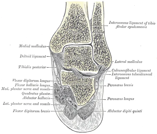
The ankle joint is where the talus meets the lower ends of the tibia (shinbone) and the fibula (the thin outer lower-leg bone)1,2. The tibia and fibula form a socket around the talus, called the mortise1,2. The part where the talus meets the tibia carries body weight between the leg and the foot1. The tibia’s lower end forms the medial malleolus (the bony bump on the inner side of the ankle)1.
Experimental studies found about 83% of the load at the ankle passes between the tibia and talus3. The other 17% passes through the fibula, and more of it does so when the foot is pulled up3. The talus passes body weight and forces between the lower leg and the foot5. Yet no muscle attaches directly to it3,5.
A review of ankle biomechanics describes an “ankle joint complex” of three joints3. Besides the ankle joint itself, they include the subtalar joint and one other joint of the foot3. The subtalar joint lies between the talus and the calcaneus (heel bone)1,3.
A hinge, mostly
An anatomy textbook describes the medial malleolus and the lateral malleolus (outer ankle bone) holding the sides of the talus1. This stops side-to-side motion, so the ankle works as a hinge1. It allows only dorsiflexion (pulling the foot and toes up toward the shin) and plantar flexion (pointing the foot down)1. The review of ankle biomechanics sees it differently3. The top of the talus is cone-shaped and the joint turns about a slanted axis, which suggests it may not be a simple hinge3.
The talus is widest at the front3. So the ankle is more stable when the foot is pulled up3. Turning the sole inward (inversion) and outward (eversion) happens mainly below the ankle, at the subtalar joint1,3.
How far it moves
Studies put the ankle’s total up-and-down range at 65 to 75 degrees, varying between people3. That is 10 to 20 degrees of dorsiflexion and 40 to 55 degrees of plantar flexion3. Everyday tasks use much less of that range, at most about this much3:
| Activity | Up-and-down range used, degrees |
|---|---|
| Walking | 303 |
| Climbing stairs | 373 |
| Going down stairs | 563 |
What holds it together
Three groups of ligaments (tough bands of tissue that hold bones together at a joint) steady the ankle3:
| Group | Where it sits | What it does |
|---|---|---|
| Lateral ankle ligaments | The outer side: the anterior and posterior talofibular and the calcaneofibular ligaments1 | Support the ankle and resist too much inversion1 |
| Deltoid ligament | The inner side, a broad ligament1 | Supports the joint and resists too much eversion1 |
| Syndesmosis | Just above the ankle, a strong fibrous link between the tibia and fibula1 | Keeps the two bones from separating and the talus locked between them1 |
The calcaneofibular ligament is the only connective tissue that directly links the ankle joint and the subtalar joint3. While the foot is on the ground, the joint’s shape alone is enough to resist eversion3. Otherwise, its stability comes from the soft tissues around it3.
The loads it carries
The review of ankle biomechanics reports that the ankle complex carries about five times body weight while the foot is down in normal walking3. In activities such as running, it carries up to thirteen times body weight3. The bone surfaces fit closely, giving a large load-bearing area of 11 to 13 square centimetres3. Some have proposed that this means lower stress than in the hip or knee3.
In walking, the ankle makes its greatest power about halfway through each stride, as the calf muscles push the body forward3.
Breakdown
The most injured joint in many sports
A review gathered sports-injury studies from 38 countries, published from 1977 to 20054. Across them, the ankle was the second most often injured body site, after the knee4. Sprain (a stretched or torn ligament) was its most common injury4. Among 70 sports, the ankle was the most often injured site in 244.
A meta-analysis (a study that pools the results of several earlier studies) looked at 181 prospective studies (studies that follow people forward in time)6. Sprains were more frequent in females than males, and in children and adolescents than adults6. Indoor and court sports had the highest rate of any sport group, at 7 sprains per 1,000 exposures6. Sprains on the outer side were the most common type6. The ankle sprain page covers who has sprains and how they run their course.
How the ligaments give way
In an outer ankle sprain, the anterior talofibular ligament gives way first, and may be the only one hurt7,8. The calcaneofibular ligament is most often next7,8.
A high ankle sprain is a sprain of the ligaments that join the lower shinbone and fibula, just above the ankle joint9. An anatomy review reports that the weakest of them is the anterior inferior tibiofibular ligament9. It is the first to give way when the fibula is twisted outward9.
The biomechanics review says the tight binding of the lower tibia and fibula makes that joint highly prone to injury3. More on each group is on the lateral ankle ligaments, deltoid ligament and syndesmosis pages.
Damage to the talus
A consensus working group, a panel of experts, looked at cartilage-and-bone lesions (areas of damage) of the talus10. Most affect the inner or outer part of its top10. In one study it cites, an earlier injury came before 98% of outer lesions and 70% of inner ones10. Another study it reports found such lesions in 42% of 37 professional soccer players who had no symptoms10.
Osteoarthritis usually follows injury
Compared with the hip or knee, the ankle seems far less prone to osteoarthritis unless it has been injured before3. At two centres, most ankles with advanced arthritis had been injured before11,12. The table compares injury-related cases with primary osteoarthritis (osteoarthritis that develops without a known earlier cause)11,12:
| Centre | Ankles studied | Following an injury | Primary osteoarthritis |
|---|---|---|---|
| One US specialist centre | 639 with advanced arthritis11 | 70%11 | 7%11 |
| One other centre | 406 with painful end-stage osteoarthritis12 | 78%12 | 9%12 |
At the US centre, the injury-related cases most often followed rotational (twisting) ankle fractures11. At the other centre, people with injury-related osteoarthritis were younger than those with primary osteoarthritis12.
A narrative review (a summary of research without a set search method) reports that outer ankle sprains lie behind 13% to 22% of ankle osteoarthritis13. It reports the same sprains lie behind 80% of injury-related ankle osteoarthritis13. It reports that injury-related ankle osteoarthritis may start younger than primary osteoarthritis, on average in people’s forties13.
A Korean national insurance cohort (a group of people followed over time) took in 195,393 people14. Those with a sprain had a 46% higher rate of later ankle osteoarthritis than those without14. A case series (a report on a group of patients, without a comparison group) studied 30 people with end-stage osteoarthritis after a ligament injury15. On average, it appeared 34.3 years after the injury15. It came sooner after a single severe sprain than after repeated injuries, at 25.7 against 38.0 years15.
Recovery
How the outer ligaments heal
A systematic review (a study that searches for research in a set, checkable way) looked at 7 randomised trials16. These trials assign people to groups by chance, and they measured ankle looseness after a fresh sprain16. It concluded that the outer ligaments needed 6 weeks to 3 months or more to heal16. Looseness improved over 6 weeks to 1 year16.
In two of the trial reports in that review, 3% to 31% of people still showed a sign of ligament looseness at 6 months16. Between 7% and 42% still felt the ankle was unstable up to a year after injury16.
Chronic ankle instability involves looseness and instability of the ankle that interfere with activity13. It can follow one sprain or repeated injuries to the outer ligaments13. One prospective cohort study found it in 40% of people a year after a first outer ankle sprain13. The lateral ankle ligaments page covers that tissue in more detail.
How the cartilage copes
In one study, 10 healthy adults had MRI scans (scans that show soft tissue in detail) before and after single-leg hopping17. The cartilage became measurably thinner, by about 3% on the tibia and 2% on the talus17.
A review compared cartilage in different joints18. Ankle articular cartilage holds more water and proteoglycans (large sugar-and-protein molecules in cartilage) than knee cartilage, and renews its proteoglycans faster18. These differences make it stiffer and less permeable, so fluid passes through it less easily18.
The same review reports that ankle cartilage cells respond less to breakdown signals than knee cartilage cells18. After damage, they make proteoglycans faster, which suggests a greater capacity for repair18.
The consensus working group concluded that talus lesions with few symptoms are unlikely to progress to ankle osteoarthritis10. It reports one study that followed 22 people with talus lesions for 11 to 20 years10. On X-ray, 73% of the cases assessed showed no worsening of osteoarthritis, and 27% worsened by one grade10. Also, 38% of the people had cut back on sport because of symptoms10.
Connections
The muscles and tendons that cross it
Most motion of the foot and ankle comes from twelve muscles that start in the lower leg and attach in the foot3. They sit in four compartments3:
- the gastrocnemius, soleus and plantaris at the back of the leg, which point the foot down3
- three deeper muscles at the back (tibialis posterior, flexor digitorum longus, flexor hallucis longus), which point the foot down and turn the sole in3
- the tibialis anterior, extensor digitorum longus, extensor hallucis longus and peroneus tertius at the front of the shin, which pull the foot up3
- the peroneus longus and peroneus brevis on the outer side, which point the foot down and turn the sole outward3
Judging by the force it can make, the authors of one dissection study consider the soleus likely the most important muscle acting at the ankle19. The gastrocnemius crosses two joints, the knee and the ankle20,21. The superficial calf muscles all end on the Achilles tendon, which attaches to the heel bone1. At the front, the tibialis anterior tendon belongs to the muscle that mainly lifts the foot and turns its sole inward22.
Exercises and stretches that load it
Related exercise pages: calf stretch, drop landing and single-leg squat. In 20 volunteers without symptoms, standing dorsiflexion averaged 25 degrees with the knee straight and 39 degrees with it bent 75 degrees23. A systematic review of 6 studies suggests landing changes when dorsiflexion is restricted, though the studies disagree on which measures change24. A meta-analysis of 21 studies found people with chronic ankle instability landed from a one-leg jump with more plantar flexion than controls25.
One study followed 306 young basketball and floorball players for 12 months26. Those whose knee angled far inward in a single-leg squat were 2.4 times more likely to injure an ankle26.
Ankle joint is formed by
Ankle joint is held together by
Ankle joint is lined by
Ankle joint is moved by
Ankle joint is crossed by
Ankle joint is compared with
Ankle joint is injured in
Ankle joint is loaded by
Sources
- Betts JG, Young KA, Wise JA, Johnson E, Poe B, Kruse DH, Korol O, Johnson JE, Womble M, DeSaix P (2022). Anatomy and Physiology 2e. OpenStax. Link
- Ficke J, Fakoya AO, Byerly DW (2026). Anatomy, Bony Pelvis and Lower Limb: Foot and Ankle. StatPearls. PMID 31536304
- Brockett CL, Chapman GJ (2016). Biomechanics of the ankle.. Orthopaedics and Trauma. doi:10.1016/j.mporth.2016.04.015 PMID 27594929
- Fong DT, Hong Y, Chan LK, Yung PS, Chan KM (2007). A systematic review on ankle injury and ankle sprain in sports.. Sports Medicine. doi:10.2165/00007256-200737010-00006 PMID 17190537
- Khan IA, Varacallo MA (2023). Anatomy, Bony Pelvis and Lower Limb, Foot Talus. StatPearls. PMID 31082130
- Doherty C, Delahunt E, Caulfield B, Hertel J, Ryan J, Bleakley C (2014). The incidence and prevalence of ankle sprain injury: a systematic review and meta-analysis of prospective epidemiological studies.. Sports Medicine. doi:10.1007/s40279-013-0102-5 PMID 24105612
- Hertel J (2002). Functional Anatomy, Pathomechanics, and Pathophysiology of Lateral Ankle Instability.. Journal of Athletic Training. PMID 12937557
- Golanó P, Vega J, de Leeuw PA, Malagelada F, Manzanares MC, Götzens V, van Dijk CN (2010). Anatomy of the ankle ligaments: a pictorial essay.. Knee Surgery, Sports Traumatology, Arthroscopy. doi:10.1007/s00167-010-1100-x PMID 20309522
- Hermans JJ, Beumer A, de Jong TA, Kleinrensink GJ (2010). Anatomy of the distal tibiofibular syndesmosis in adults: a pictorial essay with a multimodality approach. Journal of anatomy. doi:10.1111/j.1469-7580.2010.01302.x PMID 21108526
- Walther M, Gottschalk O, Madry H, Müller PE, Steinwachs M, Niemeyer P, et al (2023). Etiology, Classification, Diagnostics, and Conservative Management of Osteochondral Lesions of the Talus. 2023 Recommendations of the Working Group "Clinical Tissue Regeneration" of the German Society of Orthopedics and Traumatology.. Cartilage. doi:10.1177/19476035231161806 PMID 37082983
- Saltzman CL, Salamon ML, Blanchard GM, Huff T, Hayes A, Buckwalter JA, Amendola A (2005). Epidemiology of ankle arthritis: report of a consecutive series of 639 patients from a tertiary orthopaedic center.. The Iowa Orthopaedic Journal. PMID 16089071
- Valderrabano V, Horisberger M, Russell I, Dougall H, Hintermann B (2009). Etiology of ankle osteoarthritis.. Clinical Orthopaedics and Related Research. doi:10.1007/s11999-008-0543-6 PMID 18830791
- Herzog MM, Kerr ZY, Marshall SW, Wikstrom EA (2019). Epidemiology of Ankle Sprains and Chronic Ankle Instability.. Journal of Athletic Training. doi:10.4085/1062-6050-447-17 PMID 31135209
- Lee S, Song K, Lee SY (2022). Epidemiological study of post-traumatic ankle osteoarthritis after ankle sprain in 195,393 individuals over middle age using the National Health Insurance Database: A retrospective design.. Journal of Science and Medicine in Sport. doi:10.1016/j.jsams.2021.08.018 PMID 34565662
- Valderrabano V, Hintermann B, Horisberger M, Fung TS (2006). Ligamentous posttraumatic ankle osteoarthritis.. The American Journal of Sports Medicine. doi:10.1177/0363546505281813 PMID 16303875
- Hubbard TJ, Hicks-Little CA (2008). Ankle ligament healing after an acute ankle sprain: an evidence-based approach.. Journal of Athletic Training. doi:10.4085/1062-6050-43.5.523 PMID 18833315
- Cher WL, Utturkar GM, Spritzer CE, Nunley JA, DeFrate LE, Collins AT (2016). An analysis of changes in in vivo cartilage thickness of the healthy ankle following dynamic activity.. Journal of Biomechanics. doi:10.1016/j.jbiomech.2016.05.030 PMID 27289415
- Kuettner KE, Cole AA (2005). Cartilage degeneration in different human joints.. Osteoarthritis and Cartilage. doi:10.1016/j.joca.2004.11.006 PMID 15694570
- Ward SR, Eng CM, Smallwood LH, Lieber RL (2009). Are current measurements of lower extremity muscle architecture accurate?. Clinical Orthopaedics and Related Research. doi:10.1007/s11999-008-0594-8 PMID 18972175
- Pedret C, Palermi S, Mechó S, Frutos EJ, Rovira J, Vicente O, Balius R (2026). Calf Injuries: An Anatomy-Based Approach to Diagnosis, Management and Return to Sport.. Current reviews in musculoskeletal medicine. doi:10.1007/s12178-026-10059-2 PMID 42799963
- Bryan Dixon J (2009). Gastrocnemius vs. soleus strain: how to differentiate and deal with calf muscle injuries.. Current reviews in musculoskeletal medicine. doi:10.1007/s12178-009-9045-8 PMID 19468870
- Juneja P, Hubbard JB (2023). Anatomy, Bony Pelvis and Lower Limb: Tibialis Anterior Muscles.. StatPearls. PMID 30020676
- Baumbach SF, Brumann M, Binder J, Mutschler W, Regauer M, Polzer H (2014). The influence of knee position on ankle dorsiflexion - a biometric study.. BMC musculoskeletal disorders. doi:10.1186/1471-2474-15-246 PMID 25053374
- Mason-Mackay AR, Whatman C, Reid D (2017). The effect of reduced ankle dorsiflexion on lower extremity mechanics during landing: A systematic review.. Journal of science and medicine in sport. doi:10.1016/j.jsams.2015.06.006 PMID 26117159
- Chan LYT, Sim YTN, Gan FK, Bin Abd Razak HR (2022). Effect of chronic ankle instability on lower extremity kinematics, dynamic postural stability, and muscle activity during unilateral jump-landing tasks: A systematic review and meta-analysis.. Physical therapy in sport : official journal of the Association of Chartered Physiotherapists in Sports Medicine. doi:10.1016/j.ptsp.2022.04.005 PMID 35462322
- Räisänen AM, Pasanen K, Krosshaug T, Vasankari T, Kannus P, Heinonen A, Kujala UM, Avela J, Perttunen J, Parkkari J (2018). Association between frontal plane knee control and lower extremity injuries: a prospective study on young team sport athletes.. BMJ Open Sport & Exercise Medicine. doi:10.1136/bmjsem-2017-000311 PMID 29387448
Changes
- Revised. Corrects a statement that the gastrocnemius is the only lower-leg muscle crossing both the knee and the ankle.
- A supporting claim was narrowed to match its source; the page text is unchanged.