Calcaneus
Also called heel bone
The heel bone, the largest bone of the foot. How it carries body weight and the pull of the calf, how it breaks down, how its growth plate closes, and what attaches to it.
- Region
- Foot & ankle
- Muscles
- gastrocnemius, soleus, abductor hallucis, flexor digitorum brevis, quadratus plantae
- Updated
- October 6, 2026
- Reviewed
- October 6, 2026 · how entries are checked
Function
Where it sits and what it carries
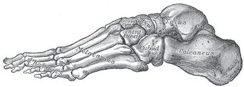
The calcaneus, or heel bone, is the largest bone of the foot1,2. It forms the heel and sits under the talus (the ankle bone that sits under the shinbone)1,2. Body weight passes from the tibia (shinbone) to the talus, then to the calcaneus, the bone resting on the ground1,2.
The calcaneus is the rear support of the foot’s lengthwise arches, where they meet the ground1. The front support is the heads of the metatarsals (the long bones in the middle of the foot)1. Besides passing body weight to the ground, the calcaneus forms a strong lever for the calf muscles3. The Achilles tendon inserts into it1.
Its shape and its joints
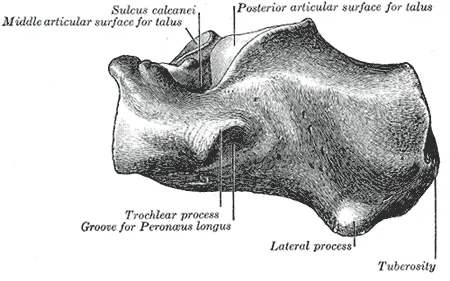
The calcaneus forms joints with two bones: the talus above and a bone called the cuboid in front1,2,3. Its surfaces carry several named landmarks3,8:
- Top. Three facets (small flat areas on a bone) meet the talus8.
- Inner side. The sustentaculum tali is a bony shelf that supports the inner side of the talus1.
- Outer side. A ridge, the peroneal tubercle, separates two grooves for tendons3.
- Underside. It ends at the back in the calcaneal tuberosity, a ridge with a process (a bony projection) at each end3. The inner process is the broader and larger of the two3.
The heel pad
Under the bone lies the heel fat pad, a cushion that absorbs shock and spreads force across the sole in walking and running9,10.
In 16 adults walking barefoot at their chosen speed, the heel pad squashed by up to 10.3 mm11. That approached the 10.7 mm predicted for the limit of pain tolerance11.
An X-ray film study followed two people running at 4.5 metres a second12. At heel strike, the pad deformed by about 60.5% barefoot and about 35.5% in shoes12. Vertical ground reaction forces (the push of the ground back up on the body) did not differ notably12.
The loads it takes in walking and running
One modelling study (a study that estimates forces inside the body with a model) used a model of the foot driven by measured gait data4. It predicted higher peak loads for running than for walking4:
| Predicted peak load | Walking | Running |
|---|---|---|
| Pull of the Achilles tendon | 3.9 body weights4 | 7.7 body weights4 |
| Joint with the talus | 5.4 body weights4 | 11.1 body weights4 |
| Joint with the cuboid | 4.2 body weights4 | 7.9 body weights4 |
In that model, the largest forces in the calcaneus came late in stance (the part of each step when the foot is on the ground)4. They came at about 70% of stance in walking and about 60% in running, for the speeds analysed4.
Breakdown
Stress fractures
A review describes calcaneal stress fractures as a frequently unrecognised source of heel pain13. It calls them common among active people such as athletes and military personnel13. A second review attributes them to repeated heel strike on uncushioned surfaces14. The Achilles tendon, pulling the other way, is also thought to contribute14.
The second review describes fractures in the tuberosity at the back as crossing the bone’s trabecular pattern (its honeycomb of thin struts) at right angles14. In an MRI series of 34 calcaneal stress injuries in military recruits, 19 were at the back, 6 in the middle and 9 in front15. The stress fracture page covers these injuries across bones.
Who gets them
Several counts of these injuries come from military recruits5,15:
| Group studied | What was found |
|---|---|
| Marine recruits, 1977 report | Calcaneal stress fractures were about 25% of the stress fractures seen, and occurred in 10.5% of 114 women recruits5 |
| US Marine recruits, prospective study | The tibia was the bone most often fractured (73%); the back of the calcaneal tuberosity was the single most common site (21%)16 |
| Military recruits, MRI study | 30 recruits had calcaneal stress injuries over 96 months, or 2.6 per 10,000 person-years across the recruit population15 |
| 97 patients under 18 with stress injuries at a children’s hospital | Those under 14 were more likely to have cuboid and calcaneal stress injuries; the mean age for calcaneal injuries was 8.3 years17 |
The authors of the prospective Marine study warned against applying their figures to other groups16. Person-years are a count of people multiplied by the years each was followed.
The 1977 report found that incidence could be correlated with physical condition at the start of training5. It found no such link with height, weight, age or the build of the foot5. In one MRI-based study, affected and unaffected recruits did not differ significantly in body mass index, weight, height, age or physical fitness15. That finding was with the numbers available15.
The growth plate at the back of the heel
The calcaneal apophysis is a secondary centre of bone formation at the back of the body of the calcaneus6. An apophysis is a bony bump where a muscle attaches that grows from its own growth plate while the bone is still growing. This one develops in early childhood6.
A study of 568 CT scans of Australian children timed its stages, and other work adds X-ray ages6:
| Stage | Girls | Boys |
|---|---|---|
| Apophysis begins turning to bone, mean age on CT | 5.2 years6 | 7.2 years6 |
| Fusion to the rest of the bone begins, mean age on CT | 11.2 years6 | 13.3 years6 |
| Earliest complete fusion seen on CT | 10 years6 | 14 years6 |
| Complete fusion reported in plain X-ray studies | 15 to 16 years6 | 18 to 20 years6 |
In the CT study, the apophysis developed and fused significantly earlier in girls than in boys6. A CT scan builds detailed pictures of the inside of the body from X-rays.
Another study followed 94 healthy children with yearly X-rays18. The apophysis first appeared a mean of 4.7 years before peak height velocity, the point of fastest growth in height18. Fusion was usually finished a mean of 2.1 years after that peak18.
A 2014 study describes Sever’s disease, or calcaneal apophysitis, as a cause of heel pain that is frequent in children19.
Heel spurs
A plantar calcaneal spur, or heel spur, is an outgrowth of bone from the calcaneal tuberosity20. On weight-bearing X-rays of 216 people aged 62 to 94, 55% had at least one21. In that group, people with spurs were more likely to be obese and to report osteoarthritis21. They were also more likely to have current or previous heel pain, with an odds ratio of 4.621. An odds ratio is how many times higher the odds of an outcome are in one group than in another.
A review describes the cause of these spurs as contentious20. It names several competing theories, among them traction, repeated trauma and vertical compression20. The plantar heel pain page covers that condition.
The bursa behind the heel
A bursa (a small fluid-filled sac that cushions tissues where they rub) lies between the Achilles tendon and the upper back corner of the calcaneus22. Inflammation of it is called retrocalcaneal bursitis22. The bursa is squeezed between bone and tendon each time the foot is pulled up toward the shin, especially in uphill running23.
Breaks from a single injury
One trauma unit recorded 752 calcaneal fractures from 1995 to 2005, an annual incidence of 11.5 per 100,00024. In that series, 71.5% of the fractures happened in falls from a height24.
Recovery
How the heel bone compares in athletes
A review describes bone as adapting to the loads it regularly carries25. Several studies compared the heel bones of athletes with those of other people7,26,27:
- In 30 young men, national-level jumpers had 52% higher calcaneal bone mineral content (the amount of mineral in a bone) than sedentary men7.
- Male athletes aged 70 to 81 had 19% to 28% higher calcaneal bone mineral content than a population sample of similar age26.
- Among 174 girls aged 11 to 17, gymnasts had higher ultrasound values at the calcaneus than non-athletic girls at the start27.
- A CT study of 18 men aged 20 to 41 found no statistically significant differences between runners and non-runners in the back of the calcaneus28.
What changed over time
One study followed girls aged 11 to 17 for one year27. The speed of sound through the calcaneus rose in runners who kept training and in non-athletic girls, and fell in runners who stopped27. The authors concluded that the effect of exercise on these measures is at least partly temporary27.
A meta-analysis (a study that pools the results of several earlier studies) covered 12 exercise studies29. It found that 4 to 36 months of exercise significantly improved an ultrasound measure of the calcaneus across age groups29.
How injuries to it heal
Stress fractures of the calcaneus are classed as low-risk30. The reason given is that they usually heal with a change in activity or a short period without weight bearing30. A 2023 systematic review noted that sorting bone stress injuries into low-risk and high-risk by location had not been sufficiently validated31.
Connections
The calf and the Achilles tendon
The Achilles tendon attaches to the middle of the back surface of the calcaneus3. In a dissection study of 12 leg specimens, fibres from the gastrocnemius and the soleus each attached to their own part of the tuberosity32.
The sole of the foot
The inner process of the tuberosity gives attachment to the plantar fascia (a band of tough tissue along the sole of the foot)3. Two muscles of the foot, abductor hallucis and flexor digitorum brevis, attach there too3.
Ligaments, tendons and nerves at the sides
Several structures attach to or cross the sides of the bone3,8:
- A tubercle near the centre of the outer surface anchors the calcaneofibular ligament, one of the lateral ankle ligaments3,33,34.
- Of the peroneal tendons, the peroneus brevis tendon passes above the peroneal tubercle and the peroneus longus tendon below it35.
- The sustentaculum tali anchors the spring ligament (a ligament under the inner arch of the foot) at its front edge3. Part of the deltoid ligament (the fan of ligaments on the inner side of the ankle) attaches at its inner edge3.
- Three tendons slide on the underside of the sustentaculum tali: flexor hallucis longus, tibialis posterior and flexor digitorum longus8.
- The medial calcaneal nerve, a branch of the tibial nerve, supplies the back and underside of the heel36,37.
Related exercise pages: running, with model-predicted joint loads of up to 11.1 body weights4.
Calcaneus is attached to by
Calcaneus is pulled on through the Achilles tendon by
Calcaneus gives origin to
Calcaneus gives origin to part of
Calcaneus is crossed on its outer side by
Calcaneus is crossed on its inner side by
Calcaneus is attached to by the calcaneofibular ligament of
Calcaneus is attached to by part of
Calcaneus is supplied at the heel by a branch of
Calcaneus receives body weight through the talus from
Calcaneus is a site of
Calcaneus has the growth plate involved in
Calcaneus is the site of
Calcaneus is loaded by
Sources
- Betts JG, Young KA, Wise JA, Johnson E, Poe B, Kruse DH, Korol O, Johnson JE, Womble M, DeSaix P (2022). Anatomy and Physiology 2e. OpenStax. Link
- Gupton M, Özdemir M, Terreberry RR (2023). Anatomy, Bony Pelvis and Lower Limb: Calcaneus. StatPearls. PMID 30137829
- Gray H; Lewis WH (editor) (1918). Anatomy of the Human Body. Lea & Febiger, Philadelphia (20th edition). Link
- Giddings VL, Beaupré GS, Whalen RT, Carter DR (2000). Calcaneal loading during walking and running. Medicine and science in sports and exercise. doi:10.1097/00005768-200003000-00012 PMID 10731005
- Hopson CN, Perry DR (1977). Stress fractures of the calcaneus in women marine recruits. Clinical orthopaedics and related research. PMID 598149
- Blythe CS, Reynolds MS, Gregory LS (2022). Quantifying the ossification and fusion of the calcaneal apophysis using computed tomography. Journal of anatomy. doi:10.1111/joa.13677 PMID 35468228
- Taaffe DR, Suominen H, Ollikainen S, Cheng S (2001). Calcaneal bone mineral and ultrasound attenuation in male athletes exposed to weight-bearing and nonweight-bearing activity. A cross-sectional report. The Journal of sports medicine and physical fitness. PMID 11447369
- Krähenbühl N, Horn-Lang T, Hintermann B, Knupp M (2017). The subtalar joint: A complex mechanism. EFORT open reviews. doi:10.1302/2058-5241.2.160050 PMID 28828179
- Chang AH, Rasmussen SZ, Jensen AE, Sørensen T, Rathleff MS (2022). What do we actually know about a common cause of plantar heel pain? A scoping review of heel fat pad syndrome. Journal of foot and ankle research. doi:10.1186/s13047-022-00568-x PMID 35974398
- Maemichi T, Matsumoto M, Tsutsui T, Ichikawa S, Okunuki T, Tanaka H, Kumai T (2024). Effect of Height and Weight on Heel Fat Pad Movements Between Microchamber and Macrochamber Layers in Loading and Unloading. Foot & ankle orthopaedics. doi:10.1177/24730114241278927 PMID 39371684
- Wearing SC, Hooper SL, Dubois P, Smeathers JE, Dietze A (2014). Force-deformation properties of the human heel pad during barefoot walking. Medicine and science in sports and exercise. doi:10.1249/MSS.0000000000000281 PMID 24504425
- De Clercq D, Aerts P, Kunnen M (1994). The mechanical characteristics of the human heel pad during foot strike in running: an in vivo cineradiographic study. Journal of biomechanics. doi:10.1016/0021-9290(94)90275-5 PMID 7962009
- Weber JM, Vidt LG, Gehl RS, Montgomery T (2005). Calcaneal stress fractures. Clinics in podiatric medicine and surgery. doi:10.1016/j.cpm.2004.08.004 PMID 15555842
- Kaiser PB, Guss D, DiGiovanni CW (2023). Republication of "Stress Fractures of the Foot and Ankle in Athletes". Foot & ankle orthopaedics. doi:10.1177/24730114231195045 PMID 37590306
- Sormaala MJ, Niva MH, Kiuru MJ, Mattila VM, Pihlajamäki HK (2006). Stress injuries of the calcaneus detected with magnetic resonance imaging in military recruits. The Journal of bone and joint surgery. American volume. doi:10.2106/JBJS.E.01447 PMID 17015602
- Greaney RB, Gerber FH, Laughlin RL, Kmet JP, Metz CD, Kilcheski TS, Rao BR, Silverman ED (1983). Distribution and natural history of stress fractures in U.S. Marine recruits. Radiology. doi:10.1148/radiology.146.2.6217486 PMID 6217486
- Gremillion MJ, Martinez A, Ghanta RB, Borici N, Kushare I (2023). An assessment of the diagnosis, treatment, and outcomes of lower extremity stress fractures in pediatric and adolescent populations. The Physician and sportsmedicine. doi:10.1080/00913847.2022.2143247 PMID 36328959
- Nicholson AD, Liu RW, Sanders JO, Cooperman DR (2015). Relationship of calcaneal and iliac apophyseal ossification to peak height velocity timing in children. The Journal of bone and joint surgery. American volume. doi:10.2106/JBJS.N.00671 PMID 25609442
- Wiegerinck JI, Yntema C, Brouwer HJ, Struijs PA (2014). Incidence of calcaneal apophysitis in the general population.. European journal of pediatrics. doi:10.1007/s00431-013-2219-9 PMID 24297670
- Kirkpatrick J, Yassaie O, Mirjalili SA (2017). The plantar calcaneal spur: a review of anatomy, histology, etiology and key associations. Journal of anatomy. doi:10.1111/joa.12607 PMID 28369929
- Menz HB, Zammit GV, Landorf KB, Munteanu SE (2008). Plantar calcaneal spurs in older people: longitudinal traction or vertical compression?. Journal of foot and ankle research. doi:10.1186/1757-1146-1-7 PMID 18822162
- van Dijk CN, van Sterkenburg MN, Wiegerinck JI, Karlsson J, Maffulli N (2011). Terminology for Achilles tendon related disorders. Knee surgery, sports traumatology, arthroscopy : official journal of the ESSKA. doi:10.1007/s00167-010-1374-z PMID 21222102
- Wnuk-Scardaccione A, Mizia E, Zawojska K, Bilski J, Wojdyła J (2021). Surface Shape of the Calcaneal Tuberosity and the Occurrence of Retrocalcaneal Bursitis among Runners. International journal of environmental research and public health. doi:10.3390/ijerph18062860 PMID 33799691
- Mitchell MJ, McKinley JC, Robinson CM (2009). The epidemiology of calcaneal fractures. Foot (Edinburgh, Scotland). doi:10.1016/j.foot.2009.05.001 PMID 20307476
- Hart NH, Nimphius S, Rantalainen T, Ireland A, Siafarikas A, Newton RU (2017). Mechanical basis of bone strength: influence of bone material, bone structure and muscle action.. Journal of musculoskeletal & neuronal interactions. PMID 28860414
- Suominen H, Rahkila P (1991). Bone mineral density of the calcaneus in 70- to 81-yr-old male athletes and a population sample. Medicine and science in sports and exercise. PMID 1766337
- Lehtonen-Veromaa M, Möttönen T, Kautiainen H, Heinonen OJ, Viikari J (2001). Influence of physical activity and cessation of training on calcaneal quantitative ultrasound measurements in peripubertal girls: a 1-year prospective study. Calcified tissue international. doi:10.1007/s002230001209 PMID 11351497
- Best A, Holt B, Troy L, Hamill J (2017). Trabecular bone in the calcaneus of runners. PloS one. doi:10.1371/journal.pone.0188200 PMID 29141022
- Babatunde OO, Forsyth JJ (2013). Quantitative Ultrasound and bone's response to exercise: a meta analysis. Bone. doi:10.1016/j.bone.2012.12.011 PMID 23269404
- Paavana T, Rammohan R, Hariharan K (2024). Stress fractures of the foot - current evidence on management. Journal of clinical orthopaedics and trauma. doi:10.1016/j.jcot.2024.102381 PMID 38435398
- Hoenig T, Eissele J, Strahl A, Popp KL, Stürznickel J, Ackerman KE, Hollander K, Warden SJ, Frosch KH, Tenforde AS, Rolvien T (2023). Return to sport following low-risk and high-risk bone stress injuries: a systematic review and meta-analysis. British journal of sports medicine. doi:10.1136/bjsports-2022-106328 PMID 36720584
- Ballal MS, Walker CR, Molloy AP (2014). The anatomical footprint of the Achilles tendon. The bone & joint journal. doi:10.1302/0301-620X.96B10.33771 PMID 25274919
- Golanó P, Vega J, de Leeuw PA, Malagelada F, Manzanares MC, Götzens V, van Dijk CN (2010). Anatomy of the ankle ligaments: a pictorial essay.. Knee Surgery, Sports Traumatology, Arthroscopy. doi:10.1007/s00167-010-1100-x PMID 20309522
- Le MQT, Harper DE, Dixon M, Tiu TK (2026). Calcaneofibular Ligament Injury.. StatPearls. PMID 32491310
- Davda K, Malhotra K, O'Donnell P, Singh D, Cullen N (2017). Peroneal tendon disorders. EFORT Open Reviews. doi:10.1302/2058-5241.2.160047 PMID 28736620
- Desai SS, La Rose J (2026). Anatomy, Bony Pelvis and Lower Limb: Tibial Nerve. StatPearls. PMID 30725713
- Banik S, Guria LR (2021). Variable Branching Pattern of Tibial Nerve in the Tarsal Tunnel: A Gross Anatomical Study With Clinical Implications.. Cureus. doi:10.7759/cureus.13729 PMID 33717767
Changes
- Re-reviewed after a published correction to a cited source was read and recorded; no change to the page.
- First published.