Deltoid ligament
Also called deltoid ligament complex, medial collateral ligament of the ankle
The broad, fan-shaped ligament on the inner side of the ankle. Its two layers and six bands, the movements it resists, how and in whom it gets sprained, and what studies report after injury.
- Region
- Foot & ankle
- Updated
- September 30, 2026
- Reviewed
- September 30, 2026 · how entries are checked
Function
Where it sits and what it joins
The deltoid ligament is the broad ligament on the inner (medial) side of the ankle joint1. It is also called the medial collateral ligament of the ankle4. It runs from the medial malleolus (the bony bump on the inner side of the ankle) to the talus, calcaneus and navicular5. The talus is the ankle bone under the shinbone, the calcaneus is the heel bone and the navicular is a bone of the inner midfoot5.
The ligament spans three joints: the ankle (tibiotalar) joint, the subtalar joint and the talonavicular joint (between the talus and the navicular)4. The subtalar joint lies between the talus and the heel bone, where the sole turns inward and outward1,6. The deltoid ligaments are one of three ligament groups that steady the ankle6. The other two are the lateral ligaments on the outer side and the syndesmosis6. The syndesmosis lies between the tibia (the shinbone) and fibula (the thin outer bone of the lower leg)6.
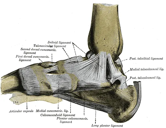
Two layers, six bands
A review of the research on its anatomy concluded that the only real agreement is that it has two layers, one superficial and one deep2. The accepted description splits it into six bands, four superficial and two deep2. In a cadaver (body donated for study) study of 10 ankles, the bands started from the front and back of the medial malleolus7.
Some bands are missing in some ankles2. A meta-analysis (a study that pools the results of several earlier studies) of 8 studies covered 142 ankle specimens8. It found each band in this share of ankles8:
| Band | Layer | Ankles where found, % |
|---|---|---|
| Deep posterior tibiotalar | Deep | 1008 |
| Tibiospring | Superficial | about 948 |
| Tibionavicular | Superficial | about 908 |
| Tibiocalcaneal | Superficial | 858 |
| Superficial posterior tibiotalar | Superficial | about 808 |
| Deep anterior tibiotalar | Deep | about 638 |
What the bands look like
One cadaver study of 10 ankles traced where the bands attach7:
- the tibionavicular band is the most forward part of the superficial layer, fanning down to the upper inner navicular7
- the tibiospring band runs from the front of the medial malleolus to the spring ligament (a ligament under the inner arch)7
- the tibiocalcaneal band attached to the sustentaculum tali, a shelf on the heel bone, in all 10 ankles studied7
- the deep posterior tibiotalar band was the thickest and largest band in all 10 ankles, over 3 mm thick7
In dissection, two tendons had to be moved aside to reach the ligament beneath7. They belong to the tibialis posterior (a deep calf muscle) and flexor digitorum longus (long toe flexor)7.
What each layer holds
The ligament supports the ankle and resists too much eversion of the foot1. There is consensus that the deep layer is the main stabiliser of the ankle joint itself7. The superficial layer mainly steadies the subtalar and talonavicular joints7. The superficial layer limits eversion of the back of the foot4. The deep layer limits outward turning of the talus and its sliding forward, backward and sideways4.
Two sources describe how the two layers share the work of stopping the talus tilting into valgus (outward)5,9. One cadaver study found the deltoid ligament the main restraint against this tilt, with its two layers equally effective9. An expert review states that the ankle tilts into valgus only when both layers are torn5. The same cadaver study suggested the deep layer is a backup restraint against sideways and forward talus movement9. In that study, the outer ankle bone and its ligaments were the main restraint9.
How foot position changes the load
The ligament is triangular and widens downward2. So its front part is tight when the foot points down, and its back part is tight when the foot pulls up2. A cadaver study of 10 ankles fits this: the tibiospring band was tense in plantar flexion (pointing the foot down)7. The tibiocalcaneal and deep posterior tibiotalar bands were tense in dorsiflexion (pulling the foot up toward the shin)7.
What happens when it is cut in the lab
Robotic cadaver studies cut the ligament, or parts of it, and measured the extra movement10,11:
| What was cut, and where | Extra movement |
|---|---|
| The whole ligament, 10 ankles | 7.6 mm forward sliding, 13.8 degrees outward rotation, 33.6 degrees eversion10 |
| The front part, 10 ankles, foot pointed down 25 degrees | 2.1 degrees outward rotation, 6.2 degrees eversion10 |
| The back part, same 10 ankles | No added looseness10 |
| Superficial posterior tibiotalar band, 8 legs, foot up | 3.3 degrees eversion11 |
| Tibiocalcaneal band, 8 legs, foot down | 4.2 degrees eversion11 |
Blood supply
In a cadaver study of 64 legs, three arteries sent branches to the ligament12. The medial tarsal artery did so in 96.7%, the posterior tibial artery in 95% and the anterior tibial artery in 38.3%12. In a cadaver study, every specimen also had blood supply entering through bone at the medial malleolus or the inner talus12.
Breakdown
How it gets hurt
The main way it is injured is eversion of the ankle, with or without outward rotation2. Typically this happens when a pronated foot (rolled inward so the arch lowers) lands on uneven ground2. A foot rolled outward with the talus twisting outward can also injure it, often along with the syndesmosis2.
One type of ankle fracture comes from an outward-twisting movement7. A cadaver study suggested that in it, the tibionavicular, tibiospring and deep anterior tibiotalar bands are at risk7. It suggested the tibiocalcaneal and especially the deep posterior tibiotalar bands are protected7. One video case report linked a non-contact inner ankle sprain to the foot pulled up at ground contact with rapid eversion13. Most tears happen where the ligament attaches to the medial malleolus5.
How often, and in whom
Most ankle sprains injure the outer ligaments2. One review reports estimates of deltoid involvement in about 5% of ankle sprains2. It adds that 3% to 4% of all ankle ligament injuries involve the deltoid ligament alone2. Among cadets at the United States Military Academy, inner ankle sprains were 5.1% of ankle sprains14. Their incidence (new cases over a set time) was 3.5 per 1,000 person-years14.
In the same cadets, men were about three times as likely as women to have an inner ankle sprain14. Sport caused 64% of them, and the rate was highest in men’s rugby and gymnastics14.
A US college sports injury database recorded 380 deltoid ligament sprains across 25 sports from 2009-10 to 2014-153. That was 0.79 sprains per 10,000 athlete-exposures, each being one athlete taking part in one practice or game3. Rates were highest in these sports, though the gymnastics figure is an imprecise estimate3:
| Sport | Sprains per 10,000 athlete-exposures |
|---|---|
| Women’s gymnastics | 2.303 |
| Men’s soccer | 1.733 |
| Women’s soccer | 1.613 |
| Men’s football | 1.403 |
In those college athletes, 49.7% of deltoid sprains came from contact with another player3. Another 19.5% involved no contact, and 18.7% came from contact with the playing surface3. Most happened in practice, but the rate in competition was 3.74 times the practice rate3.
Injured alongside other structures
The deltoid ligament is often hurt together with other parts of the ankle2,15:
- Up to 40% of ankle fractures are estimated to include a deltoid injury2
- In one arthroscopy (keyhole surgery inside a joint) study of 288 people with fresh ankle fractures, 39.6% had one2
- In an MRI (a scan that shows soft tissue in detail) study of 110 athletes with fresh outer ligament injuries, 18 also had one15
- Unstable injuries of the syndesmosis can occur with a deltoid injury, and the severity of each varies widely5
A systematic review (a study that searches for all the research on a question in a set, checkable way) looked at 14 lab studies16. In three, a deltoid injury alone significantly changed how the syndesmosis moved16.
A systematic review of 9 studies looked at people with chronic ankle instability17. MRI showed deltoid injuries in 289 of 465 people scanned, 62%17. Where the layer was named, the deep layer was involved in 146 people and the superficial layer in 6117.
In 169 freshly sprained ankles without a fracture, 48 were tender over the deltoid ligament18. Ultrasound (a scan made with sound waves) confirmed deltoid damage in 1318. Of those, 3 were superficial only, 3 were deep only and 7 involved both layers18.
Recovery
Time lost in college athletes
In the US college database, 72.9% of deltoid sprains limited participation for under a week3. In 39.5%, the limit lasted under 24 hours3. In 6.6%, the sprain was severe, meaning more than 21 days lost or a season ended3. In the same athletes, 8.2% of sprains were repeat injuries, and 2 of 380 led to surgery3.
A slower course in one man
One case report followed a recreationally active man who sprained the deltoid ligament playing basketball19. Up to 5 months later, he still had reduced ankle movement and changed nerve responses in the soleus (a wide, flat calf muscle)19. He also had poorer balance and self-reported function, and had cut back his activity19.
How ligaments heal
Tendon and ligament healing is described in three phases: inflammatory, proliferative and remodelling20. A review of knee ligament research describes injured ligament regaining function through scar tissue21. That scar is weaker, biologically and mechanically, than the tissue it replaces21. That review studied the ACL (a ligament that crosses inside the knee) and the MCL (the ligament on the inner side of the knee)21. It did not study the deltoid ligament21.
When injuries are left alone
One review states that neglected deltoid ligament injuries can lead to a worsening deformity2. It names problems with the tibialis posterior tendon, flat foot and a back of the foot angled outward2. It reports osteoarthritis (a disease in which joint cartilage wears away) after injury in 20.4% of one group2. That group had an untreated deltoid injury alone2.
The same review found no long-term studies of how often instability follows a deltoid injury2. It called the evidence on deltoid injuries limited overall2. Most ankle sprains injure the outer ligaments instead, as the ankle sprain page describes2.
Connections
The joint and bones it holds
The deltoid ligament steadies the inner side of the ankle joint, alongside the outer ligaments and the syndesmosis6. It starts on the medial malleolus at the lower end of the tibia5. It ends on the talus, heel bone and navicular5.
The other ankle ligaments
The lateral ankle ligaments on the outer side are injured in most ankle sprains2. The ankle syndesmosis lies between the tibia and fibula6. Outward-twisting injuries can hurt it and the deltoid ligament together2.
The spring ligament and tibialis posterior
The tibiospring band joins the deltoid ligament to the spring ligament5. Part of the spring ligament is so tightly attached that the two are hard to separate in dissection4. This link is described as fundamental for keeping the foot aligned5. With the tibialis posterior tendon as a second, muscle-driven support, this inner group helps hold up the inner arch5.
Deltoid ligament holds together the inner side of
Deltoid ligament attaches to
Deltoid ligament works with
Deltoid ligament is often injured with
Deltoid ligament is injured in some cases of
Sources
- Betts JG, Young KA, Wise JA, Johnson E, Poe B, Kruse DH, Korol O, Johnson JE, Womble M, DeSaix P (2022). Anatomy and Physiology 2e. OpenStax. Link
- Koris J, Calder JDF, Dalmau-Pastor M, Fernandez MA, Ramasamy A (2024). Deltoid ligament injuries: A review of the anatomy, diagnosis and treatments.. Knee Surgery, Sports Traumatology, Arthroscopy. doi:10.1002/ksa.12274 PMID 38796726
- Kopec TJ, Hibberd EE, Roos KG, Djoko A, Dompier TP, Kerr ZY (2017). The Epidemiology of Deltoid Ligament Sprains in 25 National Collegiate Athletic Association Sports, 2009-2010 Through 2014-2015 Academic Years.. Journal of Athletic Training. doi:10.4085/1062.6050-52.2.01 PMID 28318315
- Bilyy A, Win Mar WW, Al Abdeen Al Zuabi Z, Frimpong-Manso HB, Famure S, Solomou G, Brassett C, Pasapula C (2025). The Deltoid-Spring Ligament Complex: A Scoping Review and New Segmental Classification.. Cureus. doi:10.7759/cureus.81715 PMID 40322447
- Fleury C, Acker A, Dubois-Ferrière V, Assal M (2026). Deltoid ligament injury: thinking beyond the ankle - an expert opinion.. EFORT Open Reviews. doi:10.1530/EOR-2025-0268 PMID 42684953
- Brockett CL, Chapman GJ (2016). Biomechanics of the ankle.. Orthopaedics and Trauma. doi:10.1016/j.mporth.2016.04.015 PMID 27594929
- Gregersen MG, Fagerhaug Dalen A, Nilsen F, Molund M (2022). The Anatomy and Function of the Individual Bands of the Deltoid Ligament-and Implications for Stability Assessment of SER Ankle Fractures.. Foot & Ankle Orthopaedics. doi:10.1177/24730114221104078 PMID 35722175
- Yammine K (2017). The Morphology and Prevalence of the Deltoid Complex Ligament of the Ankle.. Foot & Ankle Specialist. doi:10.1177/1938640016675409 PMID 27807288
- Harper MC (1987). Deltoid ligament: an anatomical evaluation of function.. Foot & Ankle. doi:10.1177/107110078700800104 PMID 3623356
- Brady AW, Bryniarski A, Brown JR, Waltz R, Stake IK, Kreulen CD, Haytmanek CT, Clanton TO (2023). The Biomechanical Role of the Deltoid Ligament on Ankle Stability: Injury, Repair, and Augmentation.. The American Journal of Sports Medicine. doi:10.1177/03635465231181082 PMID 37449714
- Takahashi K, Teramoto A, Murahashi Y, Shiwaku K, Kamiya T, Emori M, Watanabe K, Fujie H (2025). The In Situ Force and Contribution of Each Ligamentous Band of the Deltoid Ligament in Ankle Joint Stability: A Cadaveric Biomechanical Study.. Orthopaedic Journal of Sports Medicine. doi:10.1177/23259671251327406 PMID 40182569
- Haynes JA, Gosselin M, Cusworth B, McCormick J, Johnson J, Klein S (2017). The Arterial Anatomy of the Deltoid Ligament: A Cadaveric Study.. Foot & Ankle International. doi:10.1177/1071100717702464 PMID 28447855
- Wade FE, Mok KM, Fong DT (2018). Kinematic analysis of a televised medial ankle sprain.. Asia-Pacific Journal of Sports Medicine, Arthroscopy, Rehabilitation and Technology. doi:10.1016/j.asmart.2018.02.001 PMID 29963372
- Waterman BR, Belmont PJ Jr, Cameron KL, Svoboda SJ, Alitz CJ, Owens BD (2011). Risk Factors for Syndesmotic and Medial Ankle Sprain. The American Journal of Sports Medicine. doi:10.1177/0363546510391462 PMID 21289274
- Kim JS, Kim MS, Kim DK, Lee SH (2023). Magnetic Resonance Imaging Characteristics of a Lateral Ligament Injury in Acute Ankle Sprains Among Athletes.. Orthopaedic Journal of Sports Medicine. doi:10.1177/23259671231207688 PMID 37954866
- Spennacchio P, Seil R, Gathen M, Cucchi D (2021). Diagnosing instability of ligamentous syndesmotic injuries: A biomechanical perspective. Clinical biomechanics (Bristol, Avon). doi:10.1016/j.clinbiomech.2021.105312 PMID 33756400
- Anderson A, McLellan M, Kim R, Noori N (2025). Diagnosis and Management of Deltoid Ligament Injuries in Chronic Ankle Instability: A Systematic Review.. Foot & Ankle Orthopaedics. doi:10.1177/24730114251323903 PMID 40144531
- Omodani T, Takahashi K (2023). Ultrasound findings of the deltoid ligament in patients with acute ankle sprains: A retrospective review.. Journal of Orthopaedic Science. doi:10.1016/j.jos.2022.05.010 PMID 35691876
- Terada M, Thomas AC, Pietrosimone B, Hiller CE, Bowker S, Gribble PA (2015). The Consequence of a Medial Ankle Sprain on Physical and Self-reported Functional Limitations: A Case Study Over a 5-Month Period.. The Journal of Orthopaedic and Sports Physical Therapy. doi:10.2519/jospt.2015.6097 PMID 26304642
- Leong NL, Kator JL, Clemens TL, James A, Enamoto-Iwamoto M, Jiang J (2020). Tendon and Ligament Healing and Current Approaches to Tendon and Ligament Regeneration.. Journal of Orthopaedic Research. doi:10.1002/jor.24475 PMID 31529731
- Frank CB (2004). Ligament structure, physiology and function. Journal of musculoskeletal & neuronal interactions. PMID 15615126
Changes
- A supporting claim was narrowed to match its source; the page text is unchanged.