Fibula
The thin outer bone of the lower leg. How little load it carries, how it breaks at the ankle and under repeated stress, how it responds to training and disuse, and what attaches to it.
- Region
- Lower leg
- Muscles
- peroneus longus, peroneus brevis, soleus, hamstrings
- Updated
- October 5, 2026
- Reviewed
- October 5, 2026 · how entries are checked
Function
Where it sits
The fibula is a long bone on the outer side of the tibia (shin bone)1. Of the two, the fibula is much the smaller and thinner1. It sits just behind the top of the tibia at the knee and runs down the outer side of the leg to the ankle1.
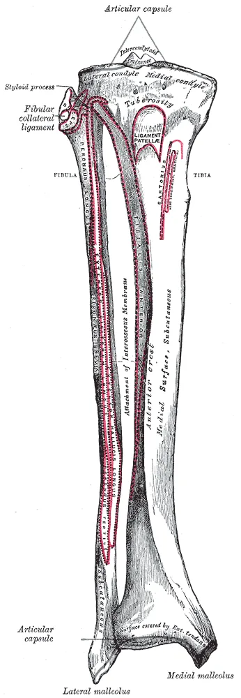
Its four parts
The fibula is described in four parts1:
- Head. The small, knob-like upper end, which meets the underside of the tibia’s outer condyle (a rounded knob at the end of a bone)8.
- Neck. The place where the head narrows1.
- Shaft. It has an outer, an inner and a back surface, and the muscles attached to it set its shape1.
- Lower end. It forms the lateral malleolus, the bony bump that is easily felt on the outer side of the ankle8.
The inner side of the lateral malleolus meets the talus (ankle bone) as part of the ankle joint8.
How it is joined to the tibia
The fibula meets the tibia at two joints, one at each end8. At the top, a capsule covers the joint, and ligaments at the front and back strengthen it9. At the bottom, the rounded lower fibula sits in a matching hollow notch in the tibia10.
Between the two ends, a ridge on the inner surface of the fibula anchors the interosseous membrane that ties it to the tibia1. This connection is a syndesmosis, a joint with very little movement1.
A slight gliding is possible at the upper joint during ankle movement9. In dorsiflexion (pulling the foot up toward the shin), the upper fibula rotates outward9.
How much load it carries
Three experiments on legs from cadavers (bodies donated for study) measured the fibula’s share of load2,3,4:
| Experiment | Fibula’s share |
|---|---|
| Autopsy specimens, ankle in a neutral position | 6.4% of the weight2 |
| Ten specimens with force sensors in both bones, ankle neutral | 7.12% of the total force, on average3 |
| 10 specimen legs under a load of 1500 newtons (a newton is a unit of force) along the leg | 17% of the load, on average4 |
In the third experiment, the fibula’s share rose as the total load rose4. In the first, the weight on the fibula rose with dorsiflexion or eversion (turning the sole of the foot outward)2. It fell with plantar flexion (pointing the foot down)2.
The authors of one bone-scan study describe the fibula’s share of the lower leg’s compressive load as small and highly variable, from -8% to +19%11. A review puts it at 10% of body weight, against 90% for the tibia12. The same review calls its role crucial to the stability of the ankle mortise12. The mortise is the socket that the lower tibia and fibula form around the talus.
Breakdown
Breaks at the ankle
In a population study of 9767 ankle fractures, a break of the lateral malleolus was the most common type in every age group5. It made up 55% of all the fractures5. In the same study, falls caused 61% of the fractures and sports 22%5.
A review of lower fibula fractures describes two mechanisms12:
- Rolling out with outward rotation, the most often reported. The talus rotates and pushes the tibia and fibula apart12. The front ligament between the lower tibia and fibula ruptures first, then the fibula breaks at an angle at the level of the syndesmosis12.
- Rolling out with the back of the foot forced inward, the second most frequent. The result is a ruptured ligament between talus and fibula, an ankle sprain, or an avulsion fracture at the lower fibula12. An avulsion is a tear in which a tendon or ligament pulls away from the bone.
The Danis-Weber classification sorts these fractures by where the main fibular break lies relative to the syndesmosis12:
| Type | Where the fibula breaks | How a review describes it |
|---|---|---|
| A | Below the syndesmosis | Generally stable12 |
| B | At its level | Unstable in some cases12 |
| C | Above it | Usually unstable12 |
Of 1756 adult ankle fractures in one Swedish county, 19.8% were type A (below the syndesmosis), 66.2% type B (across it) and 10.6% type C13.
One source reports injury to the ankle syndesmosis in about half of type B and in all type C ankle fractures10. A review reports that a shortened, outward-rotated fibula can shift the talus sideways12. That shrinks the contact area between tibia and talus and raises peak pressure in the joint cartilage12.
Breaks in sport
- In a registry of Belgian soccer players over two seasons, fibula fractures were the least common of 1600 lower leg fractures, at 9%15.
- From 2000 to 2014, 237 lower fibula fractures were reported in National Football League players16. Of the 204 that happened in games, 156 (77%) came during contact with another player16.
Stress fractures
A stress fracture is a small crack in a bone from repeated loading. A systematic review describes fibula stress fractures as moderately common among athletes and military recruits6. It adds that most of the available data on how they are managed come from case reports6. Studies have counted the fibula’s share of stress fractures6,17,18:
| Group studied | Fibula’s share of stress fractures |
|---|---|
| 320 athletes with stress fractures seen on bone scan | 6.6%17 |
| Systematic review of 3 studies, 521 stress fractures | 8.6%6 |
| 111 competitive track and field athletes over 12 months | 12%, the third most common site18 |
One review gives a range of 7% to 12% of all stress fractures, most often in the lower fibula19. It reports that those of the upper fibula are more common in jumpers than in distance runners19.
One study looked at 635 military recruits who reported leg pain during 6 weeks of basic training20. Twelve had a fibular stress fracture, with 11 of the 13 cases where the upper and middle thirds of the bone meet20. Every fracture followed repeated walking or jumping in a squatting position20.
Recovery
How the fibula responds to training and disuse
Bone adapts to the loads it regularly carries21. Several studies measured the fibula alongside the tibia7,11,22:
- Runners. Scans compared 30 long-distance runners with 30 untrained people7. The runners’ fibulas had similar or lower cortical area and lower resistance to sideways bending, in contrast to their tibias7.
- Female athletes. Among 221 premenopausal women, a tibial strength measure was higher in three athlete groups than in an active comparison group22. For the fibula, only the high-impact group differed, by 17%22.
- Soccer players and runners. In 16 female collegiate soccer players, a measure of the fibula’s structure was about 25% larger than in 16 cross-country runners23.
- Disuse. Thirteen people were scanned 5 weeks and 12 months after a spinal cord injury24. Bone mineral content near the ankle end fell by 6.9% in the fibula, against 14.8% in the tibia24.
In the study of 221 women, peak jump power tracked the tibial strength measure moderately, but not the fibular one22. The authors conclude that the two bones experience different loading environments despite sitting side by side22.
What studies found after a stress fracture
A systematic review of 3 studies included 45 fibular stress fractures6. All healed without surgery, on average by 7 weeks, and people resumed activity on average by 9 weeks6. No nonunion (a broken bone that fails to join and heal) was reported6.
A meta-analysis (a study that pools the results of several earlier studies) covered 76 studies of bone stress injuries in athletes25. Pooled time to return to sport for the fibula was 56 days, among the shortest of any site25. The fibula was also among the sites with the lowest complication rate25.
What studies found after a break
Studies of athletes reported these times16,26,27:
- Among 112 elite soccer players, median return to play was 91 days after the fibula alone, 150 after the tibia alone and 183 after both26. The differences between bones were not statistically significant26.
- Another study followed 31 athletes whose lower leg was broken by a direct blow in soccer27. Return to competitive soccer averaged 18 weeks after the fibula alone, 35 after the tibia alone and 40 after both27.
- National Football League players with a lower fibula fracture alone missed a mean 93.6 days, and those with combined fracture patterns 132.3 days16.
A systematic review of return to sport after ankle fracture included thirteen retrospective studies, generally of poor quality28. The share returning ranged from 25% to 100%, and reported times from 7 to 43 weeks28. The review found that the optimal time to return remains to be confirmed28.
Connections
Joints and ligaments
At the knee, the upper joint with the tibia is strengthened by the biceps femoris tendon in front and the fibular collateral ligament above9. In 10 cadaver knees, the fibular collateral ligament, also called the lateral collateral ligament, attached below to the head of the fibula29.
At the ankle, the lateral malleolus meets the outer side of the talus and forms part of the outer ankle1. Two of the lateral ankle ligaments start on the front border of the lower fibula, a review of cadaver studies found30. A review reports that many authors see the deltoid ligament as the most important structure holding the talus in place when the fibula is broken12. The deltoid ligament is the fan of ligaments on the inner side of the ankle.
Muscles attached to it
Several muscles start or end on the fibula31,32,33,34,35:
| Muscle | Where it attaches |
|---|---|
| Peroneus longus, one of the peroneals (the muscles on the outer side of the lower leg) | Starts from the upper two-thirds of the outer side of the fibula31 |
| Peroneus brevis, also one of the peroneals | Starts from the lower two-thirds of the outer surface of the fibula32,33 |
| Soleus (a wide, flat calf muscle) | Starts partly from the back of the fibula’s head and from the bone’s outer border34 |
| Biceps femoris, one of the hamstrings, on the outer back of the thigh | Ends on the top of the fibula and the outer tibia35 |
The nerve at its neck
Where it wraps around the neck of the fibula, the common fibular nerve lies just under the skin and is vulnerable to compression36.
Exercise
Related exercise pages: running. The authors of the scan study of 30 long-distance runners suggest the greater sideways flexibility of their upper fibulas could improve energy storage7.
Fibula is joined by the interosseous membrane to
Fibula forms part of
Fibula is held to the tibia by
Fibula gives origin to
Fibula is attached to by
Fibula gives origin to part of
Fibula is attached to by the biceps femoris of
Fibula is wrapped at its neck by
Fibula is a site of
Fibula is loaded by
Sources
- Gupton M, Munjal A, Kang M (2023). Anatomy, Bony Pelvis and Lower Limb: Fibula.. StatPearls. PMID 29261984
- Takebe K, Nakagawa A, Minami H, Kanazawa H, Hirohata K (1984). Role of the fibula in weight-bearing.. Clinical orthopaedics and related research. PMID 6705357
- Goh JC, Mech AM, Lee EH, Ang EJ, Bayon P, Pho RW (1992). Biomechanical study on the load-bearing characteristics of the fibula and the effects of fibular resection.. Clinical orthopaedics and related research. PMID 1600659
- Wang Q, Whittle M, Cunningham J, Kenwright J (1996). Fibula and its ligaments in load transmission and ankle joint stability.. Clinical orthopaedics and related research. doi:10.1097/00003086-199609000-00034 PMID 8804301
- Elsoe R, Ostgaard SE, Larsen P (2018). Population-based epidemiology of 9767 ankle fractures.. Foot and ankle surgery : official journal of the European Society of Foot and Ankle Surgeons. doi:10.1016/j.fas.2016.11.002 PMID 29413771
- Encinas R, Sharpe J, Bakaes Y, Mazoue C, Jackson B, Gonzalez T (2023). Fibula Stress Fractures: A Systematic Review.. Foot & ankle specialist. doi:10.1177/19386400231184124 PMID 37491891
- Lüscher SH, Nocciolino LM, Pilot N, Pisani L, Ireland A, Rittweger J, Ferretti JL, Cointry GR, Capozza RF (2019). Differences in the Cortical Structure of the Whole Fibula and Tibia Between Long-Distance Runners and Untrained Controls. Toward a Wider Conception of the Biomechanical Regulation of Cortical Bone Structure. Frontiers in endocrinology. doi:10.3389/fendo.2019.00833 PMID 31827461
- Betts JG, Young KA, Wise JA, Johnson E, Poe B, Kruse DH, Korol O, Johnson JE, Womble M, DeSaix P (2022). Anatomy and Physiology 2e. OpenStax. Link
- Sarma A, Borgohain B, Saikia B (2015). Proximal tibiofibular joint: Rendezvous with a forgotten articulation.. Indian journal of orthopaedics. doi:10.4103/0019-5413.164041 PMID 26538753
- Hermans JJ, Beumer A, de Jong TA, Kleinrensink GJ (2010). Anatomy of the distal tibiofibular syndesmosis in adults: a pictorial essay with a multimodality approach. Journal of anatomy. doi:10.1111/j.1469-7580.2010.01302.x PMID 21108526
- Ireland A, Capozza RF, Cointry GR, Nocciolino L, Ferretti JL, Rittweger J (2017). Meagre effects of disuse on the human fibula are not explained by bone size or geometry.. Osteoporosis international : a journal established as result of cooperation between the European Foundation for Osteoporosis and the National Osteoporosis Foundation of the USA. doi:10.1007/s00198-016-3779-0 PMID 27734100
- Canton G, Sborgia A, Maritan G, Fattori R, Roman F, Tomic M, Morandi MM, Murena L (2021). Fibula fractures management.. World journal of orthopedics. doi:10.5312/wjo.v12.i5.254 PMID 34055584
- Juto H, Nilsson H, Morberg P (2018). Epidemiology of Adult Ankle Fractures: 1756 cases identified in Norrbotten County during 2009-2013 and classified according to AO/OTA.. BMC musculoskeletal disorders. doi:10.1186/s12891-018-2326-x PMID 30545314
- Bartoníček J, Fojtík P, Bunganičová E, Tuček M (2023). Maisonneuve ankle fracture.. Rozhledy v chirurgii : mesicnik Ceskoslovenske chirurgicke spolecnosti. doi:10.33699/PIS.2023.102.2.48-59 PMID 37185026
- Vanlommel L, Vanlommel J, Bollars P, Quisquater L, Van Crombrugge K, Corten K, Bellemans J (2013). Incidence and risk factors of lower leg fractures in Belgian soccer players.. Injury. doi:10.1016/j.injury.2013.07.002 PMID 23916900
- Werner BC, Mack C, Franke K, Barnes RP, Warren RF, Rodeo SA (2017). Distal Fibula Fractures in National Football League Athletes.. Orthopaedic journal of sports medicine. doi:10.1177/2325967117726515 PMID 28959699
- Matheson GO, Clement DB, McKenzie DC, Taunton JE, Lloyd-Smith DR, MacIntyre JG (1987). Stress fractures in athletes. The American journal of sports medicine. doi:10.1177/036354658701500107 PMID 3812860
- Bennell KL, Malcolm SA, Thomas SA, Wark JD, Brukner PD (1996). The incidence and distribution of stress fractures in competitive track and field athletes. The American journal of sports medicine. doi:10.1177/036354659602400217 PMID 8775123
- Kahanov L, Eberman LE, Games KE, Wasik M (2015). Diagnosis, treatment, and rehabilitation of stress fractures in the lower extremity in runners.. Open access journal of sports medicine. doi:10.2147/OAJSM.S39512 PMID 25848327
- Hong SH, Chu IT (2009). Stress fracture of the proximal fibula in military recruits.. Clinics in orthopedic surgery. doi:10.4055/cios.2009.1.3.161 PMID 19885052
- Hart NH, Nimphius S, Rantalainen T, Ireland A, Siafarikas A, Newton RU (2017). Mechanical basis of bone strength: influence of bone material, bone structure and muscle action.. Journal of musculoskeletal & neuronal interactions. PMID 28860414
- Rantalainen T, Nikander R, Heinonen A, Suominen H, Sievänen H (2010). Direction-specific diaphyseal geometry and mineral mass distribution of tibia and fibula: a pQCT study of female athletes representing different exercise loading types.. Calcified tissue international. doi:10.1007/s00223-010-9358-z PMID 20383493
- Sventeckis AM, Surowiec RK, Fuchs RK, Warden SJ (2024). Cross-sectional size, shape, and estimated strength of the tibia, fibula and second metatarsal in female collegiate-level cross-country runners and soccer players.. Bone. doi:10.1016/j.bone.2024.117233 PMID 39127437
- Abdelrahman S, Purcell M, Rantalainen T, Coupaud S, Ireland A (2022). Fibula response to disuse: a longitudinal analysis in people with spinal cord injury.. Archives of osteoporosis. doi:10.1007/s11657-022-01095-9 PMID 35305185
- Hoenig T, Eissele J, Strahl A, Popp KL, Stürznickel J, Ackerman KE, Hollander K, Warden SJ, Frosch KH, Tenforde AS, Rolvien T (2023). Return to sport following low-risk and high-risk bone stress injuries: a systematic review and meta-analysis. British journal of sports medicine. doi:10.1136/bjsports-2022-106328 PMID 36720584
- Lavoie-Gagne O, Gong MF, Patel S, Cohn MR, Korrapati A, Forlenza EM, Barmonyallah M, Parvaresh KC, Wolfson TS, Forsythe B (2021). Traumatic Leg Fractures in UEFA Football Athletes: A Matched-Cohort Analysis of Return to Play, Reinjury, Player Retention, and Performance Outcomes.. Orthopaedic journal of sports medicine. doi:10.1177/23259671211024218 PMID 34527753
- Boden BP, Lohnes JH, Nunley JA, Garrett WE (1999). Tibia and fibula fractures in soccer players.. Knee surgery, sports traumatology, arthroscopy : official journal of the ESSKA. doi:10.1007/s001670050160 PMID 10462219
- Sinha A, Robertson G, Maffulli N (2022). Doctor, I fractured my ankle. When can I return to play? An updated systematic review.. British medical bulletin. doi:10.1093/bmb/ldac016 PMID 35511134
- LaPrade RF, Ly TV, Wentorf FA, Engebretsen L (2003). The Posterolateral Attachments of the Knee. The American Journal of Sports Medicine. doi:10.1177/03635465030310062101 PMID 14623649
- Matsui K, Takao M, Tochigi Y, Ozeki S, Glazebrook M (2017). Anatomy of anterior talofibular ligament and calcaneofibular ligament for minimally invasive surgery: a systematic review.. Knee Surgery, Sports Traumatology, Arthroscopy. doi:10.1007/s00167-016-4194-y PMID 27295109
- Davda K, Malhotra K, O'Donnell P, Singh D, Cullen N (2017). Peroneal tendon disorders. EFORT Open Reviews. doi:10.1302/2058-5241.2.160047 PMID 28736620
- Olewnik Ł, Landfald IC, Domosławska D, Ruzik K, Triantafyllou G, Piagkou M, Vázquez T (2025). Comparative and developmental anatomy of the fibularis brevis muscle: morphological variants and their clinical significance. Frontiers in cell and developmental biology. doi:10.3389/fcell.2025.1647407 PMID 40894923
- Basit H, Shah J, Siccardi MA (2023). Anatomy, Bony Pelvis and Lower Limb, Foot Peroneus Brevis Muscle.. StatPearls. PMID 30571048
- Trybulski R, Gałęziok K, Matuszczyk F, Halski T, Muracki J (2025). Problems in Diagnosis and Treatment of Soleus Muscle Injuries-Narrative Review and Case Report.. Journal of Clinical Medicine. doi:10.3390/jcm14061955 PMID 40142763
- Koolmees DS, Klott JD, Poppe TR, Bernholt DL (2025). Distal Hamstring Injuries and Disorders.. Journal of the American Academy of Orthopaedic Surgeons: Global Research & Reviews. doi:10.5435/JAAOSGlobal-D-25-00248 PMID 40972536
- Nori SL, Stretanski MF (2025). Foot Drop.. StatPearls. PMID 32119280
Changes
- First published.