Ankle syndesmosis
Also called high ankle ligaments, distal tibiofibular syndesmosis, tibiofibular syndesmosis, distal tibiofibular joint, syndesmotic ligaments
The ligaments that bind the lower ends of the shinbone and fibula just above the ankle. What they hold, how high ankle sprains happen and in whom, and what studies found about recovery.
- Region
- Foot & ankle
- Updated
- September 30, 2026
- Reviewed
- September 30, 2026 · how entries are checked
Function
How the two bones fit together
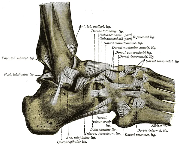
The distal (lower) tibiofibular syndesmosis is a fibrous joint, where four ligaments join the lower ends of the tibia and fibula1. The lower fibula is rounded and sits in a matching hollow notch in the tibia1. The notch is hollowed in about 60–75% of people and shallow in 25–40%1. Its depth ranges from 1.0 to 7.5 mm, and it is slightly shallower in women than in men1.
The four ligaments
Four ligaments (tough bands of tissue that hold bones together at a joint) hold the syndesmosis together2:
| Ligament | Where it runs | What stands out |
|---|---|---|
| Anterior inferior tibiofibular (front) | Slants down and out from the front of the lower tibia to the front of the fibula1 | The weakest of the four1 |
| Posterior inferior tibiofibular (back) | Slants down and out from the back of the lower tibia to the back of the fibula1 | Strong; the thickest in pooled cadaver (donated body) studies1,6 |
| Inferior transverse | Runs straight across from the fibula to the back rim of the tibia1 | Deepens the back of the socket like a labrum (a rim of tough tissue around a joint socket)1 |
| Interosseous | The thickened lower end of the interosseous membrane (the sheet joining the shinbone and fibula)1,2 | The widest where it meets the fibula, in pooled cadaver studies6 |
The front ligament crosses the front outer corner of the talus, and about 20% of it lies inside the ankle joint1. A meta-analysis (a study that pools the results of several earlier studies) took in 10 cadaver studies of 265 ankles6. It found the front, back and interosseous ligaments in every joint, and the inferior transverse ligament in 96%6. A systematic review (a study that searches for all the research on a question and sums it up) took in 12 cadaver studies7. They named and described these ligaments in quite different ways7.
What it does as the ankle moves
The syndesmosis keeps the ankle mortise stable and lets load pass through the ankle normally in walking2. It stops the tibia and fibula from spreading apart, which keeps the talus locked between them3. Its ligaments have been described as loaded through the whole stance phase of walking, when the foot is on the ground1.
The front ligament is slack when the foot points down1. It is stretched in dorsiflexion (pulling the foot up toward the shin), because the talus then pushes the two bones apart1. The back ligament behaves the same way1. The interosseous ligament is thought to work like a spring, letting the bones part slightly so the talus can wedge into the mortise1.
One study cut the ligaments of eight cadaver specimens one at a time8. It measured how much each resisted 2 mm of separation between the tibia and fibula8:
| Ligament, as the study named it | Share of the resistance (%) |
|---|---|
| Anterior inferior tibiofibular | 358 |
| Deep posterior inferior tibiofibular | 338 |
| Interosseous | 228 |
| Superficial posterior inferior tibiofibular | 98 |
An anatomy review cites an early study in which a 1 mm sideways shift of the talus cut the ankle joint’s contact area by 42%1. In 14 healthy people filmed with moving X-ray images during single-leg landings, the joint mainly turned and rotated outward after touchdown9. When they landed wearing a weighted vest, the joint rotated more9.
Breakdown
What an injury here means
Although the syndesmosis is a joint, “syndesmotic injury” in research means injury to its ligaments1. An anatomy textbook calls such an injury a syndesmotic or “high” ankle sprain3. The joint is often involved in ankle fractures and in injuries that turn the foot outward10. A European consensus panel (ESSKA-AFAS), drawing on a systematic review, recommends sorting acute (recent) injuries of the syndesmosis alone into stable or unstable11.
The front ligament is the weakest of the four, and the first to give way when a force turns the fibula outward1. The back ligament is thick and strong1. So too much stress more often pulls off a piece of the back of the tibia than tears it1. A systematic review took in 14 lab studies of how the joint moves12. In eight, cutting only the front ligament significantly changed that movement under an outward twist12.
In three of those studies, an injury to the deltoid ligament alone, on the inner ankle, significantly changed how the syndesmosis moved12. Unstable syndesmosis injuries can occur together with a deltoid ligament injury13.
An anatomy review reports syndesmotic injury in about half of Weber B fractures (the fibula breaks at the level of the ankle joint)1. It reports it in all Weber C fractures (the fibula breaks above that level)1. Studies cited in an anatomy review suggest a shallow notch may predispose to repeat ankle sprains1. It may also predispose to syndesmotic injury with a fracture-dislocation (a broken bone with the joint also forced out of place)1.
How it happens in sport
Video studies of professional players show the positions and contact involved14,15:
- In 13 injuries in male professional rugby union players, the knee most often fell inward, the ankle was pulled up and the foot turned out14.
- In the same 13, six came from a direct blow to the leg, and seven from the body collapsing over the ankle in a tackle14.
- In 12 professional soccer players, higher-grade injuries happened with the ankle pulled up and twisted outward15. The authors add that an inversion sprain (turning the sole inward) that tears the anterior talofibular ligament may spread upward into a lower-grade injury15. That ligament runs from the outer ankle bone to the talus16.
How often, and in whom
A 2010 anatomy review estimated that injury to the syndesmosis occurs in 1–11% of all ankle sprains1. One study read 261 MRI scans (scans that show soft tissue in detail) of athletes after a recent sprain17. Of these athletes, about 20% had this injury17.
A meta-analysis found outer-side sprains were the most common type, covered on the ankle sprain page18. In US hospital and emergency department data from eight states, syndesmotic injuries occurred at 2.09 per 100,000 person-years (people multiplied by years followed)19. These injuries tended to occur in younger patients19.
A study of 25 US college (NCAA) sports counted rates per 10,000 athlete-exposures (one athlete taking part in one practice or game)4:
| Group | High ankle sprains per 10,000 athlete-exposures |
|---|---|
| All 25 sports | 1.004 |
| Men’s football | 2.424 |
| Wrestling | 2.114 |
| Ice hockey | 1.194 |
In NCAA sports played by both sexes, men had a higher rate than women (rate ratio 1.77, one group’s rate divided by the other’s)4. Player contact was the most common cause (60.4%)4. Other studies add detail20,21:
- NCAA football: high ankle sprains were 24.6% of all ankle sprains, and nearly 14 times more likely in games than in practice20.
- Professional soccer: in 3677 male players, isolated injuries came to about one per team every three seasons21. Their rate rose each year from 2001 to 201621.
Recovery
Time away from sport
Studies report time lost in different groups5,23:
| Group studied | What was found |
|---|---|
| 63 young Australian sport participants | Median recovery of 62 days without surgery, against 15 days for outer-ankle sprains5 |
| US Military Academy cadets, incomplete injuries | 55 days, against 28 days for severe (third-degree) ankle sprains24 |
| US college (NCAA) football | Complete injuries cost significantly more time than partial ones, 31.3 against 15.8 days20 |
| Male professional soccer players | A mean of 39 days away after an isolated injury21 |
| Six case series of young, active patients | 0 to 137 days lost23 |
Return to sport
Two pooled analyses of elite athletes reached different figures25,26:
- A meta-analysis of 14 studies (901 elite athletes) found 96% returned to sport, after about 58 days on average25.
- A meta-analysis of 10 studies (440 elite college or professional athletes) estimated 99% returned, after a mean of 38 days26.
In the National Football League, 89.7% of players returned after a high ankle sprain, at an average of 80.5 days27. Older age at injury raised the risk of not returning27.
How ligaments heal
Two reviews of ligaments in general, not of the syndesmosis, describe how they heal28,29. After injury, scar tissue restores function, but it is weaker, biologically and mechanically, than the ligament it replaces29. The scar is also larger, and stretches more under a steady load (creep)28.
Blood reaches the syndesmosis mainly from the fibular (peroneal) artery, which runs down the back of the lower leg close to the fibula2. Branches of the tibial arteries help to a varying degree2. A cadaver study of 50 legs traced a branch of the peroneal artery30. It was the main supply to the front ligaments in 27 of the 43 specimens (63%) where the front blood supply could be seen clearly30. The authors conclude that an injury reaching about 3 cm above the ankle puts this branch at risk30.
Bone in the membrane, and the long view
A retrospective study (one that looks back at past cases) took in 44 football players31. Of the 22 with follow-up X-rays, 11 had grown bone in the interosseous membrane31. This is called heterotopic ossification, and those with it needed 11 more days to recover on average31. In US Military Academy cadets with incomplete injuries, 9 of 10 showed bone forming in the syndesmosis on X-rays about 20 months later24. None had developed lasting instability, loss of motion or arthritis24.
One case series (a report on a group of patients without a comparison group) followed 31 former school and college athletes32. They were followed a mean of 25 years after a high ankle sprain managed without surgery32. Of the 11 with X-rays, 27% showed osteoarthritis (a joint disease in which the cartilage lining the joint wears away)32. It was beyond Kellgren-Lawrence grade 2 (a scale for rating how worn a joint looks on an X-ray)32.
In the same series, sprains without widening on imaging were linked with acceptable self-reported function32. Arthritis was common but mostly not clinically significant32.
Connections
The bones and joint it belongs to
The tibia meets the fibula at two joints, and the syndesmosis is the lower one3. Three groups of ligaments steady the ankle joint10. They are the syndesmosis, the deltoid ligament on the inner side, and the outer ankle ligaments10.
The deep fibular nerve (a nerve of the lower leg) supplies the syndesmosis2. So does the sural nerve (a nerve that runs down the back of the calf to the outer foot)2. The tibial nerve (the main nerve at the back of the lower leg) does too2.
Exercises that load it
In 14 healthy people doing single-leg landings, the joint turned and rotated outward after touchdown9. Related exercise pages: drop landing9.
Ankle syndesmosis joins
Ankle syndesmosis holds together
Ankle syndesmosis shares the work of steadying the ankle with
Ankle syndesmosis can be injured together with
Ankle syndesmosis is injured in a share of
Ankle syndesmosis is loaded by
Sources
- Hermans JJ, Beumer A, de Jong TA, Kleinrensink GJ (2010). Anatomy of the distal tibiofibular syndesmosis in adults: a pictorial essay with a multimodality approach. Journal of anatomy. doi:10.1111/j.1469-7580.2010.01302.x PMID 21108526
- Lezak B, Varacallo MA (2026). Anatomy, Bony Pelvis and Lower Limb: Distal Tibiofibular Joint (Tibiofibular Syndesmosis).. StatPearls. PMID 31613435
- Betts JG, Young KA, Wise JA, Johnson E, Poe B, Kruse DH, Korol O, Johnson JE, Womble M, DeSaix P (2022). Anatomy and Physiology 2e. OpenStax. Link
- Mauntel TC, Wikstrom EA, Roos KG, Djoko A, Dompier TP, Kerr ZY (2017). The Epidemiology of High Ankle Sprains in National Collegiate Athletic Association Sports. The American journal of sports medicine. doi:10.1177/0363546517701428 PMID 28423285
- Sman AD, Hiller CE, Rae K, Linklater J, Black DA, Refshauge KM (2014). Prognosis of ankle syndesmosis injury. Medicine and science in sports and exercise. doi:10.1249/MSS.0000000000000151 PMID 24002346
- Yammine K, Jalloul M, Assi C (2022). Distal tibiofibular syndesmosis: A meta-analysis of cadaveric studies. Morphologie : bulletin de l'Association des anatomistes. doi:10.1016/j.morpho.2021.10.004 PMID 34801386
- Anand Prakash A (2020). Anatomy of Ankle Syndesmotic Ligaments: A Systematic Review of Cadaveric Studies. Foot & ankle specialist. doi:10.1177/1938640019897214 PMID 31904271
- Ogilvie-Harris DJ, Reed SC, Hedman TP (1994). Disruption of the ankle syndesmosis: biomechanical study of the ligamentous restraints. Arthroscopy : the journal of arthroscopic & related surgery : official publication of the Arthroscopy Association of North America and the International Arthroscopy Association. doi:10.1016/s0749-8063(05)80014-3 PMID 7999167
- Luo Y, Li F, Huang X, Wu D, Zhao Y, Wang S (2025). In Vivo Kinematics of the Distal Tibiofibular Syndesmosis With Different Loading Weights. Foot & ankle international. doi:10.1177/10711007251315163 PMID 39932276
- Brockett CL, Chapman GJ (2016). Biomechanics of the ankle.. Orthopaedics and Trauma. doi:10.1016/j.mporth.2016.04.015 PMID 27594929
- van Dijk CN, Longo UG, Loppini M, Florio P, Maltese L, Ciuffreda M, Denaro V (2016). Classification and diagnosis of acute isolated syndesmotic injuries: ESSKA-AFAS consensus and guidelines. Knee surgery, sports traumatology, arthroscopy : official journal of the ESSKA. doi:10.1007/s00167-015-3942-8 PMID 26704800
- Spennacchio P, Seil R, Gathen M, Cucchi D (2021). Diagnosing instability of ligamentous syndesmotic injuries: A biomechanical perspective. Clinical biomechanics (Bristol, Avon). doi:10.1016/j.clinbiomech.2021.105312 PMID 33756400
- Fleury C, Acker A, Dubois-Ferrière V, Assal M (2026). Deltoid ligament injury: thinking beyond the ankle - an expert opinion.. EFORT Open Reviews. doi:10.1530/EOR-2025-0268 PMID 42684953
- Delahunt E, Farrell G, Boylan A, Kerin F, Tierney P, Hogan H, Boreham C (2021). Mechanisms of acute ankle syndesmosis ligament injuries in professional male rugby union players: a systematic visual video analysis. British journal of sports medicine. doi:10.1136/bjsports-2020-102281 PMID 33051214
- Jain N, Murray D, Kemp S, Calder J (2023). Republication of "High-Speed Video Analysis of Syndesmosis Injuries in Soccer-Can It Predict Injury Mechanism and Return to Play? A Pilot Study". Foot & ankle orthopaedics. doi:10.1177/24730114231195048 PMID 37578852
- Golanó P, Vega J, de Leeuw PA, Malagelada F, Manzanares MC, Götzens V, van Dijk CN (2010). Anatomy of the ankle ligaments: a pictorial essay.. Knee Surgery, Sports Traumatology, Arthroscopy. doi:10.1007/s00167-010-1100-x PMID 20309522
- Roemer FW, Jomaah N, Niu J, Almusa E, Roger B, D'Hooghe P, Geertsema C, Tol JL, Khan K, Guermazi A (2014). Ligamentous Injuries and the Risk of Associated Tissue Damage in Acute Ankle Sprains in Athletes. The American journal of sports medicine. doi:10.1177/0363546514529643 PMID 24753239
- Doherty C, Delahunt E, Caulfield B, Hertel J, Ryan J, Bleakley C (2014). The incidence and prevalence of ankle sprain injury: a systematic review and meta-analysis of prospective epidemiological studies.. Sports Medicine. doi:10.1007/s40279-013-0102-5 PMID 24105612
- Vosseller JT, Karl JW, Greisberg JK (2014). Incidence of syndesmotic injury. Orthopedics. doi:10.3928/01477447-20140225-53 PMID 24762148
- Hunt KJ, George E, Harris AH, Dragoo JL (2013). Epidemiology of syndesmosis injuries in intercollegiate football. Clinical journal of sport medicine : official journal of the Canadian Academy of Sport Medicine. doi:10.1097/JSM.0b013e31827ee829 PMID 23339895
- Lubberts B, D'Hooghe P, Bengtsson H, DiGiovanni CW, Calder J, Ekstrand J (2017). Epidemiology and return to play following isolated syndesmotic injuries of the ankle: a prospective cohort study of 3677 male professional footballers in the UEFA Elite Club Injury Study. British journal of sports medicine. doi:10.1136/bjsports-2017-097710 PMID 29269487
- Prakash AA (2020). Epidemiology of High Ankle Sprains: A Systematic Review. Foot & ankle specialist. doi:10.1177/1938640020916266 PMID 32924583
- Jones MH, Amendola A (2007). Syndesmosis sprains of the ankle. Clinical orthopaedics and related research. doi:10.1097/BLO.0b013e31802eb471 PMID 17146360
- Hopkinson WJ, St Pierre P, Ryan JB, Wheeler JH (1990). Syndesmosis sprains of the ankle. Foot & ankle. doi:10.1177/107110079001000607 PMID 2113510
- Li R, Sun C, Qi P, Xu Z (2025). Return-to-sport rate and time in elite athletes after ankle syndesmosis injuries: a systematic review and meta-analysis. Journal of orthopaedic surgery and research. doi:10.1186/s13018-025-06566-6 PMID 41476226
- Bolia IK, Bogdanov J, Schoell K, Ouyang Y, Petrigliano FA, Weber AE, Tan EW (2023). Elite Athletes Successfully Return to the Preinjury Level of Sport Following Ankle Syndesmosis Injuries: A Systematic Review and Meta-Analysis. Clinical journal of sport medicine : official journal of the Canadian Academy of Sport Medicine. doi:10.1097/JSM.0000000000001019 PMID 36599363
- DeFroda SF, Bodendorfer BM, Hartnett DA, Milner JD, Yang DS, Silber ZS, Forsythe B (2022). Defining the contemporary epidemiology and return to play for high ankle sprains in the National Football League. The Physician and sportsmedicine. doi:10.1080/00913847.2021.1924046 PMID 33906554
- Hildebrand KA, Frank CB (1998). Scar formation and ligament healing. Canadian journal of surgery. Journal canadien de chirurgie. PMID 9854530
- Frank CB (2004). Ligament structure, physiology and function. Journal of musculoskeletal & neuronal interactions. PMID 15615126
- McKeon KE, Wright RW, Johnson JE, McCormick JJ, Klein SE (2012). Vascular anatomy of the tibiofibular syndesmosis. The Journal of bone and joint surgery. American volume. doi:10.2106/JBJS.K.00604 PMID 22617922
- Taylor DC, Englehardt DL, Bassett FH 3rd (1992). Syndesmosis sprains of the ankle. The American journal of sports medicine. doi:10.1177/036354659202000209 PMID 1558241
- Nussbaum ED, Silver J, Rozenberg A, Mazzeferro N, Buckley PS, Gatt CJ Jr (2024). Nonoperative Management of High Ankle Sprains: A Case Series With ≥18-Year Follow-up. The American journal of sports medicine. doi:10.1177/03635465241271593 PMID 39235770
Changes
- A supporting claim was narrowed to match its source; the page text is unchanged.