Common fibular nerve
Also called common peroneal nerve, peroneal nerve, fibular nerve, superficial fibular nerve, superficial peroneal nerve, deep fibular nerve, deep peroneal nerve
The nerve that wraps around the outer side of the knee and works the muscles that lift the foot and turn it out. Where it runs, why it is exposed at the fibula, and what studies report about recovery.
- Region
- Lower leg
- Updated
- October 5, 2026
- Reviewed
- October 5, 2026 · how entries are checked
Function
Where it starts and what it does
The common fibular nerve and the common peroneal nerve are the same nerve, and “peroneal” is the older name2. It is one of the two major branches of the sciatic nerve, the large nerve that runs down the back of the thigh1. Its fibres come from the spinal levels labelled L4 to S21. The sciatic nerve typically splits behind the knee into this nerve and the tibial nerve7.
The nerve has two main branches, and each powers different movements3:
- the deep branch lifts the foot and straightens the toes3;
- the superficial branch turns the sole outward, a movement called eversion3.
Its path around the knee
The nerve leaves the sciatic nerve above the back of the knee1. It runs down the outer back of the leg under the long head of the biceps femoris, one of the hamstring muscles1. The biceps femoris is the only muscle it supplies above the knee8. It then passes behind the head of the fibula, the bone’s small, knob-like upper end1,9.
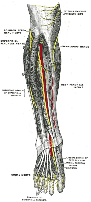
About 2 cm below the fibular head, the nerve wraps around the neck of the bone3. At the top of the fibula it runs through the fibular tunnel, a passage with bone for its floor8. The tunnel’s roof is an arch formed by two lower-leg muscles, the peroneus longus and the soleus8. At the fibular neck the nerve divides into its superficial and deep branches1.
The two branches
The superficial fibular nerve is the motor supply of the peroneus longus and peroneus brevis, two of the peroneal muscles10. About 12 cm above the ankle joint it leaves these muscles through a gap in the deep fascia, the tough sheet wrapping the leg10. The branch’s skin fibres supply the front outer leg, the top of the foot and the tops of the toes, apart from one patch10.
The deep fibular nerve runs down the leg beside the anterior tibial artery, between two muscles1. It works the four muscles in the anterior compartment, the space at the front of the lower leg8. One of them is the tibialis anterior, the muscle at the front of the shin that pulls the foot up8. Its skin fibres supply that patch, the first web space, where the skin spans the great and second toes11.
Breakdown
Why the nerve is exposed at the fibula
At the neck of the fibula, only skin and the tissue just beneath it cover the nerve4. That leaves it open to being pressed against the bone4. Trauma can also stretch it there8. An imaging review adds that the nerve has little supporting tissue of its own, which makes compression more likely8.
The nerve can be injured anywhere along its course, but compression is most common at the fibular head12. The usual result is a sudden foot drop, complete or partial12. One medical reference text defines foot drop as being unable to lift the front of the foot because the muscles that lift it are weak2. The same text notes that foot drop has diverse causes, which may include muscle, nerve, spine and autoimmune disorders2.
Pressure from outside
Causes of compression without an injury include tight bandages, long spells of leg crossing and positioning under anaesthesia3. A problem in this nerve at the fibular head that follows weight loss is known as slimmer’s paralysis4. With weight loss the fat around the fibular head shrinks, so the nerve loses its padding against the bone4.
Knee injuries
The common peroneal nerve is the lower limb’s most often injured nerve in people with trauma13. Stretch injuries can follow ankle sprains where the sole turns inward, upper fibula fractures and dislocation of the knee8. The more the bones and ligaments are damaged, the more likely the nerve is damaged too8.
Reported rates after injuries to several knee ligaments at once range from 4.5% to 40%14. The spread probably reflects how much energy the accidents in each series involved14. Several studies tie the nerve injury to the posterolateral corner, the ligaments and tendons at the outer back of the knee15,16. That group includes the lateral collateral ligament.
| Group studied | Share with a peroneal nerve injury |
|---|---|
| 43,869 knee dislocations, in a systematic review of 132 studies | 15.3%5 |
| 221 multiligament knee injuries at two centres | 19.5%, every one with a posterolateral corner injury15 |
| 61 posterolateral corner injuries that had surgery | 26% when first seen17 |
| 100 multiligament knee injuries, in a case series at one centre | 31% with a posterolateral corner injury, 4% without one16 |
A chart review covered 17 adults with a traumatic dislocation of the joint between the tops of the tibia (shinbone) and fibula18. Seven had a peroneal nerve deficit before surgery, and six of those had a lasting deficit or an amputation18.
In sport
One centre reviewed 448 peroneal nerve injuries seen between 1970 and 2010, and 84 were sports related19. The same review concluded that the injury at the knee in sport is usually a stretch or a contusion (bruise)19.
| Sport | Cases among the 84 |
|---|---|
| Skiing | 4219 |
| Football | 2319 |
| Soccer | 819 |
| Basketball | 619 |
In competitive athletes, a trapped peroneal nerve is a less common cause of leg pain brought on by exercise20. One sports practice reported seven runners and one soccer player with exercise-related compression of the nerve at the fibular neck21.
At the ankle and foot
Studies of ankle sprains give different pictures of how often the nerve is involved22,23. In 66 patients tested two weeks after a sprain, 17% of grade II and 86% of grade III sprains showed peroneal nerve injury22. The test was electromyography, which picks up a muscle’s electrical signals22. Another paper calls peroneal palsy with an ankle sprain rare, with few reported cases and no known true rate23.
The deep branch passes under a retinaculum, a band that holds tendons in place, just above the ankle joint24. An anatomy paper lists reasons it gets compressed there25:
- direct trauma25;
- the foot pointed far down in high-heeled shoes and high boots25;
- constant mechanical irritation, such as from tightly laced shoes25;
- a clot in the dorsalis pedis artery, an artery on the top of the foot25.
One 1979 report described the tongue of a ski boot pressing on the peroneal nerve at the ankle26. Trapping of the superficial branch is relatively rare27. Its pain and tingling tend to be made worse by walking and exercise27.
Recovery
After pressure on the nerve
A review of nerve tissue injury describes neurapraxia, the mildest grade, as a short-term signal block that clears fully within 12 weeks28. It describes injury to the fibres themselves as followed by slow regrowth, about 1 mm a day, often with incomplete recovery28. Two studies of pressure palsies in this nerve report recovery over weeks to months29,30:
- In 30 patients with sudden palsy after weight loss, a signal block at the fibular head cleared in 29 within three weeks to three months29.
- Sixteen farmworkers with palsy from squatting at work all recovered without surgery, in 7.4 weeks on average30.
After knee dislocation
A systematic review of 13 articles covered 214 palsies after knee dislocation6. Functional muscle recovery followed 38.4% of complete palsies, and full recovery followed 87.3% of partial ones6. In the same review, younger age predicted recovery of the nerve6.
In 61 posterolateral corner injuries, 10 complete nerve injuries were stretches, not cuts; all had an operation to free the nerve, and 5 of the 10 regained full function17. The deep branch often recovers less well than the superficial one after a stretch injury at the knee14. A review relates this to its closeness to the joint and its tether further down, which give a more severe stretch14.
What surgery series reported
Decompression is surgery to take pressure off a nerve. Neurolysis is an operation that frees a nerve from the tissue around it. A graft is a piece of tissue moved to rebuild a damaged part. In a systematic review of traumatic injuries, better outcomes went with a shorter wait before surgery, a shorter graft and younger age13.
| Group studied | What was reported |
|---|---|
| 60 patients who had decompression for a trapped nerve, followed 42 months on average | Muscle recovery was good in 33 of the 38 with both feeling and muscle symptoms beforehand31 |
| 368 patients, in a meta-analysis (a study pooling earlier studies) of neurolysis | 53.9% reached full strength32 |
| 42 studies of traumatic injury, in a systematic review | Useful muscle strength in 81.4% after neurolysis, 78.8% after end-to-end stitching, 49.0% after grafting and 62.9% after nerve transfer13 |
| 1,577 nerve repairs, in a literature review of 28 studies | Good muscle outcomes in 44% of repairs done within 6 months, against 12% after 12 months33 |
A scoping review, which maps what research exists, looked at trapped peroneal nerves with foot drop34. Good outcomes ranged from 0% to 100% without surgery and from 40% to 100% after neurolysis34. It rested mostly on case series, and no study compared the two34.
Connections
Nerves, bones and the knee
The tibial nerve is the other branch of the sciatic nerve7. In knee dislocation the common peroneal nerve is injured more often than the tibial nerve14. A review puts this down to its course around the outer back of the knee, held close to the neck of the fibula14.
In 303 patients with knee dislocations, the odds of injury to this nerve were 42 times higher when the posterolateral corner was injured35. That corner includes the lateral collateral ligament. In 16 cadaver legs loaded to mimic an ankle sprain, strain (stretch as a share of resting length) in the superficial branch rose with load36.
Muscles and exercises
The superficial branch supplies the peroneus longus and brevis, described on the peroneals page37,38,39. The deep branch supplies the anterior compartment muscles, including the tibialis anterior, whose tendon has its own page: tibialis anterior tendon40. In an electromyography study of 13 runners, the front lower-leg muscles contracted while shortening at first ground contact41. After 12 weeks of fast foot-lifting training in five people, tibialis anterior motor units switched on earlier in fast contractions42. A motor unit is a nerve cell and the muscle fibres it switches on.
Related exercise pages: tibialis raise, running41,42.
Common fibular nerve leaves the sciatic nerve beside
Common fibular nerve wraps around the neck of
Common fibular nerve supplies, through its superficial branch,
Common fibular nerve supplies, through its deep branch, the muscle of
Common fibular nerve supplies the biceps femoris, one of the
Common fibular nerve is often injured together with the outer back corner of the knee, which includes
Common fibular nerve can be stretched in a dislocation of
Common fibular nerve can be stretched in
Common fibular nerve supplies the muscles worked in
Common fibular nerve supplies the shin muscles used in
Sources
- Hardin JM, Devendra S (2022). Anatomy, Bony Pelvis and Lower Limb: Calf Common Peroneal Nerve (Common Fibular Nerve).. StatPearls. PMID 30422563
- Nori SL, Stretanski MF (2025). Foot Drop.. StatPearls. PMID 32119280
- Capodici A, Hagert E, Darrach H, Curtin C (2022). An overview of common peroneal nerve dysfunction and systematic assessment of its relation to falls. International orthopaedics. doi:10.1007/s00264-022-05593-w PMID 36169699
- Cucu LE, Popescu G, Maștaleru A, Ignat EB, Grosu C, Bîrsanu L, Leon MM (2025). Common Peroneal Nerve Paralysis Following Rapid Weight Loss-A Case Report and Literature Review. Nutrients. doi:10.3390/nu17111782 PMID 40507050
- Randall ZD, Strok MJ, Mazzola JW, Agrawal R, Yaeger LH, Berkes MB (2024). The known and unknown reality of knee dislocations: A systematic review. Injury. doi:10.1016/j.injury.2024.111904 PMID 39357194
- Woodmass JM, Romatowski NP, Esposito JG, Mohtadi NG, Longino PD (2015). A systematic review of peroneal nerve palsy and recovery following traumatic knee dislocation. Knee surgery, sports traumatology, arthroscopy : official journal of the ESSKA. doi:10.1007/s00167-015-3676-7 PMID 26115847
- Liyew WA, Abebe S (2026). Anatomical variation of the sciatic nerve division in the cadaver studies: a systematic review and meta-analysis.. Anatomy & cell biology. doi:10.5115/acb.25.139 PMID 41851007
- Van den Bergh FR, Vanhoenacker FM, De Smet E, Huysse W, Verstraete KL (2013). Peroneal nerve: Normal anatomy and pathologic findings on routine MRI of the knee. Insights into imaging. doi:10.1007/s13244-013-0255-7 PMID 23709403
- Betts JG, Young KA, Wise JA, Johnson E, Poe B, Kruse DH, Korol O, Johnson JE, Womble M, DeSaix P (2022). Anatomy and Physiology 2e. OpenStax. Link
- Garrett A, Black AC, Launico MV, Geiger Z (2023). Anatomy, Bony Pelvis and Lower Limb: Superficial Peroneal Nerve (Superficial Fibular Nerve).. StatPearls. PMID 30521214
- Palmisano LL, Launico MV (2025). Anatomy, Bony Pelvis and Lower Limb: Calf Deep Peroneal Nerve (Deep Fibular Nerve).. StatPearls. PMID 30252289
- Poage C, Roth C, Scott B (2016). Peroneal Nerve Palsy. The Journal of the American Academy of Orthopaedic Surgeons. doi:10.5435/JAAOS-D-14-00420 PMID 26700629
- Mackay MJ, Ayres JM, Harmon IP, Tarakemeh A, Brubacher J, Vopat BG (2022). Traumatic Peroneal Nerve Injuries. JBJS reviews. doi:10.2106/JBJS.RVW.20.00256 PMID 35020680
- Samson D, Ng CY, Power D (2016). An evidence-based algorithm for the management of common peroneal nerve injury associated with traumatic knee dislocation. EFORT open reviews. doi:10.1302/2058-5241.160012 PMID 28461914
- Markus DH, Mojica ES, Bi A, Kahan JB, Moran J, Mannino BJ, Alaia EF, Jazrawi LM, Medvecky MJ, Alaia MJ (2022). Relationship Between Peroneal Nerve and Anterior Cruciate Ligament Involvement in Multiligamentous Knee Injury: A Multicenter Study. The Journal of the American Academy of Orthopaedic Surgeons. doi:10.5435/JAAOS-D-21-01252 PMID 36326829
- Kahan JB, Li D, Schneble CA, Huang P, Bullock J, Porrino J, Medvecky MJ (2020). The Pathoanatomy of Posterolateral Corner Ligamentous Disruption in Multiligament Knee Injuries Is Predictive of Peroneal Nerve Injury. The American journal of sports medicine. doi:10.1177/0363546520962503 PMID 33074710
- Ridley TJ, McCarthy MA, Bollier MJ, Wolf BR, Amendola A (2018). The incidence and clinical outcomes of peroneal nerve injuries associated with posterolateral corner injuries of the knee. Knee Surgery, Sports Traumatology, Arthroscopy. doi:10.1007/s00167-016-4417-2 PMID 28160014
- Rajan PV, Ramski DE, Romeo NM (2022). Traumatic Dislocation of the Proximal Tibiofibular Joint: A Systematic Review and 10-year Experience From a Level 1 Trauma Center. Journal of the American Academy of Orthopaedic Surgeons. Global research & reviews. doi:10.5435/JAAOSGlobal-D-21-00105 PMID 35503776
- Cho D, Saetia K, Lee S, Kline DG, Kim DH (2011). Peroneal nerve injury associated with sports-related knee injury. Neurosurgical focus. doi:10.3171/2011.9.FOCUS11187 PMID 22044100
- Mitra A, Stern JD, Perrotta VJ, Moyer RA (1995). Peroneal nerve entrapment in athletes. Annals of plastic surgery. doi:10.1097/00000637-199510000-00006 PMID 8585678
- Leach RE, Purnell MB, Saito A (1989). Peroneal nerve entrapment in runners. The American journal of sports medicine. doi:10.1177/036354658901700224 PMID 2757134
- Nitz AJ, Dobner JJ, Kersey D (1985). Nerve injury and grades II and III ankle sprains. The American journal of sports medicine. doi:10.1177/036354658501300306 PMID 4014532
- Mitsiokapa E, Mavrogenis AF, Drakopoulos D, Mauffrey C, Scarlat M (2017). Peroneal nerve palsy after ankle sprain: an update. European journal of orthopaedic surgery & traumatology : orthopedie traumatologie. doi:10.1007/s00590-016-1845-0 PMID 27581424
- Gonzalez J, Bergman R (2026). Anterior Tarsal Tunnel Syndrome.. StatPearls. PMID 30860723
- Banu J, Dhakshnamoorthy N, Sakthivel S (2024). Unravelling the Anatomy of the Anterior Tarsal Tunnel and Its Clinical Implications. Cureus. doi:10.7759/cureus.64282 PMID 39130935
- Lindenbaum BL (1979). Ski boot compression syndrome. Clinical orthopaedics and related research. PMID 113156
- Matsumoto J, Isu T, Kim K, Iwamoto N, Yamazaki K, Isobe M (2018). Clinical Features and Surgical Treatment of Superficial Peroneal Nerve Entrapment Neuropathy. Neurologia medico-chirurgica. doi:10.2176/nmc.oa.2018-0039 PMID 29925720
- Grinsell D, Keating CP (2014). Peripheral nerve reconstruction after injury: a review of clinical and experimental therapies.. BioMed Research International. doi:10.1155/2014/698256 PMID 25276813
- Cruz-Martinez A, Arpa J, Palau F (2000). Peroneal neuropathy after weight loss. Journal of the peripheral nervous system : JPNS. doi:10.1046/j.1529-8027.2000.00007.x PMID 10905469
- Sipahioğlu S, Zehir S, Aşkar H, Işıkan UE (2015). Peroneal nerve palsy secondary to prolonged squatting in seasonal farmworkers. Acta orthopaedica et traumatologica turcica. PMID 25803253
- Fabre T, Piton C, Andre D, Lasseur E, Durandeau A (1998). Peroneal nerve entrapment. The Journal of bone and joint surgery. American volume. doi:10.2106/00004623-199801000-00009 PMID 9469308
- Chow AL, Levidy MF, Luthringer M, Vasoya D, Ignatiuk A (2021). Clinical Outcomes After Neurolysis for the Treatment of Peroneal Nerve Palsy. Annals of plastic surgery. doi:10.1097/SAP.0000000000002833 PMID 34397520
- George SC, Boyce DE (2014). An evidence-based structured review to assess the results of common peroneal nerve repair. Plastic and reconstructive surgery. doi:10.1097/PRS.0000000000000318 PMID 25068351
- Oosterbos C, Decramer T, Rummens S, Weyns F, Dubuisson A, Ceuppens J, Schuind S, Groen J, van Loon J, Rasulic L, Lemmens R, Theys T (2022). Evidence in peroneal nerve entrapment: A scoping review. European journal of neurology. doi:10.1111/ene.15145 PMID 34662481
- Moatshe G, Dornan GJ, Løken S, Ludvigsen TC, LaPrade RF, Engebretsen L (2017). Demographics and Injuries Associated With Knee Dislocation: A Prospective Review of 303 Patients. Orthopaedic Journal of Sports Medicine. doi:10.1177/2325967117706521 PMID 28589159
- O'Neill PJ, Parks BG, Walsh R, Simmons LM, Miller SD (2007). Excursion and strain of the superficial peroneal nerve during inversion ankle sprain. The Journal of bone and joint surgery. American volume. doi:10.2106/JBJS.F.00440 PMID 17473134
- Davda K, Malhotra K, O'Donnell P, Singh D, Cullen N (2017). Peroneal tendon disorders. EFORT Open Reviews. doi:10.1302/2058-5241.2.160047 PMID 28736620
- Basit H, Shah J, Siccardi MA (2023). Anatomy, Bony Pelvis and Lower Limb, Foot Peroneus Brevis Muscle.. StatPearls. PMID 30571048
- Olewnik Ł, Landfald IC, Domosławska D, Ruzik K, Triantafyllou G, Piagkou M, Vázquez T (2025). Comparative and developmental anatomy of the fibularis brevis muscle: morphological variants and their clinical significance. Frontiers in cell and developmental biology. doi:10.3389/fcell.2025.1647407 PMID 40894923
- Lezak B, Launico MV (2026). Anatomy, Bony Pelvis and Lower Limb: Leg Anterior Compartment.. StatPearls. PMID 30969547
- Mann RA, Hagy J (1980). Biomechanics of walking, running, and sprinting.. The American journal of sports medicine. doi:10.1177/036354658000800510 PMID 7416353
- Van Cutsem M, Duchateau J, Hainaut K (1998). Changes in single motor unit behaviour contribute to the increase in contraction speed after dynamic training in humans.. The Journal of Physiology. doi:10.1111/j.1469-7793.1998.295by.x PMID 9782179
Changes
- First published.