Tibia
Also called shin bone, shinbone
The shin bone, the main weight-bearing bone of the lower leg. How it carries load, how it breaks down under repeated stress, how bone adapts and heals, and what attaches to it.
- Region
- Lower leg
- Muscles
- soleus, tibialis anterior, hamstrings, quadriceps, gracilis, Peroneals, Hip flexors
- Updated
- September 30, 2026
- Reviewed
- September 30, 2026 · how entries are checked
Function
Where it sits and what it carries
The tibia, or shin bone, is the inner and larger of the two lower-leg bones; its partner is the fibula1. Of the two, it is the main weight-bearing bone1. It is the second-longest bone in the body, after the femur (thigh bone)1.
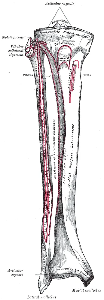
Its shape, from knee to ankle
At the top, the tibia widens into two condyles (rounded knobs at the end of a bone)1. The flat tops of these condyles meet the femur, and together they form the knee joint1. Between these two surfaces is a raised area, the intercondylar eminence, where two knee ligaments attach1.
On the front of the upper tibia is the tibial tuberosity (a bump just below the knee where the patellar ligament attaches)1.
Below the knee, the shaft becomes three-sided in cross-section1. Its front edge, the anterior border, runs from the tibial tuberosity down the length of the bone1. The front edge and the inner surface both sit just beneath the skin1.
At the bottom, the tibia forms the medial malleolus, the bony bump on the inner ankle1. The malleolus and the tibia’s lower end meet the talus (ankle bone) as part of the ankle joint1.
How it is joined to the fibula
The tibia meets the fibula at two joints1:
- At the top, the head of the fibula sits against the underside of the tibia’s outer condyle1.
- At the bottom, the lower fibula sits in a notch in the tibia1.
Between them, a ridge on the outer side of the tibial shaft anchors the interosseous membrane1. This is a sheet of dense connective tissue joining the two bones1.
The loads it takes in running
Two people had strain gauges (small sensors that measure stretch) fixed to the tibia8. Strain is how far something stretches as a share of its resting length. In some cases, it neared 2000 microstrain (millionths of the resting length) in zigzag running uphill and downhill8. That was about three times the strain of walking8.
A modelling study (one that estimates forces with a model) looked at 10 male runners2. At 3.5 to 4 metres a second, the lower tibia was squeezed with a peak force of about 9 times body weight2. In the same runners, the pull of muscles was the main source of squeezing on the lower tibia2. A review notes that, in modelling of running with a heel-first landing, muscle forces and tibial squeezing far exceed the push of the ground9.
Breakdown
Bone stress injuries
An international expert consensus describes bone stress injuries as overuse injuries to bone10. They are commonly called stress reactions and stress fractures10.
A review explains that ordinary loading causes tiny damage in bone, which the bone repairs9. The review argues that a bone stress injury follows when damage builds faster than the bone can repair or resist it9. Citing earlier work, a meta-analysis of runners describes tibial stress fracture the same way11. It also cites a finding that runners are five times more likely to have another after a first one11.
Who gets them
The tibia is a common site of stress fracture in several groups studied3,4:
| Group studied | What was found |
|---|---|
| Military populations, 70 studies | The tibia, the fibula and the metatarsals (long bones in the middle of the foot) were the most common stress fracture sites3 |
| 111 competitive track and field athletes, 12 months | The tibia was the most common site: 12 injuries, 46% of the total4 |
One systematic review (a study that gathers and sums up the research) found far more stress fractures in military recruits than qualified personnel3. Older and female personnel were at higher risk3.
Factors linked with stress fractures
A 2006 review sorts risk factors for stress fracture into two groups5. Extrinsic factors include the type of activity, training, equipment and surroundings5. Intrinsic factors include bone, muscle, joint and movement factors, fitness and sex5.
In military recruits, one systematic review lists further factors3. These include being underweight, starting training without regular prior exercise, low vitamin D and heavy drinking3. A review notes distance runners have some of the highest rates of bone stress injury, with poor bone health a contributor9.
One cross-sectional study (a group looked at once) compared 36 male distance runners12. Those with a past bone stress injury had tibias about 9% more slender12. Their tibias also had lower estimated bending strength12.
A meta-analysis (pooling several studies into one answer) of 14 studies compared runners with tibial stress fracture and others11. Peak ground forces did not differ significantly11. Most of those studies were small and looked back in time11. Single studies reported larger tibial stress and tibial acceleration in runners with the fracture, but many of these findings came from a single study, and the samples were small11.
Shin splints
Medial tibial stress syndrome, often called shin splints, is a common condition in active people13. It is felt as spread-out pain along the back inner edge of the tibia13. Two meta-analyses looked at factors linked with it13,14:
- One found five: female sex, more body weight, more navicular drop (how far the inner arch sinks when standing), a past running injury, and more outward hip turning when bent13.
- The other, in physically active people, found higher body mass index (a measure of weight for height) and greater navicular drop were risk factors14. So was more range in pointing the foot down and turning the hip outward14. Dorsiflexion (pulling the foot up toward the shin) range and the Q-angle (the angle between thigh and shin, seen from the front) were not14.
A systematic review of 17 studies found higher peak soleus (a wide, flat calf muscle) activity during push-off among the factors most strongly linked with developing it15. The same review said it is still unknown whether this is a cause or an effect15.
A review looked at 11 trials of approaches to shin splints16. It rated every randomised trial (one that assigns people to groups by chance) at high risk of bias, meaning flaws may have skewed the results16. It judged no approach free enough of bias to recommend, and named shockwave therapy (pulses of sound energy aimed through the skin) the most promising16.
The same review pooled trials of a lower-leg brace and found no significant difference in outcomes with it than without one16.
Breaks from a single injury
In a population study of 198 breaks of the tibial shaft, there were 16.9 breaks per 100,000 people a year17.
Recovery
How bone adapts to load
A review describes bone as adapting to the loads it regularly carries6. The review adds that loading too light and too rare may lead to bone being broken down6. In 14 crew members on 4- to 6-month space station missions, hip bone density fell by 1.4 to 1.5% a month18.
In 90 male British Army recruits, tibial bone density rose at every site measured over the first 10 weeks of training7. The shaft also gained thickness and estimated strength7. The changes in density and shape were mostly in the front and inner-front of the bone, and along its front-to-back axis7. The authors conclude the bone grew stronger through higher density and new bone on its outer surface7.
Studies of athletes compared their tibias with those of less active people19,20,21:
- In 30 trained distance runners, scans showed greater strength measures in the tibia than in 30 untrained people, unlike their fibulas19.
- Among 113 national-level female athletes, those in impact sports had larger tibial shafts than 30 non-athletes; swimmers did not20.
- In master athletes aged 35 to 94, tibial shaft bone mineral and bending resistance were highest in sprinters, then longer-distance runners, race-walkers and inactive people21.
The authors of the master athletes study note a second possibility: people with larger bones may be more drawn to athletics in the first place21.
How damage is repaired, and how long it takes
A review explains that two kinds of bone cell do the repair: osteoclasts remove damaged bone and osteoblasts replace it9. According to one review, removing damaged compact bone takes about 4 weeks, replacing it can take three months, and full mineralisation up to a year9. While the gaps left by removal remain, the bone resists fatigue less well9.
A review reports that repair usually restores a broken bone’s structure and function, but about 10% of fractures do not heal normally22. A physiology textbook sets out the stages. After a break, clotting blood forms a mass, then cartilage-forming and bone-forming cells build calluses that steady the break1. Over the following weeks, the callus cartilage is replaced by bone, and remodelling often leaves no outward sign1.
What studies found on return to sport
Across 76 studies of bone stress injuries in the legs and pelvis, more than 90% of athletes returned to sport23. Time to return depended on the site23. In the same pooled data, injuries on the back inner tibial shaft had the shortest return time, 44 days, and among the lowest complication rates23. Injuries on the front of the shaft had among the highest23.
Another meta-analysis compared sites by bone type24. Injuries where spongy (trabecular) bone is common, such as the pelvis, healed more slowly than where compact (cortical) bone is common, such as the tibia24.
For breaks of the shaft from a single injury, a review of 16 studies found 92% returned to sport after surgery and 67% without it25. Most returned to sport, but a limited share reached their earlier level25.
Connections
The knee
The anterior and posterior cruciate ligaments (the two ligaments that cross inside the knee) are anchored to the tibia between its condyles1. The patella (kneecap) forms a joint only with the femur, not with the tibia1. Instead, the patellar ligament runs from the kneecap to the front of the tibia1. It continues the tendon of the quadriceps (the four muscles on the front of the thigh) and attaches at the tibial tuberosity1.
Muscles attached to it
Several muscles start or end on the tibia26,27,28:
| Muscle | Where it attaches |
|---|---|
| Soleus | Starts partly from the soleal line on the back of the tibia and from its inner border1,26 |
| Tibialis anterior (the muscle at the front of the shin that pulls the foot up) | Starts from the outer condyle and upper two-thirds of the outer surface27 |
| Semimembranosus and semitendinosus, two of the hamstrings | End on the inner side of the tibia at the knee28 |
| Biceps femoris (a hamstring on the outer back of the thigh) | Ends on the top of the fibula and the outer tibia28 |
| Gracilis (a long, thin inner thigh muscle), one of the hip adductors (the inner thigh muscles) | Ends on the upper inner tibia29 |
The soleal line is a rough diagonal ridge across the upper back of the tibia, where muscles of the back of the leg attach1.
Tibia forms the knee with
Tibia forms part of
Tibia is joined by the patellar ligament to
Tibia is attached to by
Tibia is pulled on through the patellar ligament by
Tibia gives origin to
Tibia gives origin to part of
Tibia is attached to by the gracilis of
Sources
- Betts JG, Young KA, Wise JA, Johnson E, Poe B, Kruse DH, Korol O, Johnson JE, Womble M, DeSaix P (2022). Anatomy and Physiology 2e. OpenStax. Link
- Sasimontonkul S, Bay BK, Pavol MJ (2007). Bone contact forces on the distal tibia during the stance phase of running. Journal of biomechanics. doi:10.1016/j.jbiomech.2007.05.024 PMID 17662295
- Campbell PG, Pope R, Simas V, Canetti EFD, Schram B, Orr RM (2025). Incidence and Risk Factors for the Development of Stress Fractures in Military Recruits and Qualified Personnel: A Systematic Review. International journal of environmental research and public health. doi:10.3390/ijerph22111760 PMID 41302705
- Bennell KL, Malcolm SA, Thomas SA, Wark JD, Brukner PD (1996). The incidence and distribution of stress fractures in competitive track and field athletes. The American journal of sports medicine. doi:10.1177/036354659602400217 PMID 8775123
- Warden SJ, Burr DB, Brukner PD (2006). Stress fractures: pathophysiology, epidemiology, and risk factors. Current osteoporosis reports. doi:10.1007/s11914-996-0029-y PMID 16907999
- Hart NH, Nimphius S, Rantalainen T, Ireland A, Siafarikas A, Newton RU (2017). Mechanical basis of bone strength: influence of bone material, bone structure and muscle action.. Journal of musculoskeletal & neuronal interactions. PMID 28860414
- Izard RM, Fraser WD, Negus C, Sale C, Greeves JP (2016). Increased density and periosteal expansion of the tibia in young adult men following short-term arduous training. Bone. doi:10.1016/j.bone.2016.03.015 PMID 27046087
- Burr DB, Milgrom C, Fyhrie D, Forwood M, Nyska M, Finestone A, Hoshaw S, Saiag E, Simkin A (1996). In vivo measurement of human tibial strains during vigorous activity. Bone. doi:10.1016/8756-3282(96)00028-2 PMID 8739897
- Warden SJ, Edwards WB, Willy RW (2021). Preventing Bone Stress Injuries in Runners with Optimal Workload. Current osteoporosis reports. doi:10.1007/s11914-021-00666-y PMID 33635519
- Hoenig T, Hollander K, Popp KL, Fredericson M, Kraus EA, Warden SJ, Tenforde AS, Bone Stress Injury Authorship Group (2025). International Delphi consensus on bone stress injuries in athletes. British journal of sports medicine. doi:10.1136/bjsports-2024-108616 PMID 39638438
- Milner CE, Foch E, Gonzales JM, Petersen D (2023). Biomechanics associated with tibial stress fracture in runners: A systematic review and meta-analysis. Journal of sport and health science. doi:10.1016/j.jshs.2022.12.002 PMID 36481573
- Popp KL, Frye AC, Stovitz SD, Hughes JM (2020). Bone geometry and lower extremity bone stress injuries in male runners. Journal of science and medicine in sport. doi:10.1016/j.jsams.2019.09.009 PMID 31594711
- Reinking MF, Austin TM, Richter RR, Krieger MM (2017). Medial Tibial Stress Syndrome in Active Individuals: A Systematic Review and Meta-analysis of Risk Factors. Sports health. doi:10.1177/1941738116673299 PMID 27729482
- Hamstra-Wright KL, Bliven KC, Bay C (2015). Risk factors for medial tibial stress syndrome in physically active individuals such as runners and military personnel: a systematic review and meta-analysis. British journal of sports medicine. doi:10.1136/bjsports-2014-093462 PMID 25185588
- Mattock JPM, Steele JR, Mickle KJ (2021). Are Leg Muscle, Tendon and Functional Characteristics Associated with Medial Tibial Stress Syndrome? A Systematic Review.. Sports Medicine - Open. doi:10.1186/s40798-021-00362-2 PMID 34626247
- Winters M, Eskes M, Weir A, Moen MH, Backx FJ, Bakker EW (2013). Treatment of medial tibial stress syndrome: a systematic review. Sports medicine (Auckland, N.Z.). doi:10.1007/s40279-013-0087-0 PMID 23979968
- Larsen P, Elsoe R, Hansen SH, Graven-Nielsen T, Laessoe U, Rasmussen S (2015). Incidence and epidemiology of tibial shaft fractures. Injury. doi:10.1016/j.injury.2014.12.027 PMID 25636535
- Lang T, LeBlanc A, Evans H, Lu Y, Genant H, Yu A (2004). Cortical and trabecular bone mineral loss from the spine and hip in long-duration spaceflight.. Journal of Bone and Mineral Research. doi:10.1359/JBMR.040307 PMID 15125798
- Lüscher SH, Nocciolino LM, Pilot N, Pisani L, Ireland A, Rittweger J, Ferretti JL, Cointry GR, Capozza RF (2019). Differences in the Cortical Structure of the Whole Fibula and Tibia Between Long-Distance Runners and Untrained Controls. Toward a Wider Conception of the Biomechanical Regulation of Cortical Bone Structure. Frontiers in endocrinology. doi:10.3389/fendo.2019.00833 PMID 31827461
- Nikander R, Sievänen H, Uusi-Rasi K, Heinonen A, Kannus P (2006). Loading modalities and bone structures at nonweight-bearing upper extremity and weight-bearing lower extremity: a pQCT study of adult female athletes. Bone. doi:10.1016/j.bone.2006.04.005 PMID 16731064
- Wilks DC, Winwood K, Gilliver SF, Kwiet A, Chatfield M, Michaelis I, Sun LW, Ferretti JL, Sargeant AJ, Felsenberg D, Rittweger J (2009). Bone mass and geometry of the tibia and the radius of master sprinters, middle and long distance runners, race-walkers and sedentary control participants: a pQCT study. Bone. doi:10.1016/j.bone.2009.03.660 PMID 19332164
- Einhorn TA, Gerstenfeld LC (2015). Fracture healing: mechanisms and interventions.. Nature reviews. Rheumatology. doi:10.1038/nrrheum.2014.164 PMID 25266456
- Hoenig T, Eissele J, Strahl A, Popp KL, Stürznickel J, Ackerman KE, Hollander K, Warden SJ, Frosch KH, Tenforde AS, Rolvien T (2023). Return to sport following low-risk and high-risk bone stress injuries: a systematic review and meta-analysis. British journal of sports medicine. doi:10.1136/bjsports-2022-106328 PMID 36720584
- Hoenig T, Tenforde AS, Strahl A, Rolvien T, Hollander K (2022). Does Magnetic Resonance Imaging Grading Correlate With Return to Sports After Bone Stress Injuries? A Systematic Review and Meta-analysis.. The American journal of sports medicine. doi:10.1177/0363546521993807 PMID 33720786
- Robertson GA, Wood AM (2016). Return to Sport After Tibial Shaft Fractures. Sports health. doi:10.1177/1941738115601425 PMID 27340245
- Trybulski R, Gałęziok K, Matuszczyk F, Halski T, Muracki J (2025). Problems in Diagnosis and Treatment of Soleus Muscle Injuries-Narrative Review and Case Report.. Journal of Clinical Medicine. doi:10.3390/jcm14061955 PMID 40142763
- Zielinska N, Tubbs RS, Paulsen F, Szewczyk B, Podgórski M, Borowski A, Olewnik Ł (2021). Anatomical Variations of the Tibialis Anterior Tendon Insertion: An Updated and Comprehensive Review.. Journal of clinical medicine. doi:10.3390/jcm10163684 PMID 34441980
- Koolmees DS, Klott JD, Poppe TR, Bernholt DL (2025). Distal Hamstring Injuries and Disorders.. Journal of the American Academy of Orthopaedic Surgeons: Global Research & Reviews. doi:10.5435/JAAOSGlobal-D-25-00248 PMID 40972536
- Jeno SH, Launico MV, Schindler GS (2023). Anatomy, Bony Pelvis and Lower Limb: Thigh Adductor Magnus Muscle.. StatPearls. PMID 30521263
Changes
- First published.