Medial tibial stress syndrome
Also called shin splints, MTSS, load-induced medial-leg pain
Exercise pain along the inner edge of the shin bone, often called shin splints. How studies define it, the two accounts of its cause, who it affects, how long it lasts, and what trials comparing approaches found.
- Region
- Lower leg
- Muscles
- soleus
- Updated
- October 22, 2026
- Reviewed
- October 22, 2026 · how entries are checked
What it is
How studies define it
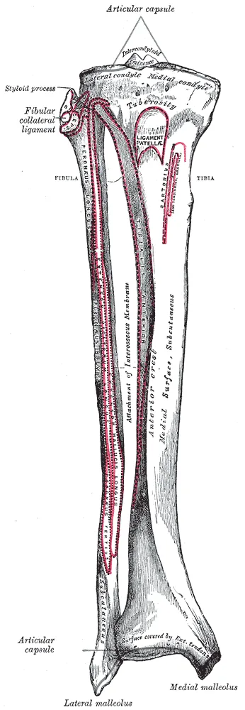
Pain near the back-inner border of the tibia in running and jumping athletes has had many names2. “Shin splints” and “medial tibial stress syndrome” are the most common2. A 2020 overview article defines the condition as exercise-induced pain along the lower back-inner border of the tibia1. The definition adds recognisable pain when that border is pressed over a length of 5 cm or more1.
One randomised trial in athletes defined its cases by the Yates criteria: exercise-induced pain on that border, diffuse over at least 5 cm when pressed5. They exclude suspected tibial stress fracture and exertional compartment syndrome, a pressure rise inside a closed space of muscle5.
The authors of a 2025 article proposed renaming it “load-induced medial-leg pain”2. They argued that “tibial stress” is often misread as meaning a bone stress injury (overuse damage to bone)2.
Two accounts of the cause
Two popular theories have been proposed for how it arises: tibial bending and fascial traction3. Its mechanics remain controversial3. A 2015 review describes it as a diffuse periostitis (inflammation of the bone lining) of the tibial surface7. In most cases, the review says, it also involves microtrauma of the cortical bone (the dense outer layer) beneath7. It says it is not clear whether the soft-tissue or the bone reaction comes first7.
Bone bending. One of the two popular theories holds that the condition arises from bending of the tibia3. Studies of bone in athletes with it found:
- Biopsies, six athletes: bone samples from the painful area showed linear microcracks in four, one an artefact of the biopsy8. There was almost no sign of a repair reaction8.
- 18 adult male athletes with long-standing symptoms: the painful region had about 15% lower bone mineral density than matched controls9. This density is how much mineral is packed into bone.
- 14 such athletes, followed for a mean of 5.7 years: density in the painful region rose by about 19% after recovery10.
- 11 athletes with symptoms for 3 to 10 weeks: tibial bone density was not lower than in 11 regularly exercising controls11.
Traction. One explanation offered for the condition in runners involves three muscles4. These are the tibialis posterior, the soleus and the flexor digitorum longus (the long toe-bending muscle)4. Their repeated contraction would put excessive stress on the tibia4. Studies of cadavers (bodies donated for study) found:
- Three fresh legs: strain in the fascia where it attaches to the tibia’s inner crest rose steadily as tension on those three tendons increased3.
- Fifty legs: the soleus, flexor digitorum longus and deep crural fascia most often attached where symptoms occur12. The tibialis posterior did not attach there in any specimen12.
- Sixteen legs: the three muscles attached above the tibia’s lower third, where the pain is commonly reported13. The authors judged that this did not support muscle-traction theories13. The deep crural fascia attached along the whole inner tibia in all but three legs, which they took as support for traction through it13.
What scans show
A 2009 critical review judged bone, CT and MRI scans of limited value here, because abnormal findings are also prevalent in people without symptoms14.
- CT, distance runners: cortical abnormalities appeared in 14 of 14 painful tibias of runners with the condition, and in 8 of 48 painless tibias15.
- Ultrasound, 15 cases and 27 control athletes: bone-lining swelling showed in 53% of cases and 37% of controls, not a significant difference16.
- MRI, 52 athletes with it: bone marrow or bone-lining oedema (swelling from fluid) showed in 43.5% of the symptomatic legs17.
How it is set against stress fracture
A 1995 MRI study of fourteen runners noted that inner shin pain had traditionally been labelled either shin splints or a stress fracture18. A stress fracture is a small crack in a bone from repeated loading. The authors suggested instead a graded scale: swelling of the bone lining, then growing marrow involvement, then a frank cortical stress fracture18.
In an MRI study of 22 athletes with leg pain, the eight with stress fractures showed a wide bright signal in one area of marrow19. Those with shin splints showed a linear signal along the tibia’s back-inner surface or inner marrow, without the wide signal19.
Who it affects
How common it is
A review article reports an incidence of 4% to 35% in military personnel and athletes14. Single studies and reviews give these figures, where prevalence means the share of a group who have a condition at a given time:
| Group studied | What was found |
|---|---|
| Runners, in a systematic review of eight studies (3,500 runners) | Prevalence 9.5%; incidence from 13.6% to 20.0%4 |
| 933 novice runners, of whom 254 were injured | The most common injury, at 15% of those injured20 |
| 6,608 British Army recruits over 26 weeks of training | 5.67%, the second most common injury21; 19.8% of recovery days, more than any other injury21 |
Factors studied
Several meta-analyses, studies that pool the results of earlier studies, have looked for traits linked with developing it22,23.
- Sex: a meta-analysis of ten papers on runners found female sex significantly associated with it, at a risk ratio of 1.7123. A risk ratio is how many times more often something happens in one group than in another.
- A previous episode: the runners’ meta-analysis gave a risk ratio of 3.7423.
- Body weight: two meta-analyses, one in physically active people, found greater body weight or higher body mass index (weight for height) to be risk factors22,24.
- Arch height: both also found greater navicular drop, how far the inner arch sinks when a person stands22,24. In high school cross-country runners, navicular drop showed no significant association25.
What studies found
How long it lasts
| Group studied | Time reported |
|---|---|
| 38 novice runners in a cohort study | Median (middle value) of 72 days to recover; 97.4% recovered20 |
| Recreational runners injured in a 24-week trial | Median of 70 days from first pain to complete pain relief, against 56 for all injury types26 |
| 74 athletes in a randomised trial | Average of 102 to 118 days to complete a graded running programme5 |
| Novice and recreational runners, in a 2020 systematic review of eleven studies | About sixteen to eighteen weeks27 |
The 2020 review warned that the studies’ poor quality stops its figure being solid evidence27. The athletes’ trial was planned on earlier army studies that suggested recovery in about 17 days5. Its authors concluded that 60 to 100 days is likely to be more realistic for athletes5.
Approaches compared in people who have it
Three reviews reached cautious conclusions6,14,28:
- A 2009 critical review found three randomised studies, in which rest was equal to any intervention tested14.
- A systematic review of 11 trials judged no intervention free enough of bias to recommend, with shockwave therapy the most promising6. Its pooled trials found no significant difference in outcomes with a lower-leg brace6.
- A 2026 systematic review of three randomised trials (130 runners) called the evidence for non-invasive approaches limited and mixed, with none clearly superior28.
Single trials tested these additions5,29:
- Calf exercises or a stocking, 74 athletes: a graded running programme was done alone, with calf stretching and strengthening, or with a sports compression stocking5. Neither time to complete the programme nor satisfaction differed significantly between groups5.
- Arch supports, 50 female recreational runners: some had arch-support shoe inserts and others flat sham (dummy) ones29. The arch-support group had lower pain scores at 6 and 12 weeks, but not at 1829.
Shockwave therapy
Shockwave therapy sends pulses of sound energy into a painful area through the skin. A 2024 systematic review concluded it may be effective as an addition to exercise in this condition, and called for further high-level research30. Four studies differed:
- Cohort comparison, 94 people with long-standing symptoms: 47 had it alongside a home training programme, and 47 did the programme alone31. At 15 months, 76% against 37% were rated completely recovered or much improved31.
- Observational study, 42 athletes at two hospitals: those given five sessions with graded running reached full recovery in a mean of 60 days32. Those at the other hospital, on graded running alone, took 9232. The authors said the finding needs a randomised, double-blind trial32.
- Randomised trial, 42 military cadets with long-standing symptoms in one leg: one group had one session plus an exercise programme33. At four weeks they ran a mean of 17 min 33 s without symptoms, against 4 min 48 s on exercise alone33.
- Sham-controlled pilot trial, 28 active adults: a standard dose was no more effective than a sham dose for pain or running distance at 10 weeks34. The authors noted the sham dose may itself have had an effect34.
Trials aimed at new cases
Two reviews differ on shock-absorbing insoles35,36.
- A 2002 systematic review found four controlled trials among military recruits35. It found no strong support for any method, with the most encouraging evidence for shock-absorbing insoles35.
- A 2025 meta-analysis of randomised trials found shock-absorbing insoles showed no significant effect, in two studies of 1,571 people36. Insoles for overpronation, the foot rolling too far inward, were associated with fewer cases in four studies of 1,047 people36.
The 2025 review also covered neuromuscular training, exercise aimed at how nerves and muscles work together to control movement36. People given it were less likely to develop the condition, in four studies of 3,887 people36. In a randomised trial of 1,020 soldiers, a 15-minute strength, flexibility and coordination programme did not change the risk against a placebo (dummy) programme37.
Connections
The pain runs along the back-inner border of the tibia22. A systematic review of 17 studies found higher peak soleus activity during push-off among the factors most strongly correlated with developing it38. The review said it is still unknown whether such differences are a cause or an effect38.
The fifty-leg cadaver study bears on the tibialis posterior tendon: its muscle did not attach at the site of symptoms12. For the navicular, one meta-analysis found strong evidence that greater navicular drop is a risk factor, with a small pooled effect39. Limited evidence from two studies in it showed no link with a drop over 10 mm39.
In running, studies pooled by one review put it at 9.1% of injuries, behind patellofemoral pain syndrome at 16.7%40. The stress fracture page covers the 1995 MRI scale of tibial stress injury18.
Medial tibial stress syndrome involves
Medial tibial stress syndrome has studies of muscle activity in
Medial tibial stress syndrome has cadaver studies of
Medial tibial stress syndrome has studies of arch height at
Medial tibial stress syndrome is compared with
Medial tibial stress syndrome is common in
Sources
- Winters M (2020). The diagnosis and management of medial tibial stress syndrome. Der Unfallchirurg. doi:10.1007/s00113-019-0667-z PMID 31098646
- Anderson LM, Warden SJ, Willy RW, Bonanno DR, Menz HB (2025). Medial Tibial Stress Syndrome Needs a New Name-Make No Bones About It. The Journal of orthopaedic and sports physical therapy. doi:10.2519/jospt.2025.13411 PMID 40985462
- Bouché RT, Johnson CH (2007). Medial tibial stress syndrome (tibial fasciitis). Journal of the American Podiatric Medical Association. doi:10.7547/0970031 PMID 17218623
- Lopes AD, Hespanhol Júnior LC, Yeung SS, Costa LO (2012). What are the Main Running-Related Musculoskeletal Injuries?. Sports Medicine. doi:10.1007/BF03262301 PMID 22827721
- Moen MH, Holtslag L, Bakker E, Barten C, Weir A, Tol JL, Backx F (2012). The treatment of medial tibial stress syndrome in athletes; a randomized clinical trial. Sports medicine, arthroscopy, rehabilitation, therapy & technology : SMARTT. doi:10.1186/1758-2555-4-12 PMID 22464032
- Winters M, Eskes M, Weir A, Moen MH, Backx FJ, Bakker EW (2013). Treatment of medial tibial stress syndrome: a systematic review. Sports medicine (Auckland, N.Z.). doi:10.1007/s40279-013-0087-0 PMID 23979968
- Franklyn M, Oakes B (2015). Aetiology and mechanisms of injury in medial tibial stress syndrome: Current and future developments. World journal of orthopedics. doi:10.5312/wjo.v6.i8.577 PMID 26396934
- Winters M, Burr DB, van der Hoeven H, Condon KW, Bellemans J, Moen MH (2019). Microcrack-associated bone remodeling is rarely observed in biopsies from athletes with medial tibial stress syndrome. Journal of bone and mineral metabolism. doi:10.1007/s00774-018-0945-9 PMID 30066165
- Magnusson HI, Westlin NE, Nyqvist F, Gärdsell P, Seeman E, Karlsson MK (2001). Abnormally decreased regional bone density in athletes with medial tibial stress syndrome. The American journal of sports medicine. doi:10.1177/03635465010290060701 PMID 11734482
- Magnusson HI, Ahlborg HG, Karlsson C, Nyquist F, Karlsson MK (2003). Low regional tibial bone density in athletes with medial tibial stress syndrome normalizes after recovery from symptoms. The American journal of sports medicine. doi:10.1177/03635465030310042001 PMID 12860551
- Ozgürbüz C, Yüksel O, Ergün M, Işlegen C, Taskiran E, Denerel N, Karamizrak O (2011). Tibial bone density in athletes with medial tibial stress syndrome: a controlled study. Journal of sports science & medicine. PMID 24149568
- Beck BR, Osternig LR (1994). Medial tibial stress syndrome. The location of muscles in the leg in relation to symptoms.. The Journal of bone and joint surgery. American volume. doi:10.2106/00004623-199407000-00015 PMID 8027114
- Stickley CD, Hetzler RK, Kimura IF, Lozanoff S (2009). Crural fascia and muscle origins related to medial tibial stress syndrome symptom location. Medicine and science in sports and exercise. doi:10.1249/MSS.0b013e3181a6519c PMID 19812520
- Moen MH, Tol JL, Weir A, Steunebrink M, De Winter TC (2009). Medial tibial stress syndrome. Sports medicine (Auckland, N.Z.). doi:10.2165/00007256-200939070-00002 PMID 19530750
- Gaeta M, Minutoli F, Vinci S, Salamone I, D'Andrea L, Bitto L, Magaudda L, Blandino A (2006). High-resolution CT grading of tibial stress reactions in distance runners. AJR. American journal of roentgenology. doi:10.2214/AJR.05.0303 PMID 16928946
- Winters M, Bon P, Bijvoet S, Bakker EWP, Moen MH (2017). Are ultrasonographic findings like periosteal and tendinous edema associated with medial tibial stress syndrome? A case-control study. Journal of science and medicine in sport. doi:10.1016/j.jsams.2016.07.001 PMID 27476374
- Moen MH, Schmikli SL, Weir A, Steeneken V, Stapper G, de Slegte R, Tol JL, Backx FJ (2012). A prospective study on MRI findings and prognostic factors in athletes with MTSS. Scandinavian journal of medicine & science in sports. doi:10.1111/j.1600-0838.2012.01467.x PMID 22515327
- Fredericson M, Bergman AG, Hoffman KL, Dillingham MS (1995). Tibial stress reaction in runners. The American journal of sports medicine. doi:10.1177/036354659502300418 PMID 7573660
- Aoki Y, Yasuda K, Tohyama H, Ito H, Minami A (2004). Magnetic resonance imaging in stress fractures and shin splints. Clinical orthopaedics and related research. doi:10.1097/01.blo.0000126333.13806.87 PMID 15123957
- Nielsen RO, Rønnow L, Rasmussen S, Lind M (2014). A prospective study on time to recovery in 254 injured novice runners. PloS one. doi:10.1371/journal.pone.0099877 PMID 24923269
- Sharma J, Greeves JP, Byers M, Bennett AN, Spears IR (2015). Musculoskeletal injuries in British Army recruits: a prospective study of diagnosis-specific incidence and rehabilitation times. BMC musculoskeletal disorders. doi:10.1186/s12891-015-0558-6 PMID 25935751
- Reinking MF, Austin TM, Richter RR, Krieger MM (2017). Medial Tibial Stress Syndrome in Active Individuals: A Systematic Review and Meta-analysis of Risk Factors. Sports health. doi:10.1177/1941738116673299 PMID 27729482
- Newman P, Witchalls J, Waddington G, Adams R (2013). Risk factors associated with medial tibial stress syndrome in runners: a systematic review and meta-analysis. Open access journal of sports medicine. doi:10.2147/OAJSM.S39331 PMID 24379729
- Hamstra-Wright KL, Bliven KC, Bay C (2015). Risk factors for medial tibial stress syndrome in physically active individuals such as runners and military personnel: a systematic review and meta-analysis. British journal of sports medicine. doi:10.1136/bjsports-2014-093462 PMID 25185588
- Plisky MS, Rauh MJ, Heiderscheit B, Underwood FB, Tank RT (2007). Medial tibial stress syndrome in high school cross-country runners: incidence and risk factors. The Journal of orthopaedic and sports physical therapy. doi:10.2519/jospt.2007.2343 PMID 17366958
- Mulvad B, Nielsen RO, Lind M, Ramskov D (2018). Diagnoses and time to recovery among injured recreational runners in the RUN CLEVER trial.. PloS one. doi:10.1371/journal.pone.0204742 PMID 30312310
- Menéndez C, Batalla L, Prieto A, Rodríguez MÁ, Crespo I, Olmedillas H (2020). Medial Tibial Stress Syndrome in Novice and Recreational Runners: A Systematic Review. International journal of environmental research and public health. doi:10.3390/ijerph17207457 PMID 33066291
- Teles AI, da Silva Carto JP, Parpa K, Thuany M, de Vasconcelos GS (2026). Load-induced medial leg pain in runners: A systematic review of non-invasive treatment approaches. Physical therapy in sport : official journal of the Association of Chartered Physiotherapists in Sports Medicine. doi:10.1016/j.ptsp.2026.101962 PMID 42462658
- Naderi A, Bagheri S, Ramazanian Ahoor F, Moen MH, Degens H (2022). Foot Orthoses Enhance the Effectiveness of Exercise, Shockwave, and Ice Therapy in the Management of Medial Tibial Stress Syndrome. Clinical journal of sport medicine : official journal of the Canadian Academy of Sport Medicine. doi:10.1097/JSM.0000000000000926 PMID 33797477
- Rhim HC, Shin J, Kang J, Dyrek P, Crockett Z, Galido P, Wade C, Hollander K, Borg-Stein J, Sampson S, Tenforde AS (2024). Use of extracorporeal shockwave therapies for athletes and physically active individuals: a systematic review. British journal of sports medicine. doi:10.1136/bjsports-2023-107567 PMID 38228375
- Rompe JD, Cacchio A, Furia JP, Maffulli N (2010). Low-energy extracorporeal shock wave therapy as a treatment for medial tibial stress syndrome. The American journal of sports medicine. doi:10.1177/0363546509343804 PMID 19776340
- Moen MH, Rayer S, Schipper M, Schmikli S, Weir A, Tol JL, Backx FJ (2012). Shockwave treatment for medial tibial stress syndrome in athletes; a prospective controlled study. British journal of sports medicine. doi:10.1136/bjsm.2010.081992 PMID 21393260
- Gomez Garcia S, Ramon Rona S, Gomez Tinoco MC, Benet Rodriguez M, Chaustre Ruiz DM, Cardenas Letrado FP, Lopez-Illescas Ruiz Á, Alarcon Garcia JM (2017). Shockwave treatment for medial tibial stress syndrome in military cadets: A single-blind randomized controlled trial. International journal of surgery (London, England). doi:10.1016/j.ijsu.2017.08.584 PMID 28882773
- Newman P, Waddington G, Adams R (2017). Shockwave treatment for medial tibial stress syndrome: A randomized double blind sham-controlled pilot trial. Journal of science and medicine in sport. doi:10.1016/j.jsams.2016.07.006 PMID 27640922
- Thacker SB, Gilchrist J, Stroup DF, Kimsey CD (2002). The prevention of shin splints in sports: a systematic review of literature. Medicine and science in sports and exercise. doi:10.1097/00005768-200201000-00006 PMID 11782644
- Marques TBT, Rangel RPS, Martins LV, Vidal APC (2025). Preventive interventions for medial tibial stress syndrome: Systematic review and meta-analysis. Gait & posture. doi:10.1016/j.gaitpost.2025.07.312 PMID 40633262
- Brushøj C, Larsen K, Albrecht-Beste E, Nielsen MB, Løye F, Hölmich P (2008). Prevention of overuse injuries by a concurrent exercise program in subjects exposed to an increase in training load. The American journal of sports medicine. doi:10.1177/0363546508315469 PMID 18337359
- Mattock JPM, Steele JR, Mickle KJ (2021). Are Leg Muscle, Tendon and Functional Characteristics Associated with Medial Tibial Stress Syndrome? A Systematic Review.. Sports Medicine - Open. doi:10.1186/s40798-021-00362-2 PMID 34626247
- Neal BS, Griffiths IB, Dowling GJ, Murley GS, Munteanu SE, Franettovich Smith MM, Collins NJ, Barton CJ (2014). Foot posture as a risk factor for lower limb overuse injury: a systematic review and meta-analysis. Journal of foot and ankle research. doi:10.1186/s13047-014-0055-4 PMID 25558288
- Kakouris N, Yener N, Fong DTP (2021). A systematic review of running-related musculoskeletal injuries in runners.. Journal of sport and health science. doi:10.1016/j.jshs.2021.04.001 PMID 33862272
Changes
- Links to the new chronic exertional compartment syndrome page
- First published.