Navicular
Also called tarsal navicular, navicular bone
A boat-shaped bone at the top of the foot's inner arch. Where it sits and how it is loaded, why its stress fractures are slow to show and slow to heal, and what studies of casts and surgery found.
- Region
- Foot & ankle
- Muscles
- tibialis posterior
- Updated
- October 5, 2026
- Reviewed
- October 5, 2026 · how entries are checked
Function
Where it sits
The navicular sits on the inner side of the midfoot1. The talus (the ankle bone that sits under the shinbone and on top of the heel bone) meets it from behind1. In front, it meets the three cuneiform bones1. It is named for its rough boat shape2.
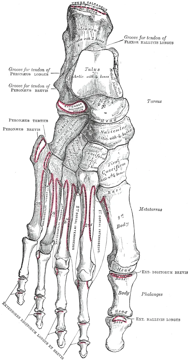
Its surfaces
- The back surface is hollowed and meets the head of the talus as a ball-and-socket joint2. It does not fully enclose the talar head, which allows movement in several directions2.
- The front surface is usually split by two faint ridges into three joint facets, one for each cuneiform2.
- The tuberosity is a blunt bony bump at the inner end, pointing down and inward, and it varies in size2.
Its place in the arch
One review calls the navicular the keystone of the medial longitudinal arch, the inner lengthwise arch of the foot3. An anatomy textbook explains that the talus receives body weight and sits at the top of the foot’s lengthwise arches1. Weight then passes to the ground through the front and back ends of those arches1.
How reviews describe its loading
Reviews describe the forces on the bone in several ways2,3:
- One review describes lengthwise compression, crosswise tension and sideways shear acting together, concentrated in the narrower, weaker middle of the bone2.
- Another describes uneven squeezing from the front3. Forces on the inner side are shared with the talus, while the navicular alone carries those on the outer side3.
- The same review describes repeated compression, as in running, as “bending” the bone at its centre3.
- A third review describes severe compression of the central third in explosive movements such as jumping and sprinting9.
A contested blood supply
In an injection study of 60 cadaver legs, branches of the dorsalis pedis artery supplied the top of the navicular5.
Sources disagree about the middle of the bone. A review describes vessels entering from each side and branching inward, leaving the central third with relatively little supply3. In the injection study, 30 of 54 specimens had vessels spread throughout the bone5. Six had a vessel-free zone in the central third reaching the top surface5. Its authors concluded that mechanical or other factors may matter more than previously thought5.
Breakdown
Stress fractures of the central third
A review names the navicular as an example of a high-risk stress fracture site3. It says fractures there carry a greater risk of delayed healing, lasting pain and nonunion (a broken bone that fails to join and heal)3. The stress fracture page covers how reviews sort sites by risk.
In a review of CT scans of 55 navicular stress fractures, every fracture involved the central third of the bone’s upper edge10. A CT scan builds detailed pictures of the inside of the body from X-rays. In the same scans, 96% of the fractures were partial, not complete10.
Who gets them
A 2021 meta-analysis states that these fractures are rare in the general population but increasingly recognised in athletes8. A meta-analysis pools the results of several earlier studies.
| Group studied | What was found |
|---|---|
| 180 stress fractures at a Melbourne sports clinic, 2 years | 26 were in the navicular, the fourth most common site11 |
| The 54 track athletes in that clinic review | The navicular was the most common site, with 19 fractures11 |
| 111 competitive track and field athletes, 12 months | The navicular was the second most common site, with 4 of 26 fractures12 |
| 315 fractures in 307 patients, pooled from 11 studies | 97% were in athletes, men and women were affected about equally, and the mean age was about 258 |
| The 155 athletes in that pooled analysis with a named sport | 52% took part in running activities including track and field, 14% in football and 7% in basketball8 |
A review reports these fractures mostly in short-distance runners and basketball players3. In the study of 111 track and field athletes, sprints, hurdles and jumps were linked with significantly more stress fractures of the foot12.
Factors that have been proposed
The authors of the 2021 meta-analysis list factors suggested to predispose the bone to stress injury8. These include the path force takes through its inner part and the pull of tibialis posterior (a deep calf muscle)8. The poor blood supply of its middle third is another8. Studies that looked back at past cases have reported features such as limited ankle dorsiflexion (pulling the foot and toes up toward the shin)13. None of those features had been shown to be statistically significant13.
One study compared 7 runners with a past navicular stress fracture and 7 matched runners without13. During running, the previously injured feet showed more and faster rearfoot eversion (turning the sole of the foot outward)13. Ground forces and foot structure did not differ13.
Slow to be recognised
In the 2021 meta-analysis, people had symptoms for a pooled mean of 9.9 months before the fracture was identified8. A review says people typically report poorly localised midfoot soreness that worsens with weight bearing3. Plain X-rays often miss these fractures, because they are often incomplete or not displaced8.
The accessory navicular
An accessory navicular is an extra small bone on the inner side of the foot14. It forms when a secondary centre of bone growth fails to fuse with the navicular14. A meta-analysis of 39 studies put its pooled prevalence at 17.5% of patients14. Most cause no symptoms, and it is usually found by chance14.
In an ultrasound survey of 875 Japanese children aged 6 to 15, 15.1% had one15. Of those children, 20.8% had pain from it15. In a two-year follow-up of 66 children aged 11 to 13, pain had gone in 15 of the 21 feet painful at the start16.
Navicular drop
Navicular drop is a measure named after the bone. It is the distance the tuberosity moves downward in standing, as the foot goes from a neutral to a relaxed position17. In 21 healthy young adults, it rose from 10.0 to 11.8 mm after the small muscles of the sole were exercised to fatigue18.
Studies have tested it against injuries elsewhere in the leg17,19:
- A meta-analysis found strong evidence that greater navicular drop is a risk factor for medial tibial stress syndrome (shin splints), with a small pooled effect19.
- The same analysis found no relationship between a pronated (rolled-inward) standing foot posture and bone stress reactions19.
- Among 287 female team-sport athletes followed over time, navicular drop was not associated with non-contact injury of the anterior cruciate ligament17.
Recovery
How bone repairs itself
A review explains that ordinary loading causes tiny damage in bone, which remodelling repairs20. The bone tissue page covers that process.
What scans show as it heals
In a review of CT scans, the earliest sign of healing was seen in three of eight fractures scanned at 6 weeks10. Firm union of the outer bone was present in 10 of 31 by 4 months, and 12 fractures did not unite10. In a follow-up of 86 fractures in athletes, how a healing fracture looked on CT did not necessarily match clinical union21.
In a case series, 7 of 9 people first found to have a navicular stress reaction later showed the features of a stress fracture22. A stress reaction is an early bone stress injury, before a crack forms.
Cast, weight bearing and surgery
In the follow-up of 86 fractures in athletes, 19 of 22 first managed with at least six weeks in a non-weight-bearing cast returned to sport21. Of those who first kept bearing weight with limited activity, 9 of 34 returned21.
Two pooled analyses of published cases, eleven years apart, point in different directions7,8.
| Pooled analysis | Successful outcomes with surgery | Successful outcomes without surgery | Difference |
|---|---|---|---|
| 2010 | 82% | 96%, counting non-weight-bearing management only | Not statistically significant7 |
| 2021 | 96% (104 of 108 fractures) | 72% (149 of 207 fractures) | Significant, in favour of surgery8 |
The 2010 analysis also found that management allowing weight bearing was significantly less effective than either of the other two7. The 2021 analysis counted an outcome as successful when the person was pain-free, back at the previous activity level and had no repeat fracture8.
The 2021 analysis reported further results8:
- Return to play followed 97 of 98 fractures managed with surgery and 152 of 207 managed without; the difference was not statistically significant8.
- Time to return had a pooled mean of 4.17 months after surgery and 4.67 months without, with no significant difference8.
- Nonunion was reported for 3 of 94 fractures managed with surgery and 33 of 153 managed without8.
- Repeat fracture was reported for 1 of 78 fractures managed with surgery and 20 of 85 managed without8.
Longer follow-up
In 62 navicular stress fractures in athletes, 57 were followed by a return to activity at the pre-injury level24. Seven fractured again, mostly in athletes under 21, none of whom had had surgery24.
One study looked at 32 fractures more than two years after a non-weight-bearing cast or surgical fixation25. It found no significant differences between the two in current pain, function or CT findings25. Some pain and loss of function remained, small but measurable25.
Connections
Joints and ligaments
The navicular forms the inner half of the midtarsal joint2. A review lists the main ligaments around it, among them the dorsal talonavicular ligament and the spring ligament complex2. The spring ligament spans the gap between the calcaneus (heel bone) and the navicular, holding up the head of the talus2. The deltoid ligament of the inner ankle also reaches the navicular26.
In front, the navicular is compressed through the joints between the cuneiforms and the first and second metatarsals3.
The tibialis posterior tendon
The tibialis posterior tendon has its main footprint on the underside of the navicular2. In a dissection study of 41 specimens, its main insertion was on the navicular in all 4127. A review describes its contraction as raising the stress on the inner part of the bone3. The spring ligament is described as working with this tendon to steady the foot in standing and walking2.
Exercise
Related exercise pages: running, in which a review describes repeated compression concentrating force at the centre of the bone3.
Navicular is attached to by
Navicular is joined by the spring ligament to
Navicular takes load from
Navicular is a site of
Navicular gives its name to a measure studied in
Navicular is held up in part by
Navicular is loaded by
Sources
- Betts JG, Young KA, Wise JA, Johnson E, Poe B, Kruse DH, Korol O, Johnson JE, Womble M, DeSaix P (2022). Anatomy and Physiology 2e. OpenStax. Link
- Liu X, Hong CC, Reina F, Carrera A, Alcolea-Madera E, Del Vecchio JJ, Dalmau-Pastor M (2026). Navicular and fifth metatarsal base stress fractures: An illustrated anatomical review. Knee surgery, sports traumatology, arthroscopy : official journal of the ESSKA. doi:10.1002/ksa.70413 PMID 42171011
- Shakked RJ, Walters EE, O'Malley MJ (2017). Tarsal navicular stress fractures. Current reviews in musculoskeletal medicine. doi:10.1007/s12178-017-9392-9 PMID 28110392
- Boden BP, Osbahr DC (2000). High-risk stress fractures: evaluation and treatment. The Journal of the American Academy of Orthopaedic Surgeons. doi:10.5435/00124635-200011000-00002 PMID 11104398
- McKeon KE, McCormick JJ, Johnson JE, Klein SE (2012). Intraosseous and extraosseous arterial anatomy of the adult navicular. Foot & ankle international. doi:10.3113/FAI.2012.0857 PMID 23050710
- Hoenig T, Eissele J, Strahl A, Popp KL, Stürznickel J, Ackerman KE, Hollander K, Warden SJ, Frosch KH, Tenforde AS, Rolvien T (2023). Return to sport following low-risk and high-risk bone stress injuries: a systematic review and meta-analysis. British journal of sports medicine. doi:10.1136/bjsports-2022-106328 PMID 36720584
- Torg JS, Moyer J, Gaughan JP, Boden BP (2010). Management of tarsal navicular stress fractures. The American journal of sports medicine. doi:10.1177/0363546509355408 PMID 20197494
- Attia AK, Mahmoud K, Bariteau J, Labib SA, DiGiovanni CW, D'Hooghe P (2021). Return to sport following navicular stress fracture: a systematic review and meta-analysis of three hundred and fifteen fractures. International orthopaedics. doi:10.1007/s00264-021-05147-6 PMID 34415421
- Fowler JR, Gaughan JP, Boden BP, Pavlov H, Torg JS (2011). The non-surgical and surgical treatment of tarsal navicular stress fractures. Sports medicine (Auckland, N.Z.). doi:10.2165/11590670-000000000-00000 PMID 21780848
- Kiss ZS, Khan KM, Fuller PJ (1993). Stress fractures of the tarsal navicular bone: CT findings in 55 cases. AJR. American journal of roentgenology. doi:10.2214/ajr.160.1.8416606 PMID 8416606
- Brukner P, Bradshaw C, Khan KM, White S, Crossley K (1996). Stress fractures: a review of 180 cases. Clinical journal of sport medicine : official journal of the Canadian Academy of Sport Medicine. PMID 8673581
- Bennell KL, Malcolm SA, Thomas SA, Wark JD, Brukner PD (1996). The incidence and distribution of stress fractures in competitive track and field athletes. The American journal of sports medicine. doi:10.1177/036354659602400217 PMID 8775123
- Becker J, James S, Osternig L, Chou LS (2018). Foot Kinematics Differ Between Runners With and Without a History of Navicular Stress Fractures. Orthopaedic journal of sports medicine. doi:10.1177/2325967118767363 PMID 29780837
- Stolarz K, Osiowski A, Preinl M, Osiowski M, Jasiewicz B, Taterra D (2024). The prevalence and anatomy of accessory navicular bone: a meta-analysis. Surgical and radiologic anatomy : SRA. doi:10.1007/s00276-024-03459-x PMID 39136746
- Sasaki T, Teraguchi M, Mure K, Asai Y, Kido Y, Onishi M, Shimoe T, Miyai N, Nakagawa Y, Hashizume H, Yamada H (2025). Prevalence of accessory navicular in Japanese children: A cross-sectional study using ultrasound-Katsuragi Integrated Defense for Locomotive Syndrome in children study. PloS one. doi:10.1371/journal.pone.0318014 PMID 39854496
- Sasaki T, Teraguchi M, Mure K, Asai Y, Kido Y, Onishi M, Minamino K, Shimoe T, Miyai N, Nakagawa Y, Hashizume H, Yamada H (2025). Investigation of accessory navicular in local pre-adolescence children: A longitudinal study with 2-year follow-up-KID Locomo Study. PloS one. doi:10.1371/journal.pone.0328339 PMID 41183048
- Pasanen K, Seppänen A, Leppänen M, Tokola K, Järvelä T, Vasankari T, Myklebust G, Krosshaug T, Parkkari J (2025). Knee laxity, joint hypermobility, femoral anteversion, hamstring extensibility and navicular drop as risk factors for non-contact anterior cruciate ligament injury in female athletes: A 4.5-year prospective cohort study. Knee surgery, sports traumatology, arthroscopy : official journal of the ESSKA. doi:10.1002/ksa.12625 PMID 39943896
- Headlee DL, Leonard JL, Hart JM, Ingersoll CD, Hertel J (2008). Fatigue of the plantar intrinsic foot muscles increases navicular drop. Journal of electromyography and kinesiology : official journal of the International Society of Electrophysiological Kinesiology. doi:10.1016/j.jelekin.2006.11.004 PMID 17208458
- Neal BS, Griffiths IB, Dowling GJ, Murley GS, Munteanu SE, Franettovich Smith MM, Collins NJ, Barton CJ (2014). Foot posture as a risk factor for lower limb overuse injury: a systematic review and meta-analysis. Journal of foot and ankle research. doi:10.1186/s13047-014-0055-4 PMID 25558288
- Warden SJ, Edwards WB, Willy RW (2021). Preventing Bone Stress Injuries in Runners with Optimal Workload. Current osteoporosis reports. doi:10.1007/s11914-021-00666-y PMID 33635519
- Khan KM, Fuller PJ, Brukner PD, Kearney C, Burry HC (1992). Outcome of conservative and surgical management of navicular stress fracture in athletes. The American journal of sports medicine. doi:10.1177/036354659202000606 PMID 1456359
- Burne SG, Mahoney CM, Forster BB, Koehle MS, Taunton JE, Khan KM (2005). Tarsal navicular stress injury. The American journal of sports medicine. doi:10.1177/0363546505278253 PMID 16157855
- Mallee WH, Weel H, van Dijk CN, van Tulder MW, Kerkhoffs GM, Lin CW (2015). Surgical versus conservative treatment for high-risk stress fractures of the lower leg (anterior tibial cortex, navicular and fifth metatarsal base): a systematic review. British journal of sports medicine. doi:10.1136/bjsports-2013-093246 PMID 25138980
- Saxena A, Behan SA, Valerio DL, Frosch DL (2017). Navicular Stress Fracture Outcomes in Athletes: Analysis of 62 Injuries. The Journal of foot and ankle surgery : official publication of the American College of Foot and Ankle Surgeons. doi:10.1053/j.jfas.2017.06.009 PMID 28842101
- Potter NJ, Brukner PD, Makdissi M, Crossley K, Kiss ZS, Bradshaw C (2006). Navicular stress fractures: outcomes of surgical and conservative management. British journal of sports medicine. doi:10.1136/bjsm.2005.022079 PMID 16611725
- Fleury C, Acker A, Dubois-Ferrière V, Assal M (2026). Deltoid ligament injury: thinking beyond the ankle - an expert opinion.. EFORT Open Reviews. doi:10.1530/EOR-2025-0268 PMID 42684953
- Willegger M, Seyidova N, Schuh R, Windhager R, Hirtler L (2020). The tibialis posterior tendon footprint: an anatomical dissection study.. Journal of foot and ankle research. doi:10.1186/s13047-020-00392-1 PMID 32430082
Changes
- First published.