Tibialis posterior tendon
Also called posterior tibial tendon, PTT, tibialis posterior, posterior tibial tendon dysfunction, PTTD, tibialis posterior tendinopathy
The tendon that turns behind the inner ankle bone and fans out under the foot. How it holds up the arch, how it wears, what studies of exercise and surgery found, and what it works with.
- Region
- Foot & ankle
- Muscles
- tibialis posterior
- Updated
- October 8, 2026
- Reviewed
- October 8, 2026 · how entries are checked
Function
The muscle behind the tendon
The tibialis posterior is the deepest muscle of the deep compartment at the back of the lower leg1. Its belly arises from three places1:
- the back of the interosseous membrane, the sheet of tough tissue joining the two lower-leg bones1;
- the upper two-thirds of the fibula, the thin outer bone of the lower leg1;
- the upper part of the tibia (shinbone)1.
The tibial nerve supplies the muscle, and the posterior tibial artery brings its blood1. Its main jobs are plantar flexion, pointing the foot down, and inversion, turning the sole inward1.
Where the tendon runs
The tendon forms in the lower third of the leg2. It bends sharply behind the medial malleolus to enter the foot2. That bend turns the line of pull from vertical to horizontal8.
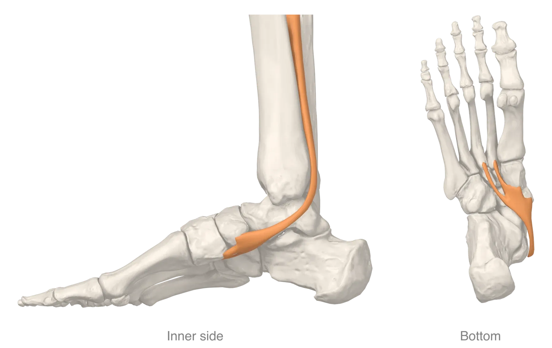
The flexor retinaculum, a band of tissue, binds the tendon into a shallow groove on the malleolus and stops it slipping forward8. The tendon of the flexor digitorum longus, a deep calf muscle, lies beside it and shares its line of pull8.
Where it attaches
In a dissection study of 41 specimens, the main attachment was on the navicular in all 413. In dissected feet, slips of tendon reached up to eight separate patches of bone3. After the navicular, the bones reached most often were these3:
| Bone | Share of dissected feet |
|---|---|
| Lateral cuneiform | 93%3 |
| Medial cuneiform | 80%3 |
| Bases of the metatarsals (long foot bones) | 80%3 |
| Cuboid | 46%3 |
| Intermediate cuneiform | 19%3 |
| Calcaneus (heel bone) | 12%3 |
What it does for the arch
The arch has static supports and dynamic ones8. The tibialis posterior and the small muscles within the foot give the dynamic support8. When the muscle contracts, it raises the inner arch and locks the midfoot bones, which makes the back and middle of the foot rigid6. That lets the gastrocnemius, a calf muscle, act much more efficiently in walking6.
In 10 anatomical preparations, the muscle’s inversion moment arm, or leverage, at the subtalar joint was 19.1 mm, the largest measured9. For the tibialis anterior, the muscle at the front of the shin, it was 3.8 mm9. The subtalar joint lies just below the ankle joint.
Two cadaver experiments point different ways10,11:
- In 13 cadaver feet loaded as in mid-step, loading this tendon moved the foot bones toward restoring arch alignment10. Loads on the other tendons moved them toward flattening10.
- In eight pairs of cadaver legs loaded as in single-leg standing, a 150 newton pull on the tendon did not restore the forefoot arch angle11.
What it does in walking and running
Stance is the part of each step when the foot is on the ground. Early in stance, the muscle fires to steady the back of the foot and slow the inward roll of the midfoot8. At mid-stance it starts inversion, which locks the midfoot and gives the stiffness needed for toe-off8. In walking, the tendon tissue is stretched early in stance and shortens late in stance12. The authors of that study conclude that it absorbs and returns elastic energy12.
In 15 injury-free runners, the muscles at the back of the ankle, this one among them, were most active in midstance13. They worked eccentrically, that is while lengthening, to control the ankle as it bent13.
Breakdown
A stretch with few blood vessels
The tendon’s main blood supply comes from the posterior tibial artery14. Three studies describe a poorly supplied stretch, in different places4,5,14:
- In 28 injected limbs, every tendon had a zone of low blood supply behind and below the medial malleolus4.
- The authors of a staining study call the front part of the tendon avascular, without blood vessels, where it passes behind the malleolus14. They note that ruptures are most frequent behind the malleolus14.
- A third study saw a low-vessel region in every tendon examined under the microscope5. It ran from about 2.2 cm to about 0.6 cm above the malleolus5.
Whether the tendon has a low-blood-supply region that leaves it vulnerable has been a matter of controversy5. Where the tendon wraps around the malleolus, its pulley, the surface facing the bone is built like fibrocartilage, a tough, fibrous cartilage15. That study’s authors state that most spontaneous ruptures happen there15.
Wear more than sudden tears
Dysfunction of this tendon was once put down to inflammation16. Later tissue studies showed tendinosis, meaning tendon wear, with a nonspecific repair response16. It cannot be determined whether such changes come before the dysfunction or after it16.
The authors of a report on two athletes under 30 state that, unlike the Achilles tendon, this tendon does not typically rupture suddenly17. The tears they saw at surgery were chronic17. The tendon may rupture, but dysfunction can develop without a rupture6.
From a sore tendon to a changed foot
Dysfunction of the tendon is linked with a collapsing inner arch, a heel that tilts outward and a forefoot that points outward16. Johnson and Strom described it as passing through stages18. Two published overviews describe the stages like this6,8:
| Stage | How the overviews describe it |
|---|---|
| I | Tendinopathy, local tendon pain with loading, and the arch is usually kept8 |
| II | A flatfoot has developed, but it can still be corrected by hand6 |
| III | The foot shape is fixed, with wear in the subtalar joint6 |
| IV | Wear is also present in the ankle joint; Myerson added this stage later6 |
A consensus group stated that these staging systems have never been validated19. In 2020 it voted to rename adult acquired flatfoot deformity “progressive collapsing foot deformity”19. The group noted that many, but not all, of these feet have a ruptured posterior tibial tendon19. The new classification leaves out the tendon-only picture of stage I and part of stage II20. A scoping review, which maps what research exists, calls that early picture tibialis posterior tendinopathy20.
Who is affected
- One overview names middle-aged women as the group most often affected6.
- In a survey of women over 40 from one English general practice group, with 582 usable replies, 3.3% had stage I or II dysfunction21.
- In 67 patients with rupture, rupture was statistically linked with obesity and, less strongly, with high blood pressure22.
- A literature review of risk factors found no clear agreement on which matter most23.
In runners and athletes
In a review of running injuries, this tendinopathy made up 1.1% of the injuries reported in prevalence studies of non-ultramarathon runners, with a range of 0.5% to 16.0% across studies24. A systematic review of movement risk factors in runners found one study on this tendon’s dysfunction25. A case-control study compared 12 runners with stage I dysfunction and 12 matched healthy runners26. The affected runners’ heels tilted outward more, and for longer, when walking, with no difference in the strength of inversion26. A review describes problems with this tendon as an uncommon source of disability in athletes27.
One explanation offered for medial tibial stress syndrome, exercise-related pain along the inner shin, in runners involves this muscle28. The idea is that repeated contraction of it, the soleus or the flexor digitorum longus overloads the tibia28. In fifty dissected legs, the muscle did not attach at the site of the symptoms in any specimen29. Those authors say this contradicts the idea29.
Recovery
What is known about the course
The natural course of the dysfunction is not fully known, but most agree it is progressive8. A scoping review of the early stage found 97 articles on surgery against 12 on non-surgical approaches20.
What exercise studies found
- In a randomised trial, which assigns people to groups by chance, 36 adults with stage I or II tendinopathy were followed for 12 weeks30. One group had orthoses (supports such as shaped shoe inserts) and stretching alone30. Two added resistance exercise, concentric (while the muscle shortens) or eccentric30. Scores on the Foot Functional Index, a questionnaire on foot pain, fell in all three groups, and the eccentric group improved most30.
- The systematic review did not pool its three trials7. Its moderate effects for eccentric exercise on pain and disability come from this trial7.
- In an uncontrolled series of 47 patients with stage I or II dysfunction, 39 met the study’s success criteria after an orthosis and exercise programme31.
That review concluded that high-quality research on non-surgical approaches is scarce7.
What the tendon looked like afterwards
Ten people with early-stage tendinopathy did a 10-week programme of eccentric loading and calf stretching, with orthoses32. Symptoms and function improved, but the tendon still looked abnormal on ultrasound afterwards32. Ultrasound is a scan made with sound waves.
A meta-analysis, which pools earlier studies, of how symptom-free human tendons adapt to more loading found 27 studies33. Each was on the Achilles or the patellar tendon, so none measured this tendon33.
What surgical series found
One series followed 31 patients for a mean of 15.2 years after surgery for stage II dysfunction34. The surgery moved the flexor digitorum longus tendon and reshaped the heel bone34. Of them, 27 were pain free and functioning well34. In six athletic patients with a full or partial rupture, surgery restored some but not all normal function35. It did not restore the flattened arch35.
Connections
Nerve, bone and neighbours
The tibial nerve supplies the muscle1. Nerve and tendon share the tarsal tunnel, a passage on the inner side of the ankle36. The tendon’s principal attachment is on the underside of the navicular37.
What shares the work of the arch
The spring ligament, under the inner arch, is described as working together with this tendon in standing and walking37. On MRI, a scan that shows soft tissue, the spring ligament complex was the ligament most often damaged in 31 people with an insufficient tendon38. The deltoid ligament, spring ligament and this tendon are named as key supports of the inner ankle39. The foot muscles share the dynamic support of the arch8.
Muscles that pull the other way
In five people with stage II dysfunction, muscle recordings suggested extra, compensating activity in three muscles40. These were the peroneals, which oppose it, the tibialis anterior and the gastrocnemius40. In eight cadaver feet, pulling on the peroneus longus tendon lowered the foot’s stiffness, and pulling on this tendon raised it41.
Exercises and stretches
Related exercise pages: running, calf raise and soleus stretch. In five healthy adults scanned by MRI, a single-leg heel raise raised the muscle’s signal by 27%, less than in the three other muscles measured42. In young adults without symptoms, the deep calf muscles, this one included, showed low stiffness as the foot was bent upward43.
Tibialis posterior tendon is loaded by
Tibialis posterior tendon has been studied in
Tibialis posterior tendon attaches to
Tibialis posterior tendon runs beside
Tibialis posterior tendon works with
Tibialis posterior tendon works opposite to
Tibialis posterior tendon is compared with
Tibialis posterior tendon has been proposed as a factor in
Sources
- Corcoran NM, Varacallo MA (2023). Anatomy, Bony Pelvis and Lower Limb: Tibialis Posterior Muscle.. StatPearls. PMID 30969735
- Semple R, Murley GS, Woodburn J, Turner DE (2009). Tibialis posterior in health and disease: a review of structure and function with specific reference to electromyographic studies.. Journal of foot and ankle research. doi:10.1186/1757-1146-2-24 PMID 19691828
- Willegger M, Seyidova N, Schuh R, Windhager R, Hirtler L (2020). The tibialis posterior tendon footprint: an anatomical dissection study.. Journal of foot and ankle research. doi:10.1186/s13047-020-00392-1 PMID 32430082
- Frey C, Shereff M, Greenidge N (1990). Vascularity of the posterior tibial tendon.. The Journal of bone and joint surgery. American volume. PMID 2195033
- Manske MC, McKeon KE, Johnson JE, McCormick JJ, Klein SE (2015). Arterial anatomy of the tibialis posterior tendon.. Foot & ankle international. doi:10.1177/1071100714559271 PMID 25411117
- Kohls-Gatzoulis J, Angel JC, Singh D, Haddad F, Livingstone J, Berry G (2004). Tibialis posterior dysfunction: a common and treatable cause of adult acquired flatfoot.. BMJ (Clinical research ed.). doi:10.1136/bmj.329.7478.1328 PMID 15576744
- Ross MH, Smith MD, Mellor R, Vicenzino B (2018). Exercise for posterior tibial tendon dysfunction: a systematic review of randomised clinical trials and clinical guidelines.. BMJ open sport & exercise medicine. doi:10.1136/bmjsem-2018-000430 PMID 30271611
- Ling SK, Lui TH (2017). Posterior Tibial Tendon Dysfunction: An Overview.. The open orthopaedics journal. doi:10.2174/1874325001711010714 PMID 28979585
- Klein P, Mattys S, Rooze M (1996). Moment arm length variations of selected muscles acting on talocrural and subtalar joints during movement: an in vitro study.. Journal of biomechanics. doi:10.1016/0021-9290(95)00025-9 PMID 8839014
- Kitaoka HB, Luo ZP, An KN (1997). Effect of the posterior tibial tendon on the arch of the foot during simulated weightbearing: biomechanical analysis.. Foot & ankle international. doi:10.1177/107110079701800109 PMID 9013114
- Dullaert K, Hagen JE, Simons P, Gras F, Gueorguiev B, Richards RG, Klos K (2017). Influence of tibialis posterior muscle activation on foot anatomy under axial loading: A biomechanical CT human cadaveric study.. Foot and ankle surgery : official journal of the European Society of Foot and Ankle Surgeons. doi:10.1016/j.fas.2016.07.003 PMID 29202983
- Maharaj JN, Cresswell AG, Lichtwark GA (2016). The mechanical function of the tibialis posterior muscle and its tendon during locomotion.. Journal of biomechanics. doi:10.1016/j.jbiomech.2016.08.006 PMID 27545079
- Reber L, Perry J, Pink M (1993). Muscular control of the ankle in running.. The American journal of sports medicine. doi:10.1177/036354659302100608 PMID 8291630
- Petersen W, Hohmann G, Stein V, Tillmann B (2002). The blood supply of the posterior tibial tendon.. The Journal of bone and joint surgery. British volume. doi:10.1302/0301-620x.84b1.11592 PMID 11837820
- Petersen W, Hohmann G, Pufe T, Tsokos M, Zantop T, Paulsen F, Tillmann B (2004). Structure of the human tibialis posterior tendon.. Archives of orthopaedic and trauma surgery. doi:10.1007/s00402-003-0500-5 PMID 12695917
- Mosier SM, Pomeroy G, Manoli A 2nd (1999). Pathoanatomy and etiology of posterior tibial tendon dysfunction.. Clinical orthopaedics and related research. doi:10.1097/00003086-199908000-00003 PMID 10627682
- Porter DA, Baxter DE, Clanton TO, Klootwyk TE (1998). Posterior tibial tendon tears in young competitive athletes: two case reports.. Foot & ankle international. doi:10.1177/107110079801900911 PMID 9763170
- Johnson KA, Strom DE (1989). Tibialis posterior tendon dysfunction.. Clinical orthopaedics and related research. PMID 2912622
- Myerson MS, Thordarson DB, Johnson JE, Hintermann B, Sangeorzan BJ, Deland JT, Schon LC, Ellis SJ, de Cesar Netto C (2020). Classification and Nomenclature: Progressive Collapsing Foot Deformity.. Foot & ankle international. doi:10.1177/1071100720950722 PMID 32856474
- Rhim HC, Dhawan R, Gureck AE, Lieberman DE, Nolan DC, Elshafey R, Tenforde AS (2022). Characteristics and Future Direction of Tibialis Posterior Tendinopathy Research: A Scoping Review.. Medicina (Kaunas, Lithuania). doi:10.3390/medicina58121858 PMID 36557060
- Kohls-Gatzoulis J, Woods B, Angel JC, Singh D (2009). The prevalence of symptomatic posterior tibialis tendon dysfunction in women over the age of 40 in England.. Foot and ankle surgery : official journal of the European Society of Foot and Ankle Surgeons. doi:10.1016/j.fas.2008.08.003 PMID 19410173
- Holmes GB Jr, Mann RA (1992). Possible epidemiological factors associated with rupture of the posterior tibial tendon.. Foot & ankle. doi:10.1177/107110079201300204 PMID 1349292
- Beeson P (2014). Posterior Tibial Tendinopathy. Journal of the American Podiatric Medical Association. doi:10.7547/0003-0538-104.5.455 PMID 25275733
- Kakouris N, Yener N, Fong DTP (2021). A systematic review of running-related musculoskeletal injuries in runners.. Journal of sport and health science. doi:10.1016/j.jshs.2021.04.001 PMID 33862272
- Mousavi SH, Hijmans JM, Rajabi R, Diercks R, Zwerver J, van der Worp H (2019). Kinematic risk factors for lower limb tendinopathy in distance runners: A systematic review and meta-analysis.. Gait & posture. doi:10.1016/j.gaitpost.2019.01.011 PMID 30658311
- Rabbito M, Pohl MB, Humble N, Ferber R (2011). Biomechanical and clinical factors related to stage I posterior tibial tendon dysfunction.. The Journal of orthopaedic and sports physical therapy. doi:10.2519/jospt.2011.3545 PMID 21765219
- Conti SF (1994). Posterior tibial tendon problems in athletes.. The Orthopedic clinics of North America. PMID 8290223
- Lopes AD, Hespanhol Júnior LC, Yeung SS, Costa LO (2012). What are the Main Running-Related Musculoskeletal Injuries?. Sports Medicine. doi:10.1007/BF03262301 PMID 22827721
- Beck BR, Osternig LR (1994). Medial tibial stress syndrome. The location of muscles in the leg in relation to symptoms.. The Journal of bone and joint surgery. American volume. doi:10.2106/00004623-199407000-00015 PMID 8027114
- Kulig K, Reischl SF, Pomrantz AB, Burnfield JM, Mais-Requejo S, Thordarson DB, Smith RW (2009). Nonsurgical management of posterior tibial tendon dysfunction with orthoses and resistive exercise: a randomized controlled trial.. Physical therapy. doi:10.2522/ptj.20070242 PMID 19022863
- Alvarez RG, Marini A, Schmitt C, Saltzman CL (2006). Stage I and II posterior tibial tendon dysfunction treated by a structured nonoperative management protocol: an orthosis and exercise program.. Foot & ankle international. doi:10.1177/107110070602700102 PMID 16442022
- Kulig K, Lederhaus ES, Reischl S, Arya S, Bashford G (2009). Effect of eccentric exercise program for early tibialis posterior tendinopathy.. Foot & ankle international. doi:10.3113/FAI.2009.0877 PMID 19755073
- Bohm S, Mersmann F, Arampatzis A (2015). Human tendon adaptation in response to mechanical loading: a systematic review and meta-analysis of exercise intervention studies on healthy adults.. Sports Medicine - Open. doi:10.1186/s40798-015-0009-9 PMID 27747846
- Chadwick C, Whitehouse SL, Saxby TS (2015). Long-term follow-up of flexor digitorum longus transfer and calcaneal osteotomy for stage II posterior tibial tendon dysfunction.. The bone & joint journal. doi:10.1302/0301-620X.97B3.34386 PMID 25737518
- Woods L, Leach RE (1991). Posterior tibial tendon rupture in athletic people.. The American journal of sports medicine. doi:10.1177/036354659101900513 PMID 1962716
- Farag JI, McDougall AN, Catapano M (2025). Common sports-related nerve injuries seen by the electrodiagnostic medical consultant.. Muscle & nerve. doi:10.1002/mus.28298 PMID 39535438
- Liu X, Hong CC, Reina F, Carrera A, Alcolea-Madera E, Del Vecchio JJ, Dalmau-Pastor M (2026). Navicular and fifth metatarsal base stress fractures: An illustrated anatomical review. Knee surgery, sports traumatology, arthroscopy : official journal of the ESSKA. doi:10.1002/ksa.70413 PMID 42171011
- Deland JT, de Asla RJ, Sung IH, Ernberg LA, Potter HG (2005). Posterior tibial tendon insufficiency: which ligaments are involved?. Foot & ankle international. doi:10.1177/107110070502600601 PMID 15960907
- Bilyy A, Win Mar WW, Al Abdeen Al Zuabi Z, Frimpong-Manso HB, Famure S, Solomou G, Brassett C, Pasapula C (2025). The Deltoid-Spring Ligament Complex: A Scoping Review and New Segmental Classification.. Cureus. doi:10.7759/cureus.81715 PMID 40322447
- Ringleb SI, Kavros SJ, Kotajarvi BR, Hansen DK, Kitaoka HB, Kaufman KR (2007). Changes in gait associated with acute stage II posterior tibial tendon dysfunction.. Gait & posture. doi:10.1016/j.gaitpost.2006.06.008 PMID 16876415
- Kokubo T, Hashimoto T, Nagura T, Nakamura T, Suda Y, Matsumoto H, Toyama Y (2012). Effect of the posterior tibial and peroneal longus on the mechanical properties of the foot arch. Foot & ankle international. doi:10.3113/FAI.2012.0320 PMID 22735204
- Kulig K, Burnfield JM, Requejo SM, Sperry M, Terk M (2004). Selective activation of tibialis posterior: evaluation by magnetic resonance imaging.. Medicine and science in sports and exercise. doi:10.1249/01.mss.0000126385.12402.2e PMID 15126722
- Le Sant G, Nordez A, Andrade R, Hug F, Freitas S, Gross R (2017). Stiffness mapping of lower leg muscles during passive dorsiflexion.. Journal of anatomy. doi:10.1111/joa.12589 PMID 28251615
Changes
- Figure replaced with a render from the AnatomyTOOL 3D model
- First published.