Calf raise
Also called heel raise, heel-rise
An exercise that works the muscles at the back of the lower leg. What the studies measured it loading, from the calf muscles to the Achilles tendon, and what is known about how tendons adapt.
- Region
- Lower leg
- Muscles
- soleus, gastrocnemius, tibialis posterior, fibularis longus, flexor digitorum longus
- Updated
- October 23, 2026
- Reviewed
- October 23, 2026 · how entries are checked
Movement
What the movement is
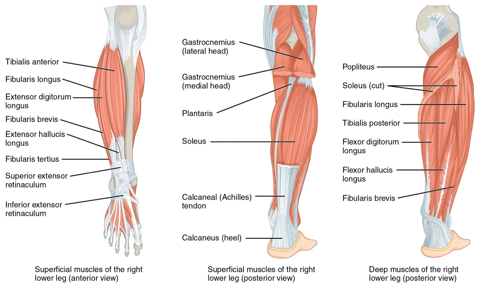
The muscles at the back of the lower leg mainly produce plantar flexion, which means pointing the foot down5. One study of the calf raise looked at the three muscles that do most of this work1. Those three are the soleus and the inner and outer heads of the gastrocnemius1.
The versions studies have used
The studies on this page tested the calf raise in several forms1,2,3,4:
- Loaded, with the heaviest weight a person could lift ten times, a 10-repetition maximum1.
- Seated, as one of a set of exercises compared for tendon load4.
- Standing, with the toes turned in or turned out2.
- With the foot straight, turned in 30 degrees, or turned out 30 degrees3.
The calf-raise test is a related use of the movement6. A review found support for it as a way to assess the calf muscles and the Achilles tendon working as one unit6.
What it loads
The calf muscles
The triceps surae is a group of three calf muscles: the soleus and the two heads of the gastrocnemius1. Together, these three do most of the work of pointing the foot down1. The gastrocnemius is the calf muscle nearest the skin5. Its two heads, inner and outer, run from the thigh bone to the heel bone, so they cross both the knee and the ankle1,2. The soleus is a wide, flat muscle that lies deeper5.
In a study of resistance-trained men, the authors suggested that calf raises can recruit these three muscles1. In 22 trained men lifting a 10-repetition maximum, soleus and gastrocnemius activity did not differ significantly between the calf raise and the leg press1.
How position shifts the work
Foot position made a difference to which gastrocnemius head was more active2. In 33 people doing a standing heel-rise with the toes turned out, the inner head was more active than the outer head2. With the toes turned in, the two heads did not differ2.
Knee angle did not single out one calf muscle in a related test7. In it, 48 healthy people pushed the foot down as hard as they could, without the ankle moving7. They did this with the knee straight, bent to 45 degrees, and bent to 90 degrees7. No knee angle of the three worked the soleus or either gastrocnemius head alone7.
The Achilles tendon
The calf muscles nearest the skin end in the Achilles tendon, which attaches to the calcaneus (heel bone)5. The tendon holds fibres from the soleus and from both heads of the gastrocnemius8. A tendon passes force from a working muscle to the bone, and that pull moves the body9.
One study of eight healthy young adults estimated peak Achilles tendon load across a range of exercises4. The seated heel raise had the lowest estimated load of the exercises tested4:
| Exercise | Estimated peak Achilles tendon load |
|---|---|
| Seated heel raise | 0.5 times body weight4 |
| Forward hop on one leg | 7.3 times body weight4 |
In that study, peak tendon load differed more than twelve-fold between the lightest and heaviest exercise4.
Deeper and outer lower-leg muscles
Three other lower-leg muscles also work during heel raises, and foot position changes how hard3. The tibialis posterior and flexor digitorum longus are two of the four deep muscles at the back of the leg5. They lie beneath the calf muscles that join the Achilles tendon5. The fibularis (peroneus) longus is one of two muscles on the outer side of the leg5. In the front and back of the leg, the muscles toward the inner and outer sides turn the foot in or out and rotate it5.
In 14 healthy men, the tibialis posterior and flexor digitorum longus were most active with the foot turned in 30 degrees3. The fibularis longus was most active with the foot turned out 30 degrees3:
| Muscle | Where it lies | Most active in heel raises with the foot |
|---|---|---|
| Tibialis posterior | Deep, back of the leg | Turned in 30 degrees3,5 |
| Flexor digitorum longus | Deep, back of the leg | Turned in 30 degrees3,5 |
| Fibularis longus | Outer side of the leg | Turned out 30 degrees3,5 |
How the body adapts
What training does to tendon in general
Research on loading in general shows how tendons respond to repeated load, though it did not single out the calf raise10. A meta-analysis pooled exercise studies of at least eight weeks in healthy adults aged 18 to 5010. It found that loading made tendons stiffer10. Its Young’s modulus also rose10. Cross-sectional area, a measure of thickness, grew by a smaller amount10.
Gains in stiffness depended on how heavy the load was, not on the type of muscle contraction10. Most programmes in that analysis ran for 8 to 14 weeks10. Those of 12 weeks or more showed larger effects, though the difference was not statistically significant10.
What happens without load
Tendon also changes when load is taken away9. A review concluded that keeping a limb still lowers tendon mechanical properties in animals, and that human data largely mirror this9. Even one to two weeks of unloading a limb seems to reduce tendon stiffness, without changing its thickness9.
Progressive overload
A 2026 position stand describes progressive overload as the need to increase the stimulus placed on the muscle throughout a training programme11. The calf-raise studies here each raised the work in their own way12,13,14,15:
| Who trained, and for how long | How the work was raised |
|---|---|
| 14 untrained adults, standing and seated calf raises twice a week, 12 weeks | Sets rose from 3 to 4 to 5 over the first three sessions. After that, 5% of one-repetition maximum was added whenever a person finished the whole session without help12 |
| 23 untrained men, standing Smith machine calf raises twice a week, 10 weeks | The bar load rose by 0.25 to 0.50 kg once a person managed 20 or more full-range repetitions on the first set13 |
| 16 untrained men, single-leg standing calf raises twice a week, 10 weeks | Sets rose from 4 to 6 in week 7 (2 to 3 in the leg that used partial repetitions). Load rose 5% to 10% once a person went past 14 repetitions to failure on the first set14 |
| 22 young men, single-leg calf raises on a seated leg-press machine three times a week, 9 weeks | 3 sets of 20 to 25 repetitions in weeks 1 to 3, then 4 sets in weeks 4 to 915 |
In the first study, each added step was 5% of one-repetition maximum, the heaviest load a person can lift once12. The second used a Smith machine, a barbell that slides up and down on fixed vertical rails13.
More load or more repetitions
One study compared two ways of progressing in 43 people with at least a year of lower-body resistance training16. Their 8-week programme included straight-leg and seated calf raises twice a week16. Increasing load and increasing repetitions produced similar growth in the soleus and gastrocnemius16. That result belongs to the whole programme, not to the calf raises on their own16.
How many sets a week
In 61 untrained young women, groups did 6, 9 or 12 weekly sets of calf raises for 6 weeks17. Each set used a 15- to 20-repetition maximum, the heaviest load a person could lift 15 to 20 times17. All three groups grew calf muscle17. The 12-set group grew the outer gastrocnemius head, the soleus and the triceps surae as a whole more than the 6-set group17. The 9-set group did not differ significantly from either17.
Light loads, with and without a cuff
A randomised trial in trained men used 4 sets of standing calf raises at 30% of one-repetition maximum, taken to failure18. They trained twice a week for 6 weeks, with 30-second rests between sets18. One group wore blood flow restriction cuffs, which partly limit blood flow to the limb. Both groups gained gastrocnemius thickness and calf-raise one-repetition maximum, with no difference between them18.
Connections
The tendon it loads
The calf raise loads the Achilles tendon, which joins the calf muscles to the heel4,5. The calf-raise test is described as a way to assess that tendon together with the calf muscles6.
The muscles it works
- The soleus, a wide, flat calf muscle beneath the gastrocnemius1,5.
- The gastrocnemius, both inner and outer heads1,2.
- The tibialis posterior, a deep muscle at the back of the leg3,5.
- The flexor digitorum longus, another deep muscle at the back of the leg3,5.
- The fibularis longus, on the outer side of the leg3,5.
A related exercise
In 22 trained men, soleus and gastrocnemius activity in the leg press did not differ significantly from the calf raise1.
Calf raise loads
Calf raise is progressed by
Sources
- Gentil P, Souza D, Santana M, Alves RR, Campos MH, Pinto R, Bottaro M (2020). Multi- and Single-Joint Resistance Exercises Promote Similar Plantar Flexor Activation in Resistance Trained Men.. International Journal of Environmental Research and Public Health. doi:10.3390/ijerph17249487 PMID 33352879
- Cibulka M, Wenthe A, Boyle Z, Callier D, Schwerdt A, Jarman D, Strube MJ (2017). Variation in medial and lateral gastrocnemius muscle activity with foot position.. International Journal of Sports Physical Therapy. PMID 28515978
- Akuzawa H, Imai A, Iizuka S, Matsunaga N, Kaneoka K (2017). The influence of foot position on lower leg muscle activity during a heel raise exercise measured with fine-wire and surface EMG.. Physical Therapy in Sport. doi:10.1016/j.ptsp.2017.08.077 PMID 28950148
- Baxter JR, Corrigan P, Hullfish TJ, O'Rourke P, Silbernagel KG (2021). Exercise Progression to Incrementally Load the Achilles Tendon.. Medicine and Science in Sports and Exercise. doi:10.1249/MSS.0000000000002459 PMID 32658037
- Betts JG, Young KA, Wise JA, Johnson E, Poe B, Kruse DH, Korol O, Johnson JE, Womble M, DeSaix P (2022). Anatomy and Physiology 2e. OpenStax. Link
- Hébert-Losier K, Schneiders AG, Newsham-West RJ, Sullivan SJ (2009). Scientific bases and clinical utilisation of the calf-raise test.. Physical Therapy in Sport. doi:10.1016/j.ptsp.2009.07.001 PMID 19897168
- Hébert-Losier K, Schneiders AG, García JA, Sullivan SJ, Simoneau GG (2011). Peak triceps surae muscle activity is not specific to knee flexion angles during MVIC.. Journal of Electromyography and Kinesiology. doi:10.1016/j.jelekin.2011.04.009 PMID 21669538
- Pękala PA, Henry BM, Ochała A, Kopacz P, Tatoń G, Młyniec A, Walocha JA, Tomaszewski KA (2017). The twisted structure of the Achilles tendon unraveled: A detailed quantitative and qualitative anatomical investigation.. Scandinavian Journal of Medicine & Science in Sports. doi:10.1111/sms.12835 PMID 28139009
- Magnusson SP, Kjaer M (2019). The impact of loading, unloading, ageing and injury on the human tendon.. The Journal of Physiology. doi:10.1113/JP275450 PMID 29920664
- Bohm S, Mersmann F, Arampatzis A (2015). Human tendon adaptation in response to mechanical loading: a systematic review and meta-analysis of exercise intervention studies on healthy adults.. Sports Medicine - Open. doi:10.1186/s40798-015-0009-9 PMID 27747846
- Currier BS, D'Souza AC, Singh MAF, Lowisz CV, Rawson ES, Schoenfeld BJ, Smith-Ryan AE, et al (2026). American College of Sports Medicine Position Stand. Resistance Training Prescription for Muscle Function, Hypertrophy, and Physical Performance in Healthy Adults: An Overview of Reviews.. Medicine and science in sports and exercise. doi:10.1249/MSS.0000000000003897 PMID 41843416
- Kinoshita M, Maeo S, Kobayashi Y, Eihara Y, Ono M, Sato M, Sugiyama T, Kanehisa H, Isaka T (2023). Triceps surae muscle hypertrophy is greater after standing versus seated calf-raise training.. Frontiers in Physiology. doi:10.3389/fphys.2023.1272106 PMID 38156065
- Larsen S, Swinton PA, Sandberg NØ, Kristiansen BS, Fredriksen AB, Falch HN, et al (2025). Resistance training beyond momentary failure: the effects of past-failure partials on muscle hypertrophy in the gastrocnemius.. Frontiers in Psychology. doi:10.3389/fpsyg.2025.1494323 PMID 39995432
- Goli A, Attarieh P, Nunes JP, Nehegadar S, Khani S, Fashi M, et al (2026). Does Performing Partial Repetitions Beyond Momentary Failure Enhance Muscle Hypertrophy in Volume-Load-Equated Calf-Raise Resistance Training?. International Journal of Exercise Science. doi:10.70252/IJES2026403 PMID 42006449
- Nunes JP, Costa BDV, Kassiano W, Kunevaliki G, Castro-E-Souza P, Rodacki ALF, et al (2020). Different Foot Positioning During Calf Training to Induce Portion-Specific Gastrocnemius Muscle Hypertrophy.. Journal of Strength and Conditioning Research. doi:10.1519/JSC.0000000000003674 PMID 32735428
- Plotkin D, Coleman M, Van Every D, Maldonado J, Oberlin D, Israetel M, et al (2022). Progressive overload without progressing load? The effects of load or repetition progression on muscular adaptations.. PeerJ. doi:10.7717/peerj.14142 PMID 36199287
- Kassiano W, Costa BDV, Kunevaliki G, Lisboa F, Tricoli I, Francsuel J, et al (2024). Bigger Calves from Doing Higher Resistance Training Volume?. International Journal of Sports Medicine. doi:10.1055/a-2316-7885 PMID 38684187
- Gavanda S, Isenmann E, Schlöder Y, Roth R, Freiwald J, Schiffer T, et al (2020). Low-intensity blood flow restriction calf muscle training leads to similar functional and structural adaptations than conventional low-load strength training: A randomized controlled trial.. PLoS One. doi:10.1371/journal.pone.0235377 PMID 32603351
Changes
- Added a Progressive overload section.
- First published.