Peroneals
Also called peroneal muscles, fibularis muscles, fibular muscles, peroneus longus, peroneus brevis, peroneus tertius, fibularis longus, fibularis brevis, fibularis tertius, evertors, ankle evertors
The muscles on the outer side of the lower leg that turn the sole of the foot outward. What each one does, how they work in walking and running, what changes after ankle sprains, and how they respond to training.
- Region
- Lower leg
- Starts on (peroneus longus)
- the upper two-thirds of the fibula's outer side, the intermuscular septum, and the lateral condyle (outer top) of the shinbone8
- Starts on (peroneus brevis)
- the lower two-thirds of the fibula's outer surface, and the intermuscular septum8,9,10
- Starts on (peroneus tertius)
- usually the lower third or half of the fibula, and the intermuscular septum12
- Ends on (peroneus longus)
- the outer side of the first metatarsal's base, and the medial cuneiform, a wedge-shaped inner midfoot bone (Peroneal tendons)11,44
- Ends on (peroneus brevis)
- the tuberosity (bump) at the base of the fifth metatarsal, by a tendon running behind the outer ankle bone (Peroneal tendons)9,44
- Ends on (peroneus tertius)
- most often the fifth metatarsal, at its base, shaft or both; in about a third, the fourth and fifth metatarsals12
- Tendons
- Peroneal tendons
- Muscles
- peroneus longus, peroneus brevis, peroneus tertius
- Updated
- October 5, 2026
- Reviewed
- October 5, 2026 · how entries are checked
Function
Which muscles make up the group
The leg has four compartments, and the lateral one holds the peroneus longus and the peroneus brevis1. Anatomical naming has replaced “peroneal” with “fibular”, so the peroneus longus is also called the fibularis longus1,2. A third muscle, the peroneus tertius, lies in the anterior compartment instead, at the front of the lower leg7,8.
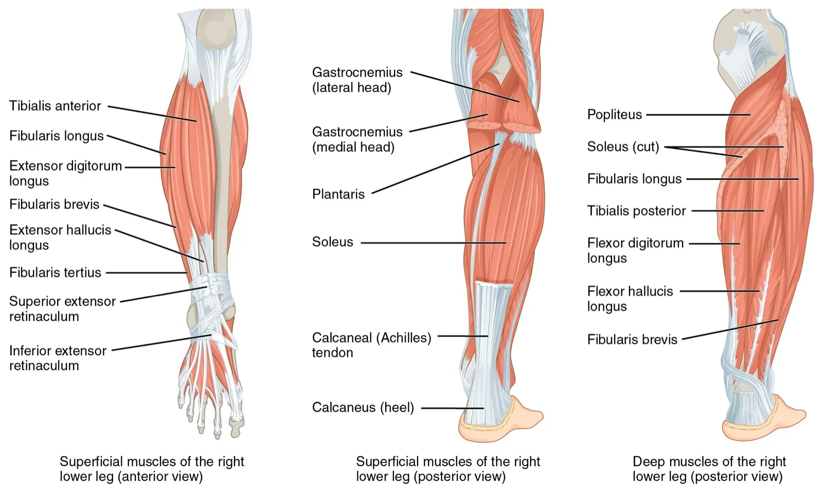
The peroneus longus arises from the upper two-thirds of the outer side of the fibula, the thin outer bone of the lower leg8. Part of it arises from the outer top of the tibia (shinbone)8. The peroneus brevis arises from the lower two-thirds of the fibula’s outer surface9,10. Both also take fibres from an intermuscular septum, a sheet of tough tissue that separates groups of muscles8,9.
Both tendons pass behind the lateral malleolus, the bony bump on the outer side of the ankle9,11. The longus tendon then wraps beneath the foot to reach its inner side11. The brevis tendon attaches at the base of the fifth metatarsal, the long foot bone behind the little toe9. The peroneal tendons have their own page.
The peroneus tertius
The peroneus tertius usually arises from the lower third or half of the fibula and from the intermuscular septum12. A systematic search of anatomical studies found it most often ends on the base of the fifth metatarsal12. Not everyone has one. A meta-analysis (a study that pools earlier results) of 25 studies of cadavers (bodies donated for study) gave a weighted prevalence of 93.2%12.
Their nerves and blood supply
The superficial fibular nerve supplies the peroneus longus and brevis8,9,10. The deep fibular nerve supplies the peroneus tertius7. The lateral compartment gets its blood from the anterior tibial and fibular arteries1,2.
What they move
The main action of the longus and brevis is eversion of the foot and ankle1. Plantar flexion (pointing the foot down) and support of the foot’s arches are secondary1. The brevis is credited with roughly 60% to 65% of the eversion force at the ankle8,9. The peroneus tertius everts the foot and dorsiflexes it, pulling it up toward the shin13. It has been described as levelling the sole while the foot swings forward, before the next touchdown12.
Whether the longus holds up the arch
One anatomical source describes the peroneus longus as stabilising the long inner arch of the foot11. Three cadaver studies tested this by pulling on its tendon, and they do not agree14,15,16:
- In seven specimens, a stronger pull rotated the inner column of the foot toward eversion, which the authors read as stabilising that column14.
- In eight feet, the pull reduced bone movement but lowered the foot’s stiffness and raised the energy it stored15.
- In eight pairs of legs, a 15 kg pull rotated the first metatarsal but did not significantly change the arch angle16.
The authors of the third study said their result calls into question how much the tendon maintains the arch16.
What they do in walking and running
In walking, the peroneals are most active in mid-stance, when one leg alone supports the body3. Stance is the part of each step when the foot is on the ground. They are also active at push-off3. One study measured 16 healthy people walking on the flat and across a sideways slope3. Average peroneus longus activity during stance, as a share of the muscle’s hardest effort with the joint still, was3:
| Walking surface | Peroneus longus activity (% of hardest effort) |
|---|---|
| Flat ground | 143 |
| Side slope, foot flat on it | 583 |
| Side slope, outer edge of the foot raised | 773 |
These figures are approximate3. In the same people, peroneus brevis activity was about 15% on the flat and about 45% to 53% across the slope3.
In 15 injury-free recreational and competitive runners tested at three paces, peroneus brevis activity rose significantly as pace increased17. Ten men ran in shoes wedged to tilt the foot inward, outward or neither way18. The activation patterns of the muscles recorded, the peroneus longus among them, did not change with the shoe18.
How fast they can react
The longus and brevis are described as integral to controlling supination (the foot rolling outward onto its outer edge) and guarding against lateral ankle sprains19. One study found a protective reflex from the peroneals would take at least 126 milliseconds after a sudden, unexpected inversion19. That figure assumes the muscles are not already active before the heel touches down19. The peroneals are in fact active before touchdown when going down stairs and landing from a jump19.
A walking study of 25 people with chronic ankle instability and 10 controls reached a more cautious view20. Its authors concluded that the peroneus longus serves balance and cannot be relied on to stop an inversion injury in normal walking20.
Breakdown
What studies find after ankle sprains
Chronic ankle instability is an ankle that stays loose or keeps giving way after one or more sprains. The ankle sprain page covers the condition itself. Reviews have compared people who have it with controls21,22:
| What was measured | What the review found |
|---|---|
| Reaction to sudden inversion | Delayed after an earlier sprain, most with chronic instability, in a review of 23 studies4 |
| Peroneus longus timing | Late activation in sudden inversion and in landing on unstable surfaces, reported by a review of 63 muscle-activity studies23 |
| Activity just before landing | Less peroneus longus activity before a single-leg jump landing, in a review of 21 studies24 |
| Eversion strength, 20 studies | Lower than controls in shortening and lengthening work, with standardised mean differences of 0.73 to 0.9521 |
| Eversion strength, search to 2007 | Ankles with functional instability were weaker in shortening work, by a small difference of 0.22425 |
| Sense of eversion force | Less accurate at 10% and 20% of hardest effort; each result rested on two small studies26 |
| Spinal reflex in the longus | Lower excitability, with a small effect, across 17 studies22 |
A standardised mean difference puts the gap between two groups on a common scale. A review of reviews found strong evidence that deficits in peroneal reaction time, eversion strength and balance contribute to ankle instability27. Only 17% of its primary studies measured a clearly defined chronic ankle instability group27.
Size and activity in unstable ankles
A study using ultrasound, a scan made with sound waves, compared 29 people with chronic ankle instability and 29 healthy controls, aged 18 to 3028. The instability group had a smaller peroneal cross-sectional area (the size of a slice cut straight across a muscle) when standing28. The groups did not differ when lying on the side28.
In treadmill walking, 15 adults with chronic ankle instability had the peroneus longus active for 36.0% of the stride29. In 15 matched healthy controls the figure was 23.3%29.
Tears and pressure in the lateral compartment
A case report describes a 23-year-old collegiate football player with a large tear in the upper peroneus longus muscle belly30. The player felt a sharp leg pain in practice, with no contact, and developed acute lateral compartment syndrome30. Compartment syndrome is a rise in pressure inside a closed space of muscle, which limits blood flow there. Rupture of this muscle after a twisting ankle injury is a known, though uncommon, cause of it30.
One institution saw 26 people over 11 years with chronic exertional compartment syndrome confined to the lateral compartment31. Their median (middle) age was 2131. Running and walking were among the activities they most often named as provoking it31. In another series of 73 people with exertional leg pain and raised compartment pressures, 5 (7%) had raised pressure in the lateral compartment alone32.
Recovery
How they respond to training in healthy people
Twenty healthy people were randomly split to train either the longus or the brevis with elastic bands for 8 weeks33. The cross-sectional area and ankle strength of the trained muscle rose significantly33. Some of the size gain was still present in the period after training stopped33.
Trials of reaction time and strength have mixed results34,35:
- In 24 healthy male recreational athletes, 6 weeks of isokinetic training shortened the peroneus longus reaction to a sudden foot tilt34.
- In 26 healthy, active college-age people, 6 weeks of neuromuscular training shortened peroneus longus reaction time during walking, compared with controls36.
- In a randomised trial of 40 people, 4 weeks of elastic-resistance training changed neither eversion strength nor peroneus longus reaction time35.
Isokinetic training uses a machine that holds a limb to a fixed speed. Neuromuscular training is exercise aimed at how nerves and muscles work together. Half of the 40 people in the third trial had a history of ankle sprains35.
How they respond in people with unstable ankles
In a randomised trial of 39 people with chronic ankle instability, 6 weeks of strength training improved eversion strength37. The untrained control group did not improve37.
Two studies of muscle size after a 4-week supervised exercise programme point different ways5,6. In 26 people with chronic ankle instability, peroneal cross-sectional area on ultrasound rose from 3.44 to 3.72 cm²5. The untrained leg showed a similar rise5. In a preliminary study of five young adults using MRI, a detailed scan, the programme did not enlarge the peroneals6. Eversion strength in those five still improved by a large amount6.
Connections
Tendons, bones and nerves
The peroneus brevis becomes tendon 2 to 3 cm above the tip of the fibula8. From there down, the peroneal tendons page takes over. The longus tendon attaches to the base of the first metatarsal and to the medial cuneiform, a wedge-shaped bone of the inner midfoot11. Both muscles start on the fibula, and the longus also on the tibia8,9,10.
At the neck of the fibula, the common fibular nerve divides into the superficial and deep fibular nerves38. The superficial one is the motor supply of the peroneus longus and brevis39.
Muscles and joints they work with
At the ankle, the longus and brevis point the foot down and turn the sole outward40. The everting pull of the peroneus tertius is described as countering the inverting pull of the tibialis anterior12. In 10 active men doing side-shuffle and cutting moves, the tibialis anterior and peroneus longus steadied the subtalar joint41. The subtalar joint sits between the talus and the heel bone, just below the ankle joint.
Exercises that load them
Related exercise pages: running, calf raise, drop landing, single-leg Romanian deadlift17,19,42,43.
| Exercise | What studies found |
|---|---|
| Calf raise | In 14 healthy men, peroneus longus activity was highest with the foot turned out 30°42 |
| Single-leg Romanian deadlift | In 28 healthy athletes holding a sandbag, it gave more peroneus longus activity than standing still on one leg43 |
| Drop landing | The peroneals are active before the foot touches down when landing from a jump19 |
Peroneals pulls through
Peroneals starts on
Peroneals starts partly on
Peroneals attaches to
Peroneals is supplied by branches of
Peroneals moves
Peroneals works alongside
Peroneals changes after
Peroneals is loaded by
Sources
- Khan IA, Mahabadi N, D'Abarno A, Varacallo MA (2023). Anatomy, Bony Pelvis and Lower Limb: Leg Lateral Compartment.. StatPearls. PMID 30137811
- Lezak B, Varacallo MA (2026). Anatomy, Bony Pelvis and Lower Limb: Calf Peroneus Longus Muscle.. StatPearls. PMID 31536256
- Bavdek R, Zdolšek A, Strojnik V, Dolenec A (2018). Peroneal muscle activity during different types of walking. Journal of foot and ankle research. doi:10.1186/s13047-018-0291-0 PMID 30202446
- Hoch MC, McKeon PO (2014). Peroneal reaction time after ankle sprain. Medicine & Science in Sports & Exercise. doi:10.1249/MSS.0b013e3182a6a93b PMID 23899892
- Jaffri A, Koldenhoven R, Lempke A, Park J, Hart J, Hertel J, Saliba S (2026). Peroneal Muscle Size and Quality Changes after Impairment-Based Rehabilitation in Patients with Chronic Ankle Instability. International journal of sports physical therapy. doi:10.26603/001c.167120 PMID 42694063
- Feger MA, Donovan L, Herb CC, Handsfield GG, Blemker SS, Hart JM, Saliba SA, Abel MF, Park JS, Hertel J (2019). Effect of Impairment-Based Rehabilitation on Lower Leg Muscle Volumes and Strength in Patients With Chronic Ankle Instability: A Preliminary Study. Journal of sport rehabilitation. doi:10.1123/jsr.2017-0136 PMID 29405809
- Palomo-López P, Losa-Iglesias ME, Calvo-Lobo C, Rodríguez-Sanz D, Navarro-Flores E, Becerro-de-Bengoa-Vallejo R, López-López D (2019). Fibularis tertius muscle in women & men: A surface anatomy cross-sectional study across countries. PloS one. doi:10.1371/journal.pone.0215118 PMID 30964928
- Davda K, Malhotra K, O'Donnell P, Singh D, Cullen N (2017). Peroneal tendon disorders. EFORT Open Reviews. doi:10.1302/2058-5241.2.160047 PMID 28736620
- Olewnik Ł, Landfald IC, Domosławska D, Ruzik K, Triantafyllou G, Piagkou M, Vázquez T (2025). Comparative and developmental anatomy of the fibularis brevis muscle: morphological variants and their clinical significance. Frontiers in cell and developmental biology. doi:10.3389/fcell.2025.1647407 PMID 40894923
- Basit H, Shah J, Siccardi MA (2023). Anatomy, Bony Pelvis and Lower Limb, Foot Peroneus Brevis Muscle.. StatPearls. PMID 30571048
- Landfald IC, Ciechanowska M, Olewnik Ł (2025). The fibularis longus muscle revisited: comparative anatomy, developmental perspectives, and clinical relevance. Frontiers in cell and developmental biology. doi:10.3389/fcell.2025.1678965 PMID 41113456
- Yammine K, Erić M (2017). The Fibularis (Peroneus) Tertius Muscle in Humans: A Meta-Analysis of Anatomical Studies with Clinical and Evolutionary Implications. BioMed research international. doi:10.1155/2017/6021707 PMID 28596965
- Witvrouw E, Borre KV, Willems TM, Huysmans J, Broos E, De Clercq D (2006). The Significance of Peroneus Tertius Muscle in Ankle Injuries. The American journal of sports medicine. doi:10.1177/0363546505286021 PMID 16493171
- Johnson CH, Christensen JC (1999). Biomechanics of the first ray. Part I. The effects of peroneus longus function: a three-dimensional kinematic study on a cadaver model. The Journal of Foot and Ankle Surgery. doi:10.1016/s1067-2516(99)80002-7 PMID 10553544
- Kokubo T, Hashimoto T, Nagura T, Nakamura T, Suda Y, Matsumoto H, Toyama Y (2012). Effect of the posterior tibial and peroneal longus on the mechanical properties of the foot arch. Foot & ankle international. doi:10.3113/FAI.2012.0320 PMID 22735204
- Dullaert K, Hagen J, Klos K, Gueorguiev B, Lenz M, Richards RG, Simons P (2016). The influence of the Peroneus Longus muscle on the foot under axial loading: A CT evaluated dynamic cadaveric model study. Clinical biomechanics (Bristol, Avon). doi:10.1016/j.clinbiomech.2016.03.001 PMID 27015031
- Reber L, Perry J, Pink M (1993). Muscular control of the ankle in running.. The American journal of sports medicine. doi:10.1177/036354659302100608 PMID 8291630
- O'Connor KM, Hamill J (2004). The role of selected extrinsic foot muscles during running. Clinical biomechanics (Bristol, Avon). doi:10.1016/j.clinbiomech.2003.09.001 PMID 14659933
- Hertel J (2002). Functional Anatomy, Pathomechanics, and Pathophysiology of Lateral Ankle Instability.. Journal of Athletic Training. PMID 12937557
- Louwerens JW, van Linge B, de Klerk LW, Mulder PG, Snijders CJ (1995). Peroneus longus and tibialis anterior muscle activity in the stance phase. A quantified electromyographic study of 10 controls and 25 patients with chronic ankle instability. Acta orthopaedica Scandinavica. doi:10.3109/17453679509002306 PMID 8553819
- Khalaj N, Vicenzino B, Heales LJ, Smith MD (2020). Is chronic ankle instability associated with impaired muscle strength? Ankle, knee and hip muscle strength in individuals with chronic ankle instability: a systematic review with meta-analysis. British journal of sports medicine. doi:10.1136/bjsports-2018-100070 PMID 31937576
- Kim KM, Kim JS, Cruz-Díaz D, Ryu S, Kang M, Taube W (2019). Changes in Spinal and Corticospinal Excitability in Patients with Chronic Ankle Instability: A Systematic Review with Meta-Analysis. Journal of clinical medicine. doi:10.3390/jcm8071037 PMID 31315231
- Labanca L, Mosca M, Ghislieri M, Agostini V, Knaflitz M, Benedetti MG (2021). Muscle activations during functional tasks in individuals with chronic ankle instability: a systematic review of electromyographical studies. Gait & posture. doi:10.1016/j.gaitpost.2021.09.182 PMID 34564008
- Chan LYT, Sim YTN, Gan FK, Bin Abd Razak HR (2022). Effect of chronic ankle instability on lower extremity kinematics, dynamic postural stability, and muscle activity during unilateral jump-landing tasks: A systematic review and meta-analysis.. Physical therapy in sport : official journal of the Association of Chartered Physiotherapists in Sports Medicine. doi:10.1016/j.ptsp.2022.04.005 PMID 35462322
- Arnold BL, Linens SW, de la Motte SJ, Ross SE (2009). Concentric evertor strength differences and functional ankle instability: a meta-analysis. Journal of athletic training. doi:10.4085/1062-6050-44.6.653 PMID 19911093
- Altun A, Dixon S, Nunns M, Rice H (2025). How do neuromuscular characteristics of the peroneal muscles differ in adults with and without chronic ankle instability? A systematic review with meta-analysis. Gait & posture. doi:10.1016/j.gaitpost.2025.01.019 PMID 39862526
- Thompson C, Schabrun S, Romero R, Bialocerkowski A, van Dieen J, Marshall P (2018). Factors Contributing to Chronic Ankle Instability: A Systematic Review and Meta-Analysis of Systematic Reviews. Sports medicine (Auckland, N.Z.). doi:10.1007/s40279-017-0781-4 PMID 28887759
- Jaffri A (2025). Functional Assessment of Peroneal Muscles Using Ultrasound Imaging in Chronic Ankle Instability. Journal of foot and ankle research. doi:10.1002/jfa2.70088 PMID 41249869
- Feger MA, Donovan L, Hart JM, Hertel J (2015). Lower extremity muscle activation in patients with or without chronic ankle instability during walking. Journal of athletic training. doi:10.4085/1062-6050-50.2.06 PMID 25562453
- Merriman J, Villacis D, Kephart C, Yi A, Romano R, Hatch GF 3rd (2015). Acute Compartment Syndrome after Non-Contact Peroneus Longus Muscle Rupture. Clinics in orthopedic surgery. doi:10.4055/cios.2015.7.4.527 PMID 26640640
- van Zantvoort AP, de Bruijn JA, Winkes MB, Dielemans JP, van der Cruijsen-Raaijmakers M, Hoogeveen AR, Scheltinga MR (2015). Isolated Chronic Exertional Compartment Syndrome of the Lateral Lower Leg. Orthopaedic journal of sports medicine. doi:10.1177/2325967115617728 PMID 26740955
- van Zantvoort APM, Hundscheid HPH, de Bruijn JA, Hoogeveen AR, Teijink JAW, Scheltinga MRM (2019). Isolated Lateral Chronic Exertional Compartment Syndrome of the Leg: A New Entity?. Orthopaedic journal of sports medicine. doi:10.1177/2325967119890105 PMID 31903402
- Arima S, Maeda N, Oda S, Tamura Y, Komiya M, Tashiro T, Urabe Y (2024). Selective Strength Training Changes the Morphology and Ankle Strength of the Peroneus Longus and the Peroneus Brevis. Journal of human kinetics. doi:10.5114/jhk/176131 PMID 38736605
- Keles SB, Sekir U, Gur H, Akova B (2014). Eccentric/concentric training of ankle evertor and dorsiflexors in recreational athletes: muscle latency and strength. Scandinavian journal of medicine & science in sports. doi:10.1111/sms.12105 PMID 23941193
- Han K, Ricard MD (2011). Effects of 4 weeks of elastic-resistance training on ankle-evertor strength and latency. Journal of sport rehabilitation. doi:10.1123/jsr.20.2.157 PMID 21576708
- Linford CW, Hopkins JT, Schulthies SS, Freland B, Draper DO, Hunter I (2006). Effects of neuromuscular training on the reaction time and electromechanical delay of the peroneus longus muscle. Archives of physical medicine and rehabilitation. doi:10.1016/j.apmr.2005.10.027 PMID 16500175
- Hall EA, Docherty CL, Simon J, Kingma JJ, Klossner JC (2015). Strength-training protocols to improve deficits in participants with chronic ankle instability: a randomized controlled trial. Journal of athletic training. doi:10.4085/1062-6050-49.3.71 PMID 25365134
- Hardin JM, Devendra S (2022). Anatomy, Bony Pelvis and Lower Limb: Calf Common Peroneal Nerve (Common Fibular Nerve).. StatPearls. PMID 30422563
- Garrett A, Black AC, Launico MV, Geiger Z (2023). Anatomy, Bony Pelvis and Lower Limb: Superficial Peroneal Nerve (Superficial Fibular Nerve).. StatPearls. PMID 30521214
- Brockett CL, Chapman GJ (2016). Biomechanics of the ankle.. Orthopaedics and Trauma. doi:10.1016/j.mporth.2016.04.015 PMID 27594929
- Neptune RR, Wright IC, van den Bogert AJ (1999). Muscle coordination and function during cutting movements. Medicine and science in sports and exercise. doi:10.1097/00005768-199902000-00014 PMID 10063820
- Akuzawa H, Imai A, Iizuka S, Matsunaga N, Kaneoka K (2017). The influence of foot position on lower leg muscle activity during a heel raise exercise measured with fine-wire and surface EMG.. Physical Therapy in Sport. doi:10.1016/j.ptsp.2017.08.077 PMID 28950148
- Rodríguez-Sanz J, Llurda-Almuzara L, López-de-Celis C, Romaní-Sánchez S, Labata-Lezaun N, Canet-Vintró M, Caballero-Martínez I, Pérez-Bellmunt A (2023). Peroneal muscle activity during stable and unstable load exercises. A cross-sectional study.. Physical Therapy in Sport. doi:10.1016/j.ptsp.2023.01.010 PMID 36738669
- Walt J, Massey P (2023). Peroneal Tendon Syndromes. StatPearls. PMID 31335074
Changes
- First published.