Tibialis anterior tendon
Also called anterior tibial tendon
The tendon at the front of the ankle that carries the pull of the muscle that lifts the foot. How it carries load, how it tears or wears, what is known about recovery, and what it works with.
- Region
- Foot & ankle
- Muscles
- tibialis anterior
- Updated
- October 22, 2026
- Reviewed
- October 22, 2026 · how entries are checked
Function
Where the tendon runs
The tibialis anterior is the largest of the four muscles in the front, or anterior, compartment of the leg1. It arises from the outer side of the tibia (shinbone), and its tendon attaches on the inner border of the foot1. The tendon begins at about the point where the middle and lower thirds of the tibia meet2. On ultrasound, a scan made with sound waves, the normal tendon is about twice as wide as the other extensor tendons at the ankle2. An extensor is a muscle that straightens a joint.
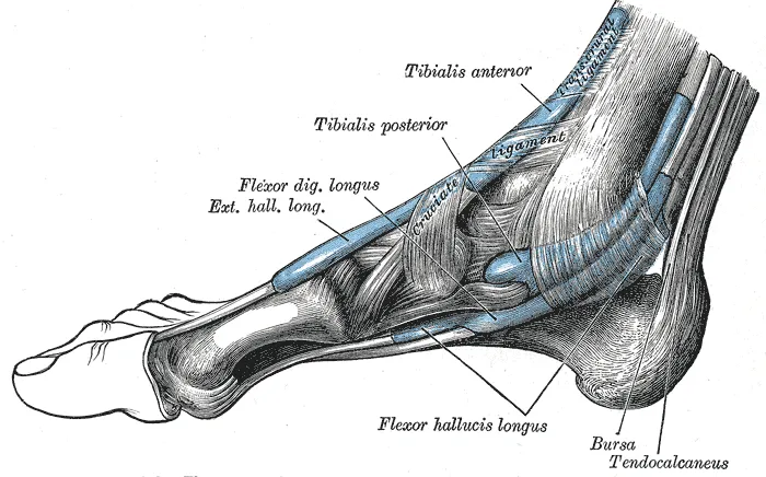
It is the tendon nearest the inner side at the front of the ankle and foot2. The extensor retinaculum holds it tightly against them along its course2. This is made of two bands of connective tissue, the superior and inferior extensor retinacula3. They keep the front-of-leg tendons in place while the foot lifts3.
How the bands hold it at the ankle
In seven cadaver feet, bodies donated for study, the tendon passed through three tunnels9. The superior band and two limbs of the inferior band formed them9. In about a quarter of cases, the tendon has a tunnel of its own, formed between two layers of the superior band2.
Where it attaches
The tendon typically attaches to the medial cuneiform and the first metatarsal, two bones on the inner side of the foot2,10. Its attachment varies between people10. A study of 41 cadaver feet sorted the attachment into three shapes11:
| Shape of the attachment | Share of 41 feet |
|---|---|
| Equal on the medial cuneiform and metatarsal | 48.8%11 |
| Wider on the medial cuneiform | 48.8%11 |
| Wider on the first metatarsal | 2.4%11 |
What the muscle and tendon do
The muscles of the front compartment are the main muscles of dorsiflexion, pulling the foot up toward the shin12. They also extend the toes12. The tibialis anterior mainly lifts the foot and turns its sole inward, called inversion1. A review describes it as playing a role in supporting the arch of the foot2.
What the tendon carries
Studies of five men measured how the tendon stretches under load13,14:
- In five men at maximum isometric load, when the muscle works but the joint does not move, the tendon carried about 530 newtons13. It stretched about 4.1 mm, a strain, or stretch as a share of resting length, of about 2.5%13.
- At maximum isometric load in the same men, its stiffness, the force to stretch it a set amount, was 161 newtons per mm13.
- In five men, the tendon strained 61 to 64% less than its aponeurosis, a broad, flat sheet of tendon14.
Under normal loads, the tendon appeared to work within the elastic “toe” region of its stretch curve13. The tendon’s moment arm is its leverage, meaning how far its line of pull sits from the ankle15. At rest, estimates put it at about 4.5 cm with the foot lifted and about 2.9 cm with the foot pointed15. Six people were scanned with MRI, which shows soft tissue in detail16. Their moment arm was 0.9 to 1.5 cm larger during a maximum isometric pull than at rest16.
What it does in walking and running
- Nine healthy people walked on a treadmill17. Early in stance, the part of a step with the foot on the ground, the muscle fibres kept their length as the ankle pointed down17.
- In 12 healthy people walking, the fibres stayed nearly the same length at foot contact while the whole muscle and tendon lengthened18. The authors read this as the tendon tissue stretching and absorbing energy18.
- In the same study, the muscle shortened early in the swing of the leg to lift the foot and clear the toes18. At fast walking speed, most of that shortening came from the tendon tissue springing back18.
Breakdown
A stretch without blood vessels
In anatomical studies, the tendon got blood mainly from the anterior tibial artery, the artery at the front of the shin, near the top19. Near the foot, branches of the medial tarsal artery, an artery on the inner side of the foot, supplied it19.
Injection and staining studies found a zone 45 to 67 mm long with no blood vessels, in the front half of the tendon19. This zone matches the site of spontaneous rupture that is reported most often19. It lies where the tendon wraps around the extensor retinacula, which act as pulleys20. A thin layer of cartilage-like cells lines it20.
Rupture is rare and often recognised late
Rupture of this tendon is uncommon, but losing the strength to lift the foot can leave a substantial loss of function21. A systematic review and meta-analysis calls it rare and often recognised late4. Pain is brief, and the other front-of-leg tendons make up for it4. After a tear, the foot can usually still be lifted, but with much less strength2. That is because the long toe extensors, extensor digitorum longus and extensor hallucis longus, remain intact2.
Who it happens to and why
- One article reports that most ruptures happen in elderly men after minor trauma22. It says they often show up as a sudden “foot drop”, difficulty lifting the front of the foot when walking22.
- In the review of 87 closed ruptures, the mean age was 63.9 in the surgery group and 72.4 in the non-surgical group5.
- A review reports that tears from open or blunt trauma usually affect younger people2. It says spontaneous ruptures usually affect middle-aged and older people2.
Sources describe the causes in different terms2,23. One paper says ruptures can follow a cut, a blow or a forced downward bend of the foot23. It calls spontaneous ruptures rare, and says most come from wear in the poorly supplied last 5 to 30 mm of the tendon23.
A review describes one kind of rupture as typically following repeated small strains on a weakened tendon2. Conditions it lists as able to weaken the tendon beforehand include diabetes, gout and ageing2. It also lists injection of a corticosteroid, a drug that copies a type of hormone the body makes2.
A review reports that tears typically sit within 5 to 30 mm of the tendon’s attachment on the foot2. In MRI scans of seven people with tears, four tears were partial9. Each sat at the level of the ankle joint, near a limb of the inferior band9. In another group of seven people with rupture, four had used systemic (whole-body) corticosteroids, and one had a local steroid injection24. Those authors report a link with both24.
Wear at the foot end
For tendinopathy at the foot end of this tendon, practice rests mainly on case reports and small retrospective case series7. There is little agreement on the best approach7. Surgeons reporting on nine patients who had surgery describe it as common but rarely written about25. They say it often affects overweight women of about 50 to 70, with pain that worsens at night25.
One study looked at 29 people with tendinosis (tendon wear) at the foot end26:
- Their mean age was 62, 27 were women and 21 were overweight26.
- The usual symptom was burning pain on the inner midfoot, often worst at night26.
- MRI showed a thickened tendon in each person26.
- Lengthwise split tears showed in 19 of 32 feet, and the pathology was typical of degenerative tendinosis26.
A review reports that in most people with tendinopathy at this end, the affected part is the band of tendon attaching to the medial cuneiform2.
In one MRI study summarised by a review, changes sat in the back part of the tendon, next to bone, in 39% of cases2. They were spread through the tendon in 54%, and in the front part in 7%2. The changes were linked with bony features of the midfoot, which suggests that rubbing against bone may play a part2. One study of 28 people, cited in a review, linked MRI changes with arthritis of nearby midfoot joints2. Whether one leads to the other is unclear2.
Recovery
After a rupture
The systematic review of 87 closed ruptures concluded that surgical repair leads to successful outcomes in many patients5.
- One study followed 19 repaired ruptures in 18 people aged 21 to 78 for an average of 53.3 months21. Lifting the foot and walking improved in 18 of 1921. The strength to lift the foot was full in 1521.
- In that group, the average hindfoot score, a measure of foot and ankle function, rose from 55.5 before surgery to 93.6 after21. Results did not appear to vary with age, sex or other illness21.
- In 10 people tested on average 27.9 months after repair, satisfaction was high27. Even so, strength in lifting and turning in the foot was lower than on the uninjured side27.
- In 20 ruptures in 19 people, 11 were managed without surgery and 9 with it28. The surgical cases regained full muscle strength, and non-surgical management worked in low-demand people with smaller deficits28.
- Seven people with rupture were followed for 12 months to 12 years24. Mean foot and ankle scores were 85 in the surgery group and 52 in those managed without surgery24.
When repair happens
On timing, the sources disagree4,23. A systematic review and meta-analysis of 217 cases looked at acute repair, done soon after an injury, and chronic repair, done later4. Acute repair had significantly better outcomes4. In a cohort of 48 people followed for a mean of 30 months, outcomes did not differ significantly between prompt and delayed repair23.
After wear at the foot end
Of 40 people with tendinopathy at the foot end, 11 had surgery after non-surgical care did not work29. After a mean of 29 months, their midfoot score had improved by a mean of 33 points29. Their mean pain was 0.9 out of 1029.
A small prospective study, one that follows people forward in time, took in 18 people with tendinopathy at the foot end7. Each had an injection of platelet-rich plasma, a part of their own blood with extra platelets, around the tendon7. Each then spent three weeks in a walking cast7. Eccentric exercises, meaning muscle work done while the muscle lengthens, and calf stretching followed7.
Pain and function scores had improved by 6 weeks, with no further significant gain at 12 weeks7. Two of the 18 later chose surgery7.
What is not known about adapting to load
A systematic review and meta-analysis of how human tendons without symptoms adapt to more loading found 27 studies8. Each was on the Achilles or the patellar tendon, so none measured the tibialis anterior tendon directly8. In those studies, the data suggested that how heavy the load was mattered more than the type of muscle contraction8.
Connections
The muscles it works with
The front-compartment muscles, including the tibialis anterior, are supplied by the deep fibular (peroneal) nerve, a nerve of the lower leg12. The anterior tibial artery also supplies them12. The tibialis anterior, extensor hallucis longus and extensor digitorum longus each help raise the front of the foot3. Those two toe extensors are why the foot can usually still lift after a tear2.
The muscles that work against it
Muscles in the front compartment generally lift the foot, and those in the back compartment generally point it down3. In the back, the soleus and the two heads of the gastrocnemius do most of the work of pointing the foot down30. Their fibres form the Achilles tendon31.
Exercises that load it
Related exercise pages: tibialis raise, standing shin stretch. Lifting the foot is one of the two main jobs of the muscle this tendon serves1.
Tibialis anterior tendon is loaded by
Tibialis anterior tendon works opposite at the ankle to
Sources
- Juneja P, Hubbard JB (2023). Anatomy, Bony Pelvis and Lower Limb: Tibialis Anterior Muscles.. StatPearls. PMID 30020676
- Varghese A, Bianchi S (2014). Ultrasound of tibialis anterior muscle and tendon: anatomy, technique of examination, normal and pathologic appearance.. Journal of ultrasound. doi:10.1007/s40477-013-0060-7 PMID 24883132
- Betts JG, Young KA, Wise JA, Johnson E, Poe B, Kruse DH, Korol O, Johnson JE, Womble M, DeSaix P (2022). Anatomy and Physiology 2e. OpenStax. Link
- Duelfer KA, McAlister JE (2023). Tibialis Anterior Tendon Rupture Surgical Treatments and Outcomes: A Systematic Review and Meta-Analysis.. The Journal of foot and ankle surgery : official publication of the American College of Foot and Ankle Surgeons. doi:10.1053/j.jfas.2023.01.007 PMID 36963479
- Christman-Skieller C, Merz MK, Tansey JP (2015). A systematic review of tibialis anterior tendon rupture treatments and outcomes.. American journal of orthopedics (Belle Mead, N.J.). PMID 25844597
- Vosoughi AR, Heyes G, Molloy AP, Mason LW, Hoveidaei AH (2020). Management of tibialis anterior tendon rupture. Foot and ankle surgery : official journal of the European Society of Foot and Ankle Surgeons. doi:10.1016/j.fas.2019.06.003 PMID 31501017
- Desomer L, van Beek N, Van Riet A, Verfaillie S (2024). Outcomes of Platelet-Rich Plasma Infiltration and Weightbearing Cast Immobilization in Distal Tibialis Anterior Tendinopathy: A Prospective Cohort Study.. Foot & ankle international. doi:10.1177/10711007231210506 PMID 37964467
- Bohm S, Mersmann F, Arampatzis A (2015). Human tendon adaptation in response to mechanical loading: a systematic review and meta-analysis of exercise intervention studies on healthy adults.. Sports Medicine - Open. doi:10.1186/s40798-015-0009-9 PMID 27747846
- Lee MH, Chung CB, Cho JH, Mohana-Borges AV, Pretterklieber ML, Trudell DJ, Resnick D (2006). Tibialis anterior tendon and extensor retinaculum: imaging in cadavers and patients with tendon tear.. AJR. American journal of roentgenology. doi:10.2214/AJR.05.0073 PMID 16861505
- Zielinska N, Tubbs RS, Paulsen F, Szewczyk B, Podgórski M, Borowski A, Olewnik Ł (2021). Anatomical Variations of the Tibialis Anterior Tendon Insertion: An Updated and Comprehensive Review.. Journal of clinical medicine. doi:10.3390/jcm10163684 PMID 34441980
- Willegger M, Seyidova N, Schuh R, Windhager R, Hirtler L (2017). Anatomical Footprint of the Tibialis Anterior Tendon: Surgical Implications for Foot and Ankle Reconstructions.. BioMed research international. doi:10.1155/2017/9542125 PMID 28660212
- Lezak B, Launico MV (2026). Anatomy, Bony Pelvis and Lower Limb: Leg Anterior Compartment.. StatPearls. PMID 30969547
- Maganaris CN, Paul JP (1999). In vivo human tendon mechanical properties.. The Journal of physiology. doi:10.1111/j.1469-7793.1999.00307.x PMID 10562354
- Maganaris CN, Paul JP (2000). Load-elongation characteristics of in vivo human tendon and aponeurosis.. The Journal of experimental biology. doi:10.1242/jeb.203.4.751 PMID 10648216
- Maganaris CN (2000). In vivo measurement-based estimations of the moment arm in the human tibialis anterior muscle-tendon unit.. Journal of biomechanics. doi:10.1016/s0021-9290(99)00188-8 PMID 10673122
- Maganaris CN, Baltzopoulos V, Sargeant AJ (1999). Changes in the tibialis anterior tendon moment arm from rest to maximum isometric dorsiflexion: in vivo observations in man.. Clinical biomechanics (Bristol, Avon). doi:10.1016/s0268-0033(99)00018-2 PMID 10521650
- Chleboun GS, Busic AB, Graham KK, Stuckey HA (2007). Fascicle length change of the human tibialis anterior and vastus lateralis during walking.. The Journal of orthopaedic and sports physical therapy. doi:10.2519/jospt.2007.2440 PMID 17710906
- Maharaj JN, Cresswell AG, Lichtwark GA (2019). Tibialis anterior tendinous tissue plays a key role in energy absorption during human walking.. The Journal of experimental biology. doi:10.1242/jeb.191247 PMID 31064856
- Petersen W, Stein V, Tillmann B (1999). Blood supply of the tibialis anterior tendon.. Archives of orthopaedic and trauma surgery. doi:10.1007/s004020050431 PMID 10613222
- Petersen W, Stein V, Bobka T (2000). Structure of the human tibialis anterior tendon.. Journal of anatomy. doi:10.1046/j.1469-7580.2000.19740617.x PMID 11197535
- Sammarco VJ, Sammarco GJ, Henning C, Chaim S (2009). Surgical repair of acute and chronic tibialis anterior tendon ruptures.. The Journal of bone and joint surgery. American volume. doi:10.2106/JBJS.G.01386 PMID 19181976
- Negrine JP (2007). Tibialis anterior rupture: acute and chronic.. Foot and ankle clinics. doi:10.1016/j.fcl.2007.07.004 PMID 17996615
- Johansen JK, Jordan M, Thomas M, Walther M, Simatira AM, Waizy H (2021). Surgical treatment of tibialis anterior tendon rupture.. Foot and ankle surgery : official journal of the European Society of Foot and Ankle Surgeons. doi:10.1016/j.fas.2020.06.011 PMID 32948441
- Gwynne-Jones D, Garneti N, Wyatt M (2009). Closed tibialis anterior tendon rupture: a case series.. Foot & ankle international. doi:10.3113/FAI.2009.0758 PMID 19735632
- Lemmens L, van Beek N, Verfaillie S (2020). Postoperative results of distal tibialis anterior tendinosis.. Foot and ankle surgery : official journal of the European Society of Foot and Ankle Surgeons. doi:10.1016/j.fas.2019.11.005 PMID 31870617
- Beischer AD, Beamond BM, Jowett AJ, O'Sullivan R (2009). Distal tendinosis of the tibialis anterior tendon.. Foot & ankle international. doi:10.3113/FAI.2009.1053 PMID 19912714
- Kopp FJ, Backus S, Deland JT, O'Malley MJ (2007). Anterior tibial tendon rupture: results of operative treatment.. Foot & ankle international. doi:10.3113/FAI.2007.1045 PMID 17923052
- Meyer C, Herrington L, Tolin JC, Badillo K, Kiefer C, Parker A, Burns P, Manway J, Cain J (2026). Evaluation of treatment options for tibialis anterior tendon ruptures.. The Journal of foot and ankle surgery : official publication of the American College of Foot and Ankle Surgeons. doi:10.1053/j.jfas.2026.04.016 PMID 42034330
- Grundy JR, O'Sullivan RM, Beischer AD (2010). Operative management of distal tibialis anterior tendinopathy.. Foot & ankle international. doi:10.3113/FAI.2010.0212 PMID 20230699
- Gentil P, Souza D, Santana M, Alves RR, Campos MH, Pinto R, Bottaro M (2020). Multi- and Single-Joint Resistance Exercises Promote Similar Plantar Flexor Activation in Resistance Trained Men.. International Journal of Environmental Research and Public Health. doi:10.3390/ijerph17249487 PMID 33352879
- Pękala PA, Henry BM, Ochała A, Kopacz P, Tatoń G, Młyniec A, Walocha JA, Tomaszewski KA (2017). The twisted structure of the Achilles tendon unraveled: A detailed quantitative and qualitative anatomical investigation.. Scandinavian Journal of Medicine & Science in Sports. doi:10.1111/sms.12835 PMID 28139009
Changes
- Links to the new standing shin stretch page
- Tidy pass: minor review findings fixed, glosses aligned with the reviewed glossary.
- First published.