Tibialis anterior
Also called tibialis anterior muscle, anterior tibial muscle
The muscle at the front of the shin that lifts the foot. How it is built, what it does in walking and running, how it is hurt, how it ages and trains, and what it works with.
- Region
- Lower leg
- Starts on
- the upper outer shinbone (its top bulge and upper two-thirds), the membrane between it and the fibula, and leg fascia1,8
- Ends on
- usually the medial cuneiform, a wedge-shaped inner midfoot bone, and the first metatarsal, on the foot's inner border (Tibialis anterior tendon)1,8
- Tendons
- Tibialis anterior tendon
- Updated
- October 28, 2026
- Reviewed
- October 28, 2026 · how entries are checked
Function
Where it sits
The tibialis anterior is the biggest of four muscles sitting in the front, or anterior, compartment of the lower leg1. It starts on the outer condyle, a rounded knob at the top of the tibia (shinbone), and the upper two-thirds of its outer surface8. It also starts on the interosseous membrane, the sheet joining the shinbone and fibula, and on the leg’s fascia (a sheet of tough tissue)8.
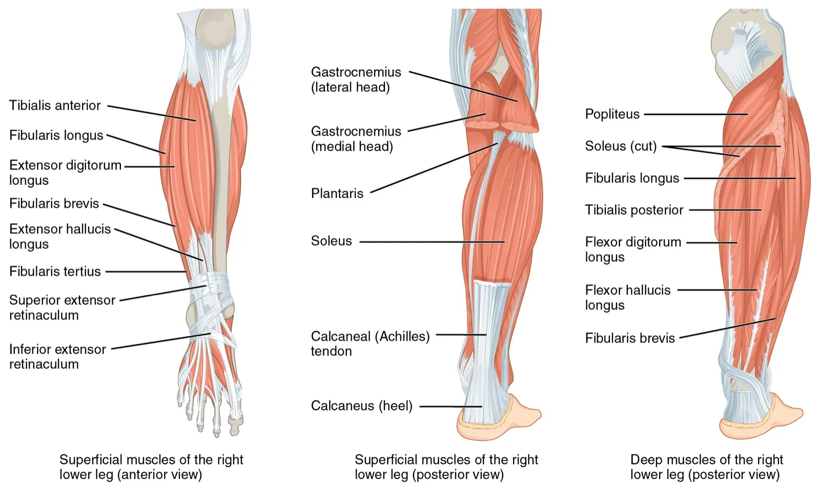
Its tendon typically attaches to the medial cuneiform (a wedge-shaped midfoot bone) and the first metatarsal (the long foot bone behind the big toe)8. That attachment varies between people8. The tendon’s course, tears and wear are covered on the tibialis anterior tendon page.
The deep fibular nerve and the anterior tibial artery supply the muscles of this compartment, including this one9. The nerve runs down beside the artery, between this muscle and the extensor digitorum longus, which lifts the smaller toes and the foot10. It is one of two branches the common fibular nerve divides into, at the neck of the fibula (the thin outer lower-leg bone)10.
What it moves
The tibialis anterior mainly lifts the foot, called dorsiflexion, and turns its sole inward, called inversion1. A review describes it as playing a role in supporting the arch of the foot11. In nine people standing quietly, its length tracked body sway closely, and the authors suggest it may sense sway better than the calf muscles12.
How it is built
A study that took apart 21 lower limbs measured its shape13. The physiological cross-sectional area is a measure of size taken straight across a muscle’s fibres. Pennation angle is the angle of the fibres against the muscle’s line of pull.
| Measure (21 limbs) | Average |
|---|---|
| Mass | About 80 g13 |
| Muscle length | About 26 cm13 |
| Fibre length | About 6.8 cm13 |
| Pennation angle | About 10 degrees13 |
| Physiological cross-sectional area | About 11 cm²13 |
Small studies looked at its fibre types6,14:
- In 30 young men and women, about 73% of fibres were type 1, the slow-contracting kind, with no difference between the sexes14.
- In 7 young men, about 67% were type 16.
- In the same 30 adults, the men had larger fibres, with the bigger gap in type 2, fast-contracting fibres14.
Twelve people pulled the foot upward isometrically, meaning the muscle works but the joint does not move15. The muscle kept its volume and bulged, mainly in thickness15.
What it does in walking
In 10 young adults walking on a treadmill at 11 speeds, its activity peaked around heel strike and around toe-off16. In a second study, peak activity came about 31 ms after heel contact4. That was earlier than a quick reflex could explain4. Its authors suggest that activity just before heel strike may anticipate how hard the foot will meet the ground4.
Studies describe what its fascicles do as the foot lands, in stance, the part of each step with the foot on the ground2,3,4:
- In 9 healthy people on a treadmill, fascicle length stayed constant early in stance as the ankle pointed down2.
- In 12 healthy people, fascicles stayed nearly the same length at foot contact while the whole muscle and tendon lengthened3.
- Tracked step by step in 11 adults, fascicles briefly lengthened about 1 mm early in stance on 96.7% of steps, with little net change4.
The authors of that last study say averaging many steps can make the fascicles look as if they hold their length4. They add that brief lengthening does not contradict the tendon stretching much more than the muscle4.
Running, sprinting and landing
- In 13 runners, the front-of-shin muscles shortened as they worked at first ground contact in running and sprinting, but lengthened in walking17.
- In 10 male students running at 6 km/h, activity before foot contact was lower when they landed on the forefoot than on the heel18.
- In 10 trained sprinters, its activity held fairly steady from the third stride and fell above about 8 to 9 metres per second19. Over the same strides, three other leg muscles grew more active19.
- Thirteen male students landed on one leg from 0.3 m20. More activity just before contact went with a higher peak ground reaction force, the push of the ground back up20.
Breakdown
Tears of the muscle
Tears of the tibialis anterior muscle itself are described as rarely seen in clinical practice5. A sudden tear during running is rare, but can badly limit function5. Tears of its tendon are also called rare, and are known mostly from case reports21,22.
Muscle hernia
In a muscle hernia, the muscle pushes through a gap in its fascia23. Case reports say that in the lower limb, hernias that cause symptoms are most often in the tibialis anterior, usually after an injury23. They are a rare cause of long-standing leg pain23. One report counts about 200 cases, and most people report vague, long-standing leg pain, which makes them hard to pin down23.
- One report says muscle hernias usually appear in athletes, often after a blow or after the muscle grows with hard exercise24.
- Reports say they are usually confirmed with ultrasound of the muscle at rest and while working23.
- In a report of four teenagers, the hernias showed as painless lumps under the skin of the lower leg25.
Fatigue and the front compartment
In 15 injury-free runners, fine-wire recordings showed this muscle working above its fatigue threshold for 85% of the time26. The authors suggest this may explain fatigue-related injuries to it in runners26. In 21 young adults, 5 sets of 30 eccentric (lowering) contractions cut maximal torque, or turning force, by 28%27. The authors read the incomplete recovery as a sign of muscle damage27.
Chronic exertional compartment syndrome is compartment syndrome that comes on with exercise and eases with rest. A systematic review, which gathers the research on a question, covered 1,596 people who had surgery for it28. The anterior compartment was the one most often involved, in 51%28. Tendinopathy is local pain in a tendon that comes with loading. In a review of running injuries, front-compartment tendinopathy was the largest share of injuries in ultramarathon runners, at 19.4%29.
For shin splints, 50 legs from bodies donated for study showed other structures attached where the pain occurs30. They were the soleus, the flexor digitorum longus (a deep calf muscle) and deep fascia30.
When the nerve fails
Foot drop is being unable to lift the front of the foot because the muscles that lift it are weak31. Pressure on the fibular nerve can weaken the muscles that lift the foot, and the person may lift the foot higher to clear the toes32. These sources describe the foot-lifting muscles as a group, not this muscle alone31,32.
Recovery
How it ages
A motor unit is a nerve cell and the muscle fibres it switches on. One study estimated how many the muscle had in men of three ages7:
| Group of men | Estimated motor units |
|---|---|
| About 25 years | 1507 |
| About 65 years | 917 |
| 80 years or older | 597 |
Strength in lifting the foot was not reduced until beyond 807. The authors suggest the loss may not limit function until it passes a threshold7.
Another study compared young men (about 26) with older men (about 71) and masters runners (about 69), who compete in age-group events33. The older men had about 45% of the young men’s estimated motor units, and the runners about 40%33. The authors read this as lifelong training not lessening the loss33. The muscle’s cross-sectional area did not differ between the three groups33.
When the leg is held still
In 7 healthy young men, one leg was held still in a brace and boot for 15 days6. The medial gastrocnemius, the inner part of a calf muscle, lost about 3% of its volume and 13% of its thickness6. The tibialis anterior’s volume and thickness did not change6.
How it responds to training
Studies that trained the foot-lifting muscles report these findings34,35:
- In 12 trained young men, 6 weeks of training thickened the muscle by about 8% on ultrasound, similarly with lifting or lowering work34.
- In the same men, fascicles grew 6% longer in the leg trained with lowering work, and not in the other leg34.
- In 44 adults training twice weekly for 8 weeks, its cross-sectional area on MRI, a detailed scan, did not change in any age group35.
- In 19 people training one leg for 5 weeks, force rose 14.7% in that leg and 8.4% in the untrained one, called cross-education36.
In 26 young adults, 200 hard lowering contractions cut torque by about 24%37. After the same bout 4 weeks later, torque came back sooner37.
After a tear
In the single case report, the 47-year-old runner with a partial tear was managed without surgery and later ran ultramarathons again5.
Connections
The muscles it works with and against
The four front-of-shin muscles lift the foot, and the tibialis anterior and extensor hallucis longus, which lifts the big toe, also turn the sole inward38. The soleus and the two heads of the gastrocnemius do most of the work of pointing the foot down39. In 10 trained sprinters, the soleus and tibialis anterior were steadily active together, which the authors read as steadying the ankle19.
Exercises that load it
| Exercise | What a study found |
|---|---|
| Tibialis raise | In 22 college students, each foot drill in a set that includes heel walking raised its activity 4.0 to 68.3% above normal walking40 |
| Deadlift | In 13 college football players, its activity was greater in the sumo style than the conventional style41 |
| Decline squat | In 26 adults holding squats, its activity was lower on a 25-degree wedge than on a flat floor42 |
| Single-leg Romanian deadlift | In 30 healthy men, a foam pad raised its activity43 |
| Pogo hops | In 7 men hopping, its activity stayed low through ground contact compared with the calf muscles44 |
| Standing shin stretch | In 16 men and 13 women, its fascicles lengthened as the foot was passively pointed down45 |
Related exercise pages: running, child’s pose. In 38 people after ankle fracture surgery, a stiffer tibialis anterior went with less foot pointing when sitting back on the heels46.
Tibialis anterior pulls through
Tibialis anterior is supplied by a branch of
Tibialis anterior works opposite at the ankle to
Tibialis anterior sits in the compartment most often involved in
Tibialis anterior is loaded by
Tibialis anterior is stretched by
Sources
- Juneja P, Hubbard JB (2023). Anatomy, Bony Pelvis and Lower Limb: Tibialis Anterior Muscles.. StatPearls. PMID 30020676
- Chleboun GS, Busic AB, Graham KK, Stuckey HA (2007). Fascicle length change of the human tibialis anterior and vastus lateralis during walking.. The Journal of orthopaedic and sports physical therapy. doi:10.2519/jospt.2007.2440 PMID 17710906
- Maharaj JN, Cresswell AG, Lichtwark GA (2019). Tibialis anterior tendinous tissue plays a key role in energy absorption during human walking.. The Journal of experimental biology. doi:10.1242/jeb.191247 PMID 31064856
- Kwak ST, Chang YH (2023). Fascicle dynamics of the tibialis anterior muscle reflect whole-body walking economy.. Scientific reports. doi:10.1038/s41598-023-31501-2 PMID 36949112
- Santos-Faria J, Malta JN, Coelho AP, Branco JP (2022). Running on the Edge: Rupture of the Tibialis Anterior Muscle During an Ultramarathon.. Cureus. doi:10.7759/cureus.31930 PMID 36582552
- Bass JJ, Hardy EJO, Inns TB, Wilkinson DJ, Piasecki M, Morris RH, Spicer A, Sale C, Smith K, Atherton PJ, Phillips BE (2021). Atrophy Resistant vs. Atrophy Susceptible Skeletal Muscles: "aRaS" as a Novel Experimental Paradigm to Study the Mechanisms of Human Disuse Atrophy.. Frontiers in physiology. doi:10.3389/fphys.2021.653060 PMID 34017264
- McNeil CJ, Doherty TJ, Stashuk DW, Rice CL (2005). Motor unit number estimates in the tibialis anterior muscle of young, old, and very old men.. Muscle & nerve. doi:10.1002/mus.20276 PMID 15685623
- Zielinska N, Tubbs RS, Paulsen F, Szewczyk B, Podgórski M, Borowski A, Olewnik Ł (2021). Anatomical Variations of the Tibialis Anterior Tendon Insertion: An Updated and Comprehensive Review.. Journal of clinical medicine. doi:10.3390/jcm10163684 PMID 34441980
- Lezak B, Launico MV (2026). Anatomy, Bony Pelvis and Lower Limb: Leg Anterior Compartment.. StatPearls. PMID 30969547
- Hardin JM, Devendra S (2022). Anatomy, Bony Pelvis and Lower Limb: Calf Common Peroneal Nerve (Common Fibular Nerve).. StatPearls. PMID 30422563
- Varghese A, Bianchi S (2014). Ultrasound of tibialis anterior muscle and tendon: anatomy, technique of examination, normal and pathologic appearance.. Journal of ultrasound. doi:10.1007/s40477-013-0060-7 PMID 24883132
- Di Giulio I, Maganaris CN, Baltzopoulos V, Loram ID (2009). The proprioceptive and agonist roles of gastrocnemius, soleus and tibialis anterior muscles in maintaining human upright posture.. The Journal of Physiology. doi:10.1113/jphysiol.2009.168690 PMID 19289550
- Ward SR, Eng CM, Smallwood LH, Lieber RL (2009). Are current measurements of lower extremity muscle architecture accurate?. Clinical Orthopaedics and Related Research. doi:10.1007/s11999-008-0594-8 PMID 18972175
- Porter MM, Stuart S, Boij M, Lexell J (2002). Capillary supply of the tibialis anterior muscle in young, healthy, and moderately active men and women.. Journal of applied physiology (Bethesda, Md. : 1985). doi:10.1152/japplphysiol.00744.2001 PMID 11896009
- Raiteri BJ, Cresswell AG, Lichtwark GA (2016). Three-dimensional geometrical changes of the human tibialis anterior muscle and its central aponeurosis measured with three-dimensional ultrasound during isometric contractions.. PeerJ. doi:10.7717/peerj.2260 PMID 27547566
- Byrne CA, O'Keeffe DT, Donnelly AE, Lyons GM (2007). Effect of walking speed changes on tibialis anterior EMG during healthy gait for FES envelope design in drop foot correction.. Journal of electromyography and kinesiology : official journal of the International Society of Electrophysiological Kinesiology. doi:10.1016/j.jelekin.2006.07.008 PMID 16990012
- Mann RA, Hagy J (1980). Biomechanics of walking, running, and sprinting.. The American journal of sports medicine. doi:10.1177/036354658000800510 PMID 7416353
- Lin JZ, Chiu WY, Tai WH, Hong YX, Chen CY (2021). Ankle Muscle Activations during Different Foot-Strike Patterns in Running.. Sensors (Basel, Switzerland). doi:10.3390/s21103422 PMID 34069061
- Yokota K, Tamaki H (2025). Application of Wireless EMG Sensors for Assessing Agonist-Antagonist Muscle Activity During 50-m Sprinting in Athletes.. Sensors (Basel, Switzerland). doi:10.3390/s25206395 PMID 41157447
- Mahaki M, Mi'mar R, Mahaki B (2015). On the relationship between lower extremity muscles activation and peak vertical and posterior ground reaction forces during single leg drop landing.. The Journal of sports medicine and physical fitness. PMID 25924564
- Duelfer KA, McAlister JE (2023). Tibialis Anterior Tendon Rupture Surgical Treatments and Outcomes: A Systematic Review and Meta-Analysis.. The Journal of foot and ankle surgery : official publication of the American College of Foot and Ankle Surgeons. doi:10.1053/j.jfas.2023.01.007 PMID 36963479
- Christman-Skieller C, Merz MK, Tansey JP (2015). A systematic review of tibialis anterior tendon rupture treatments and outcomes.. American journal of orthopedics (Belle Mead, N.J.). PMID 25844597
- Sharma N, Kumar N, Verma R, Jhobta A (2017). Tibialis Anterior Muscle Hernia: A Case of Chronic, Dull Pain and Swelling in Leg Diagnosed by Dynamic Ultrasonography.. Polish journal of radiology. doi:10.12659/PJR.900846 PMID 28638493
- Dönmez G, Evrenos MK, Cereb M, Karanfil Y, Doral MN (2015). Double layer repair of tibialis anterior muscle hernia in a soccer player: a case report and review of the literature.. Muscles, ligaments and tendons journal. doi:10.11138/mltj/2015.5.4.331 PMID 26958545
- Clinite KL, Wyble A, Sugarman JL (2019). Tibialis anterior muscle herniation in adolescents: A case series and review of the literature.. Pediatric dermatology. doi:10.1111/pde.13875 PMID 31215058
- Reber L, Perry J, Pink M (1993). Muscular control of the ankle in running.. The American journal of sports medicine. doi:10.1177/036354659302100608 PMID 8291630
- Power GA, Dalton BH, Rice CL, Vandervoort AA (2010). Delayed recovery of velocity-dependent power loss following eccentric actions of the ankle dorsiflexors.. Journal of Applied Physiology. doi:10.1152/japplphysiol.01254.2009 PMID 20576845
- Campano D, Robaina JA, Kusnezov N, Dunn JC, Waterman BR (2016). Surgical Management for Chronic Exertional Compartment Syndrome of the Leg: A Systematic Review of the Literature. Arthroscopy : the journal of arthroscopic & related surgery : official publication of the Arthroscopy Association of North America and the International Arthroscopy Association. doi:10.1016/j.arthro.2016.01.069 PMID 27020462
- Kakouris N, Yener N, Fong DTP (2021). A systematic review of running-related musculoskeletal injuries in runners.. Journal of sport and health science. doi:10.1016/j.jshs.2021.04.001 PMID 33862272
- Beck BR, Osternig LR (1994). Medial tibial stress syndrome. The location of muscles in the leg in relation to symptoms.. The Journal of bone and joint surgery. American volume. doi:10.2106/00004623-199407000-00015 PMID 8027114
- Nori SL, Stretanski MF (2025). Foot Drop.. StatPearls. PMID 32119280
- Cucu LE, Popescu G, Maștaleru A, Ignat EB, Grosu C, Bîrsanu L, Leon MM (2025). Common Peroneal Nerve Paralysis Following Rapid Weight Loss-A Case Report and Literature Review. Nutrients. doi:10.3390/nu17111782 PMID 40507050
- Piasecki M, Ireland A, Coulson J, Stashuk DW, Hamilton-Wright A, Swiecicka A, Rutter MK, McPhee JS, Jones DA (2016). Motor unit number estimates and neuromuscular transmission in the tibialis anterior of master athletes: evidence that athletic older people are not spared from age-related motor unit remodeling.. Physiological reports. doi:10.14814/phy2.12987 PMID 27694526
- Nunes JP, Nosaka K, Blazevich AJ (2025). The Training Specificity Versus Structural Adaptation Paradox: Differential Effects of Isokinetic Concentric and Eccentric Resistance Training on Muscle Architecture and Function in Young Men.. Scandinavian Journal of Medicine and Science in Sports. doi:10.1111/sms.70166 PMID 41427667
- Psatha M, Wu Z, Gammie F, Ratkevicius A, Wackerhage H, Redpath TW, Gilbert FJ, Meakin JR, Aspden RM (2017). Age-related changes in the effects of strength training on lower leg muscles in healthy individuals measured using MRI.. BMJ Open Sport and Exercise Medicine. doi:10.1136/bmjsem-2017-000249 PMID 28761720
- Dragert K, Zehr EP (2011). Bilateral neuromuscular plasticity from unilateral training of the ankle dorsiflexors.. Experimental Brain Research. doi:10.1007/s00221-010-2472-3 PMID 21069308
- Bruce CD, Ruggiero L, Dix GU, Cotton PD, McNeil CJ (2021). Females and males do not differ for fatigability, muscle damage and magnitude of the repeated bout effect following maximal eccentric contractions.. Applied Physiology, Nutrition, and Metabolism. doi:10.1139/apnm-2020-0516 PMID 32937087
- Brockett CL, Chapman GJ (2016). Biomechanics of the ankle.. Orthopaedics and Trauma. doi:10.1016/j.mporth.2016.04.015 PMID 27594929
- Gentil P, Souza D, Santana M, Alves RR, Campos MH, Pinto R, Bottaro M (2020). Multi- and Single-Joint Resistance Exercises Promote Similar Plantar Flexor Activation in Resistance Trained Men.. International Journal of Environmental Research and Public Health. doi:10.3390/ijerph17249487 PMID 33352879
- Archuleta QA, McIlvain G, Leigh S, Timmons MK (2024). Lower leg muscle activation during the ebbets foot drills.. Journal of Bodywork and Movement Therapies. doi:10.1016/j.jbmt.2024.02.014 PMID 38876639
- Escamilla RF, Francisco AC, Kayes AV, Speer KP, Moorman CT 3rd (2002). An electromyographic analysis of sumo and conventional style deadlifts.. Medicine and Science in Sports and Exercise. doi:10.1097/00005768-200204000-00019 PMID 11932579
- Lee D, Lee S, Park J (2015). Impact of decline-board squat exercises and knee joint angles on the muscle activity of the lower limbs.. Journal of physical therapy science. doi:10.1589/jpts.27.2617 PMID 26357447
- Kim J, Jeon Y, Eom K (2026). Biomechanical analysis of single leg deadlift under the effect of instability.. Scientific Reports. doi:10.1038/s41598-026-56871-1 PMID 42303734
- Hobara H, Kanosue K, Suzuki S (2007). Changes in muscle activity with increase in leg stiffness during hopping.. Neuroscience letters. doi:10.1016/j.neulet.2007.02.064 PMID 17367931
- Otsuki A, Muraoka Y, Fujita E, Kubo S, Yoshida M, Komuro Y, Ikegawa S, Ohta Y, Kuno-Mizumura M (2016). Gender differences in muscle blood volume reduction in the tibialis anterior muscle during passive plantarflexion.. Clinical physiology and functional imaging. doi:10.1111/cpf.12232 PMID 25620638
- Miyasaka H, Ebihara B, Fukaya T, Iwai K, Kubota S, Mutsuzaki H (2025). Association Between Stiffness of the Deep Fibres of the Tibialis Anterior Muscle and Seiza Posture Performance After Ankle Fracture Surgery.. Journal of functional morphology and kinesiology. doi:10.3390/jfmk10030300 PMID 40843831
Changes
- First published.