Metatarsals
Also called metatarsal, metatarsal bones
The five long bones of the forefoot. How they carry load in walking and running, where and in whom they crack under repeated stress, what studies of fifth metatarsal fractures found, and what attaches to them.
- Region
- Foot & ankle
- Muscles
- fibularis brevis, fibularis longus, tibialis anterior, dorsal interossei
- Updated
- October 22, 2026
- Reviewed
- October 22, 2026 · how entries are checked
Function
Where they sit
The front half of the foot is formed by five metatarsal bones1. They lie between the tarsal bones and the toe bones, and are numbered 1 to 5 from the inner side1. The first is shorter and thicker than the others, and the second is the longest1.
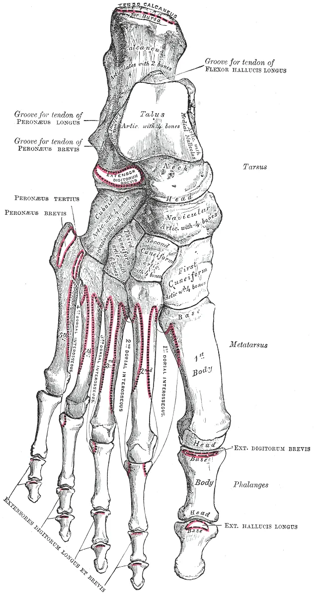
Their two ends
Each metatarsal has a base, the end nearest the ankle1. The bases form joints with the cuboid or the cuneiform bones1. The head is the expanded far end1. Each metatarsal meets the first bone of a toe at a metatarsophalangeal joint1. The heads touch the ground and make up the ball of the foot1.
The base of the fifth metatarsal widens outward, which gives muscles a place to attach1. It can be felt as a bony bump halfway along the outer border of the foot1.
Their part in the arches
The lengthwise arches of the foot are formed by the tarsal bones at the back and the metatarsals at the front1. They rest on the ground at the heel bone behind and the metatarsal heads in front1. The side-to-side arch of the midfoot is formed by the wedge-shaped cuneiform bones and the bases of the first to fourth metatarsals1.
An anatomical review describes the base of the second metatarsal as wedged deeply between the first and third cuneiforms8. It sits at the top of the side-to-side arch and acts as its keystone8. A review of the fifth metatarsal calls it a key part of the outer column of the forefoot5.
The two small bones under the first
The first metatarsophalangeal joint is made up of four bones9. They are the first metatarsal, the first bone of the big toe, and two sesamoids that lie beneath the metatarsal’s head9. Sesamoids are small bones partly or wholly embedded in a tendon10.
A review describes this joint as shallow, with little bony stability, so it relies on soft tissues such as ligaments and muscles9. Damage to its capsule and ligaments can lead to conditions such as “turf toe”9.
The strain measured in them
Strain is how far something stretches as a share of its resting length. In one study, strain was measured in the second metatarsal and the tibia of two people at the same time2. During treadmill walking and jogging, peak compression strains were significantly higher in the metatarsal than in the tibia2. Compression strain in the metatarsal passed 2500 microstrain (millionths of resting length) in barefoot treadmill walking2. In vertical and broad jumps, compression and tension strains both passed 3000 microstrain2.
Eight people had an instrumented staple placed on top of the second metatarsal11. In unloaded barefoot walking, mean peak compression strain was about 1534 microstrain11. It rose significantly when a 20 kg backpack was added, and after fatigue11.
The loads estimated in running
Three studies used models to estimate loads in the metatarsals during running12,13,14:
- In 21 male distance runners, estimated bending strain at mid-shaft was greatest in the second, 6.9 times the estimate for the first12.
- In 28 runners studied barefoot, running 10% faster than preferred raised peak stress in the second metatarsal by 4.2%13. Peak stress varied widely from runner to runner13.
- In 14 male recreational runners, minimalist shoes raised estimated strains in all metatarsals by 28.7% compared with traditional shoes14.
Breakdown
How often stress fractures land here
In military recruits, metatarsal stress fractures follow long marches15. In athletes, they follow episodes of overtraining that involve running or jumping15. Among US college athletes, the metatarsals were the most common location of stress fractures; the stress fracture page compares bones3.
| Group studied | Share of stress fractures in the metatarsals |
|---|---|
| 54 female professional ballet dancers, by survey (27 fractures) | 63%16 |
| US college athletes (671 fractures) | 37.9%3 |
| 303 athletes in Okinawa, Japan, mean age 15.6 (309 fractures) | 28%17 |
| 320 athletes with stress fractures confirmed on bone scan | 8.8%18 |
Which metatarsal
A review of lower limb stress fractures in sport says shaft stress fractures develop most often in the second metatarsal19. The third and fourth follow, and the review says these fractures are often seen in ballet dancers and distance runners19. A systematic review (a study that searches for all the research on a question and sums it up) covered 32 studies4. It found soccer associated with fractures of the fifth metatarsal4. Long-distance running and recruit training often led to fractures of the second or third4.
That review says second and third metatarsal stress fractures in recruit training are commonly known as “march fractures”4. In a prospective study (one that follows people forward in time) of 2379 professional male football players, 51 stress fractures occurred20. Of these, 78% were in the fifth metatarsal20.
A 2022 review names the second and third metatarsal shafts as typical low-risk sites, which usually heal without major complication21. A 2000 review lists the great toe sesamoids and the fifth metatarsal among high-risk sites22. There, a stress fracture carries a high risk of becoming a complete break22. The other risks it names are delayed union (a break that is slow to join) and nonunion (a broken bone that fails to join)22.
What may lie behind them
The authors of the two-person strain study suggested a reason2. Metatarsal strains may be high enough, even in moderate activity, to cause fatigue failure through the number of loading cycles alone2. That could happen before remodelling (the steady replacement of old bone with new) responds2.
The systematic review of 32 studies found three features of the foot that appear to influence risk4. These were a higher arch, abnormal inward or outward rolling, and longer metatarsals or larger metatarsal angles4. It noted that measurement is not standardised and the results remain to be examined4.
The fifth metatarsal
An anatomical review divides the near end of the fifth metatarsal into three zones5:
- The tuberosity, the enlarged bump on the base, where avulsion fractures occur5. An avulsion is a tear in which a tendon or ligament pulls away from its bone.
- The junction between the base and the shaft, the Jones fracture zone5.
- The first 1.5 cm of the shaft, typically associated with stress fractures5.
The same review describes the bone’s blood supply as uneven5. The base has a rich network of vessels, while the junction and the near shaft receive relatively limited flow5. The review calls this a zone where fractures are predisposed to delayed union or nonunion5.
Among 64 European elite football teams followed from 2001 to 2012, 67 fifth metatarsal fractures made up 0.5% of all injuries23. Of those fractures, 33% were refractures (repeat breaks)23.
Recovery
How the bone adapts and repairs
Bone adapts its structure to the loads it regularly carries25. According to one review, removing damaged compact bone takes about 4 weeks and replacing it can take three months26.
A year-long study followed 76 male elite infantry recruits27. Tibial and femoral stress fractures became less frequent after six months of training, but metatarsal stress fractures did not27. In weeks 26 to 52, 91% of the stress fractures were metatarsal27.
Return after shaft and base stress fractures
A review of lower limb stress fractures in sport reports return-to-sport times of 4 to 12 weeks after metatarsal shaft stress fractures19. One series followed 51 professional dancers with 64 stress fractures at the base of the second metatarsal28. Return to performance averaged 6.2 weeks after the fracture was identified28. Eight fractures recurred at the same site, an average of 4.3 years later28.
What studies of Jones fractures found
One randomised trial (a study that assigns people to groups by chance) covered acute Jones fractures6. It assigned 19 people to screw fixation and 18 to a cast6. Median time to bone union was 7.5 weeks in the screw group and 14.5 weeks in the cast group6. Eight of the 18 in the cast group had an unsuccessful result: five nonunions, one delayed union and two refractures6. One of the 19 in the screw group did6.
Three reviews pooled union rates with and without surgery29,30,31:
| Review | Joined after surgery | Joined without surgery |
|---|---|---|
| 26 studies of Jones fractures (acute fractures) | 96% | 76%29 |
| 22 studies of Jones fractures in athletes | 97.3% | 71.4%30 |
| 13 studies, 393 stress fractures of the fifth metatarsal’s near end | 87% | 56%31 |
Of the 26 studies in the first review, 22 were level 4 evidence29. The 13-study review rated its included studies as low in methodological quality31. A systematic review of high-risk stress fractures, the fifth metatarsal among them, found small samples and a high risk of bias7. It concluded that strong conclusions for either approach cannot be drawn7.
Among fifth metatarsal fractures in the 64 European football teams, fractures healed faster after surgery than without it23. Lay-off time did not differ significantly between players who had surgery and those who did not23. A systematic review covered 280 surgically fixed fifth metatarsal fractures in elite athletes32. It found a complication rate of 22.5% and a refracture rate of 8.6%32.
Connections
Muscles, tendons and fascia attached to them
| Structure | Where it attaches |
|---|---|
| Fibularis (peroneus) brevis, one of the peroneals, through its tendon | The bump (tuberosity) on the fifth metatarsal’s base5,33 |
| Fibularis (peroneus) longus tendon | The first metatarsal’s base, on its outer side, and the medial cuneiform34 |
| Tibialis anterior tendon | Typically the medial cuneiform and the first metatarsal, though it varies between people35 |
| Dorsal interossei, among the muscles of the foot | Start from the facing sides of the first to fifth metatarsals36 |
| Lateral band of the plantar aponeurosis, another name for the plantar fascia | The lower outer side of the fifth metatarsal’s tuberosity5,37 |
Muscles that change the strain
Nine fresh feet from cadavers (bodies donated for study) were loaded to simulate body weight and contraction of the calf muscles15. Simulated contraction of flexor hallucis longus significantly reduced strain on top of the second metatarsal shaft15.
Nerves, blood and related pages
Nerve supply to the metatarsal region comes mainly from branches of the deep fibular nerve on top of the foot38. On the sole it comes from the medial and lateral plantar nerves38. Blood arrives mainly through branches of the dorsalis pedis artery and the plantar arterial arches38.
Related exercise pages: running, where models in 21 male distance runners put the greatest bending strain in the second metatarsal12.
Metatarsals is a common site of
Metatarsals is loaded by
Metatarsals supports the foot's lengthwise arches with
Metatarsals is attached to by
Metatarsals gives origin to part of
Sources
- Betts JG, Young KA, Wise JA, Johnson E, Poe B, Kruse DH, Korol O, Johnson JE, Womble M, DeSaix P (2022). Anatomy and Physiology 2e. OpenStax. Link
- Milgrom C, Finestone A, Sharkey N, Hamel A, Mandes V, Burr D, Arndt A, Ekenman I (2002). Metatarsal strains are sufficient to cause fatigue fracture during cyclic overloading. Foot & ankle international. doi:10.1177/107110070202300307 PMID 11934065
- Rizzone KH, Ackerman KE, Roos KG, Dompier TP, Kerr ZY (2017). The Epidemiology of Stress Fractures in Collegiate Student-Athletes, 2004-2005 Through 2013-2014 Academic Years. Journal of athletic training. doi:10.4085/1062-6050-52.8.01 PMID 28937802
- Sun J, Feng C, Liu Y, Shan M, Wang Z, Fu W, Niu W (2024). Risk factors of metatarsal stress fracture associated with repetitive sports activities: a systematic review. Frontiers in bioengineering and biotechnology. doi:10.3389/fbioe.2024.1435807 PMID 39175621
- Liu X, Hong CC, Reina F, Carrera A, Alcolea-Madera E, Del Vecchio JJ, Dalmau-Pastor M (2026). Navicular and fifth metatarsal base stress fractures: An illustrated anatomical review. Knee surgery, sports traumatology, arthroscopy : official journal of the ESSKA. doi:10.1002/ksa.70413 PMID 42171011
- Mologne TS, Lundeen JM, Clapper MF, O'Brien TJ (2005). Early screw fixation versus casting in the treatment of acute Jones fractures. The American journal of sports medicine. doi:10.1177/0363546504272262 PMID 15888715
- Mallee WH, Weel H, van Dijk CN, van Tulder MW, Kerkhoffs GM, Lin CW (2015). Surgical versus conservative treatment for high-risk stress fractures of the lower leg (anterior tibial cortex, navicular and fifth metatarsal base): a systematic review. British journal of sports medicine. doi:10.1136/bjsports-2013-093246 PMID 25138980
- Liu X, Hong CC, Torrent J, Del Vecchio J, Alcolea-Madera E, Dalmau-Pastor M (2026). Anatomy of the Lisfranc joint complex: An illustrated review with surgical approach. Knee surgery, sports traumatology, arthroscopy : official journal of the ESSKA. doi:10.1002/ksa.70260 PMID 41569005
- Embaby OM, Elalfy MM (2025). First metatarsophalangeal joint: Embryology, anatomy and biomechanics. World journal of orthopedics. doi:10.5312/wjo.v16.i4.102506 PMID 40290610
- Nwawka OK, Hayashi D, Diaz LE, Goud AR, Arndt WF, Roemer FW, Malguria N, Guermazi A (2013). Sesamoids and accessory ossicles of the foot: anatomical variability and related pathology. Insights into imaging. doi:10.1007/s13244-013-0277-1 PMID 24006205
- Arndt A, Ekenman I, Westblad P, Lundberg A (2002). Effects of fatigue and load variation on metatarsal deformation measured in vivo during barefoot walking. Journal of biomechanics. doi:10.1016/s0021-9290(01)00241-x PMID 11955501
- Gross TS, Bunch RP (1989). A mechanical model of metatarsal stress fracture during distance running. The American journal of sports medicine. doi:10.1177/036354658901700514 PMID 2610283
- Frankston N, Ellison M, Riise JN, Seynnes O, Farris D, Rice H (2025). Second Metatarsal Bone Stress During Running Is Independently Influenced by Running Speed and Load Carriage. Scandinavian journal of medicine & science in sports. doi:10.1111/sms.70152 PMID 41111153
- Firminger CR, Fung A, Loundagin LL, Edwards WB (2017). Effects of footwear and stride length on metatarsal strains and failure in running. Clinical biomechanics (Bristol, Avon). doi:10.1016/j.clinbiomech.2017.08.006 PMID 28826012
- Sharkey NA, Ferris L, Smith TS, Matthews DK (1995). Strain and loading of the second metatarsal during heel-lift. The Journal of bone and joint surgery. American volume. doi:10.2106/00004623-199507000-00011 PMID 7608227
- Kadel NJ, Teitz CC, Kronmal RA (1992). Stress fractures in ballet dancers. The American journal of sports medicine. doi:10.1177/036354659202000414 PMID 1357994
- Uehara F, Higa K, Yabiku H, Toma T, Azuma C, Nishida K (2026). Incidence, Anatomic Distribution, and Return to Training After Stress Fractures in Okinawan Athletes. Orthopaedic journal of sports medicine. doi:10.1177/23259671261447186 PMID 42293039
- Matheson GO, Clement DB, McKenzie DC, Taunton JE, Lloyd-Smith DR, MacIntyre JG (1987). Stress fractures in athletes. The American journal of sports medicine. doi:10.1177/036354658701500107 PMID 3812860
- Robertson GA, Wood AM (2017). Lower limb stress fractures in sport: Optimising their management and outcome. World journal of orthopedics. doi:10.5312/wjo.v8.i3.242 PMID 28361017
- Ekstrand J, Torstveit MK (2010). Stress fractures in elite male football players. Scandinavian journal of medicine & science in sports. doi:10.1111/j.1600-0838.2010.01171.x PMID 20807388
- Hoenig T, Ackerman KE, Beck BR, Bouxsein ML, Burr DB, Hollander K, Popp KL, Rolvien T, Tenforde AS, Warden SJ (2022). Bone stress injuries. Nature reviews. Disease primers. doi:10.1038/s41572-022-00352-y PMID 35484131
- Boden BP, Osbahr DC (2000). High-risk stress fractures: evaluation and treatment. The Journal of the American Academy of Orthopaedic Surgeons. doi:10.5435/00124635-200011000-00002 PMID 11104398
- Ekstrand J, van Dijk CN (2013). Fifth metatarsal fractures among male professional footballers: a potential career-ending disease. British journal of sports medicine. doi:10.1136/bjsports-2012-092096 PMID 23467966
- Herterich V, Hofmann L, Böcker W, Polzer H, Baumbach SF (2023). Acute, isolated fractures of the metatarsal bones: an epidemiologic study. Archives of orthopaedic and trauma surgery. doi:10.1007/s00402-022-04396-3 PMID 35235028
- Hart NH, Nimphius S, Rantalainen T, Ireland A, Siafarikas A, Newton RU (2017). Mechanical basis of bone strength: influence of bone material, bone structure and muscle action.. Journal of musculoskeletal & neuronal interactions. PMID 28860414
- Warden SJ, Edwards WB, Willy RW (2021). Preventing Bone Stress Injuries in Runners with Optimal Workload. Current osteoporosis reports. doi:10.1007/s11914-021-00666-y PMID 33635519
- Finestone A, Milgrom C, Wolf O, Petrov K, Evans R, Moran D (2011). Epidemiology of metatarsal stress fractures versus tibial and femoral stress fractures during elite training. Foot & ankle international. doi:10.3113/FAI.2011.0016 PMID 21288430
- O'Malley MJ, Hamilton WG, Munyak J, DeFranco MJ (1996). Stress fractures at the base of the second metatarsal in ballet dancers. Foot & ankle international. doi:10.1177/107110079601700206 PMID 8919407
- Roche AJ, Calder JD (2013). Treatment and return to sport following a Jones fracture of the fifth metatarsal: a systematic review. Knee surgery, sports traumatology, arthroscopy : official journal of the ESSKA. doi:10.1007/s00167-012-2138-8 PMID 22956165
- Attia AK, Taha T, Kong G, Alhammoud A, Mahmoud K, Myerson M (2021). Return to Play and Fracture Union After the Surgical Management of Jones Fractures in Athletes: A Systematic Review and Meta-analysis. The American journal of sports medicine. doi:10.1177/0363546521990020 PMID 33740393
- Hollander JJ, Rikken QGH, Dahmen J, Stufkens SAS, Kerkhoffs GMMJ (2021). High union rates following surgical treatment of proximal fifth metatarsal stress fractures. Knee surgery, sports traumatology, arthroscopy : official journal of the ESSKA. doi:10.1007/s00167-021-06490-2 PMID 33615403
- Goodloe JB, Cregar WM, Caughman A, Bailey EP, Barfield WR, Gross CE (2021). Surgical Management of Proximal Fifth Metatarsal Fractures in Elite Athletes: A Systematic Review. Orthopaedic journal of sports medicine. doi:10.1177/23259671211037647 PMID 34552993
- Olewnik Ł, Landfald IC, Domosławska D, Ruzik K, Triantafyllou G, Piagkou M, Vázquez T (2025). Comparative and developmental anatomy of the fibularis brevis muscle: morphological variants and their clinical significance. Frontiers in cell and developmental biology. doi:10.3389/fcell.2025.1647407 PMID 40894923
- Landfald IC, Ciechanowska M, Olewnik Ł (2025). The fibularis longus muscle revisited: comparative anatomy, developmental perspectives, and clinical relevance. Frontiers in cell and developmental biology. doi:10.3389/fcell.2025.1678965 PMID 41113456
- Zielinska N, Tubbs RS, Paulsen F, Szewczyk B, Podgórski M, Borowski A, Olewnik Ł (2021). Anatomical Variations of the Tibialis Anterior Tendon Insertion: An Updated and Comprehensive Review.. Journal of clinical medicine. doi:10.3390/jcm10163684 PMID 34441980
- Zaottini F, Picasso R, Pistoia F, Perez MM, Möller I, Rossi F, Bruns A, Tagliafico AS, Martinoli C (2023). Ultrasound of the plantar foot: a guide for the assessment of plantar intrinsic muscles.. Journal of Ultrasonography. doi:10.15557/jou.2023.0024 PMID 37732108
- Stecco C, Corradin M, Macchi V, Morra A, Porzionato A, Biz C, De Caro R (2013). Plantar fascia anatomy and its relationship with Achilles tendon and paratenon.. Journal of anatomy. doi:10.1111/joa.12111 PMID 24028383
- Lezak B, La Rose J, Massel DH (2026). Anatomy, Bony Pelvis and Lower Limb: Metatarsal Bones. StatPearls. PMID 31751062
Changes
- Links to the new turf toe page
- First published.