Foot muscles
Also called intrinsic foot muscles, intrinsic muscles of the foot, plantar intrinsic muscles, plantar intrinsic foot muscles, foot core, sole muscles
The small muscles that start and end inside the foot. How they are layered in the sole, what they do for the arch and the toes, what studies of foot pain and flat foot found, and how they respond to training.
- Region
- Foot & ankle
- Starts on (abductor hallucis)
- the tuberosity (bump) of the heel bone, the flexor retinaculum and the plantar aponeurosis (plantar fascia)5
- Starts on (flexor digitorum brevis)
- the lower inner bump of the heel bone, the plantar aponeurosis and the intermuscular septa on either side5
- Starts on (abductor digiti minimi)
- the underside and outer side of the heel bone's tuberosity (bump), the plantar aponeurosis and an intermuscular septum5
- Starts on (quadratus plantae, medial head)
- the curved inner surface of the heel bone, below the groove for the flexor hallucis longus tendon5
- Starts on (quadratus plantae, lateral head)
- the outer edge of the underside of the heel bone, and the long plantar ligament5
- Starts on (lumbricals)
- two neighbouring slips of the flexor digitorum longus tendon for each muscle, except the first, which starts on one5
- Starts on (flexor hallucis brevis)
- the inner underside of the cuboid, the medial cuneiform and, in part, the tibialis posterior tendon5
- Starts on (adductor hallucis, oblique head)
- the bases of the second to fourth metatarsals, the cuboid and lateral cuneiform undersides, and the peroneus longus tendon sheath5
- Starts on (dorsal interossei)
- the facing sides of the first to fifth metatarsals5
- Ends on (abductor hallucis)
- the inner side of the base of the first phalanx (toe bone) and the inner sesamoid bone5
- Ends on (flexor digitorum brevis)
- the mid-shaft of the intermediate phalanx (middle toe bone) of each of the four lesser toes5
- Ends on (abductor digiti minimi)
- the outer side of the base of the first phalanx (toe bone) of the little toe5
- Ends on (quadratus plantae)
- the flexor digitorum longus tendon, through a flat sheet of tendon that both heads join5
- Ends on (lumbricals)
- the extensor hood of the toes5
- Ends on (flexor hallucis brevis)
- both sides of the base of the big toe's first phalanx (toe bone), with a sesamoid bone in each tendon5
- Ends on (adductor hallucis, oblique head)
- the outer side of the base of the first phalanx (toe bone) and the outer sesamoid bone5
- Muscles
- abductor hallucis, flexor digitorum brevis, abductor digiti minimi, quadratus plantae, lumbricals, flexor hallucis brevis, adductor hallucis, plantar interossei, dorsal interossei
- Updated
- October 5, 2026
- Reviewed
- October 5, 2026 · how entries are checked
Function
Which muscles make up the group
One anatomy reference counts 29 muscles linked with the foot4. Ten start higher up and cross the ankle, and 19 lie wholly inside the foot4. The second set are the intrinsic muscles, which start and end within the foot1. Extrinsic foot muscles, by contrast, start in the leg and end in the foot5.
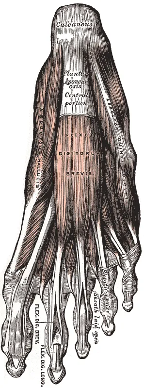
The sole (plantar) muscles lie in four layers and form inner, central and outer groups5.
| Muscle | Where sources place it | Nerve |
|---|---|---|
| Abductor hallucis | First layer, at the surface of the inner group5 | Medial plantar5 |
| Flexor digitorum brevis | At the surface of the central group, with one tendon to each of the four smaller toes5 | Medial plantar5 |
| Abductor digiti minimi | At the surface of the outer group, along the outer border of the foot5 | Lateral plantar5 |
| Quadratus plantae, lumbricals | Second layer; each has at least one attachment on the tendon of the flexor digitorum longus5 | Lateral plantar; medial plantar for the first lumbrical5 |
| Flexor hallucis brevis | Third layer, deeper than the abductor hallucis5 | Medial plantar5 |
| Adductor hallucis | Third layer, with an oblique head and a transverse head5 | Lateral plantar5 |
| Interossei | Fourth layer: three plantar and four dorsal muscles5 | Lateral plantar5 |
The flexor digitorum longus named in the table is a muscle of the deep back compartment of the leg6. The medial and lateral plantar nerves are branches of the tibial nerve, the main nerve at the back of the lower leg7.
The three largest sole muscles are the abductor hallucis, the quadratus plantae and the flexor digitorum brevis8. Each spans the length of the long arch of the foot8. The flexor digitorum brevis lies directly under the central band of the plantar fascia, a band of tough tissue along the sole5.
How they hold up the arch
A paper that proposed a new view of the foot says intrinsic and extrinsic muscles both control the arch’s movement and stability9. Its “foot core” idea likens the intrinsic foot muscles to the small trunk muscles of the core9.
Lab studies have tested this by loading the foot, switching the muscles on, or switching them off8,10:
- In nine healthy men seated with loads on the thigh, intrinsic muscle activity rose with load once the load passed certain thresholds8.
- In the same men, electrical stimulation of any of the three largest muscles shortened and raised the loaded arch8.
- In ten healthy adults, a tibial nerve block cut abductor hallucis activity to 26.8% of the control value10.
- In those ten adults, navicular drop (how far the inner arch sinks in standing) grew by 3.8 mm10.
- In 12 seated people, a block that stopped the sole muscles from contracting gave slightly more arch compression under set loads11.
Walking and running gave a different result: in healthy people with such a block, arch stiffness during ground contact did not change11.
Standing balance
In 10 healthy people, one-leg standing brought more electrical activity in the same three muscles than two-leg standing12. Their activity was moderately to strongly correlated with side-to-side shifts of the centre of pressure, the point under the foot where weight is centred12.
Walking, running and hopping
Nine people walked and ran on a treadmill while the three largest sole muscles were recorded2. The muscles slowly lengthened while active as the arch compressed2. They then shortened rapidly as the arch recoiled in the push-off phase2. Peak activity and length change both grew as speed went up2. The authors concluded that these muscles work in parallel with the plantar fascia and may limit the strain on it2.
Another study blocked the tibial nerve in healthy people, then had them walk and run11. With the block, the joints at the front of the foot could not be stiffened enough for a normal push-off11. In its 12 participants, positive work at the foot fell from 5.1 to 3.3 joules per stride in walking11. In slow running it fell from 13.6 to 9.111.
Two more lab studies recorded single sole muscles13,14:
- When feet were loaded in cycles in a rig, active flexor digitorum brevis fibres held a near-constant length, so its tendon could store elastic energy13.
- In a stepping study, going down and up a 26 cm step raised activation of two sole muscles by 69% to 410% over level steps14.
Toe strength
In nine healthy people, nerves to the toe flexors (muscles that bend the toes) were stimulated15. Stimulating extrinsic and sole muscles together gave peak torque (turning force) 208% higher at the big toe than stimulating sole muscles alone15. At the smaller toes it was 150% higher15.
Breakdown
The studies below compare muscle size and toe strength between groups of people16,17.
Foot pain
A systematic review, which searches for and sums up the research on a question, covered seven studies of plantar heel pain16. Foot muscle volume was generally smaller in people with the pain than in people without it16. Big-toe and smaller-toe flexion strength were also lower, though the studies did not agree with each other16. The review rated the strength of the evidence for both findings as very low16.
Flat foot
Studies of flat foot do not fully agree on which muscles differ17,18:
| Study | What was found in people with flat foot |
|---|---|
| Ultrasound scans of 49 adults with flat feet and 49 without | Abductor hallucis and flexor hallucis brevis significantly smaller18 |
| A meta-analysis (a study that pools earlier results) of five adult studies | Flexor hallucis brevis and abductor digiti minimi smaller in cross-sectional area17 |
| The same meta-analysis | No significant difference in abductor hallucis or flexor digitorum brevis17 |
After ankle sprains
Two small studies of chronic ankle instability after ankle sprains, an ankle that keeps giving way, point in different directions19,20. An MRI study compared five young adults who had it with five healthy ones19. The instability group had smaller volumes of the flexor hallucis brevis and of the adductor hallucis oblique head19. An ultrasound study of 80 young adults included healthy people and people with a recent sprain or chronic instability20. There, resting muscle size was set mainly by sex, body mass index and foot type, not by injury status20.
Fatigue, ageing and toe shape
In 21 healthy young adults standing still, navicular drop rose from 10.0 mm to 11.8 mm after exercise that tired the sole muscles21.
Three studies describe older adults22,23,24:
- In 17 young and 17 older adults, toe strength and the size of most foot muscles were significantly lower in the older group22.
- Across those 34 adults, toe flexor strength had a strong correlation with how large the intrinsic toe flexors were22.
- Among 44 adults over 60, those with deformed smaller toes had four significantly smaller sole muscles on ultrasound than those without a toe deformity23.
- Of 312 people aged 60 to 90 followed for 12 months, those who fell had started with weaker toe flexors than those who did not24.
Recovery
What exercise trials found
The short foot exercise was the one most often used in trials of intrinsic foot muscle training26. In it, the midfoot contracts to draw the front of the foot back toward the heel bone without the toes bending26.
| Review | What it found |
|---|---|
| Meta-analysis of 13 randomised trials | Exercise groups had lower navicular drop than controls26 |
| The same meta-analysis | Better balance and strength than controls, but not superior for reducing pain26 |
| Meta-analysis of six trials in 201 people with flat foot | The short foot exercise reduced navicular drop against controls, most of whom got something else such as insoles27 |
| The same meta-analysis | No significant difference in muscle growth; two trials measured it, both in the abductor hallucis27 |
| Systematic review of 28 randomised trials | Evidence supported foot exercises and minimalist shoes for strength and was conflicting for muscle size3 |
Reviews differ on arch motion3,28. The 28-trial review found foot exercises led to less motion of the inner arch during running, with low to very low certainty3. A review of 11 articles, mostly in younger people, found no evidence of an effect on arch motion28. It did find very low-certainty evidence of better scores on dynamic balance tests28.
Minimalist shoes
In one randomised trial, 57 runners kept up their usual running for eight weeks29. One group walked in minimalist shoes, one did foot strengthening exercises, and one was a control group29. Foot muscle size and strength rose significantly in both active groups and did not change in the controls29.
A systematic review of nine controlled studies covered at least two weeks of minimalist shoe use30. It reported strength gains of 9% to 57% and gains in sole muscle size of 7.05% to 10.6%30. Its authors concluded that such shoes may increase foot muscle size and strength in healthy people, and that research in clinical groups is still needed30.
Running and jumping after training
Two randomised studies measured running outcomes31,32:
- In a randomised study of 20 people, ten weeks of foot strengthening raised toe flexor strength but did not change running economy31.
- In a randomised trial of 118 recreational runners, one group did an eight-week foot and ankle training course, then remotely supervised training32.
- Over 12 months, the control group in that trial was 2.42 times more likely to have a running-related injury32.
Connections
The plantar fascia and the heel
A cadaver dissection study found the plantar fascia firmly joined to the muscles beneath it33. The abductor hallucis and the flexor digitorum brevis each start partly from it and partly from the calcaneus (heel bone)5.
Bones, tendons and nerves
The dorsal interossei arise from the facing sides of the metatarsals, the long bones of the midfoot5. Navicular drop, used in several studies above, is measured at the navicular, a bone on the inner side of the midfoot34.
The flexor hallucis brevis arises in part from the tibialis posterior tendon5. The oblique head of the adductor hallucis arises in part from the sheath of the peroneus longus tendon, one of the peroneal tendons5.
The tibial nerve typically divides into the medial and lateral plantar nerves, which supply the small muscles of the foot35.
Exercises that load them
Related exercise pages: running, pogo hops and the plantar fascia stretch2,36,37.
In 22 healthy people hopping on one leg, a measure of midfoot stiffness rose with hopping rate while intrinsic muscle activation during ground contact fell36. The authors say the reason remains unclear36. In 51 healthy adults, the flexor digitorum brevis was stiffer with the toes extended 45 degrees than at 0 or 15 degrees37. The authors could not say whether this came from muscle activity or from lengthening37.
Foot muscles lies under and works in parallel with
Foot muscles is supplied by branches of
Foot muscles starts on
Foot muscles attaches to
Foot muscles helps hold up the arch measured at
Foot muscles takes part of an origin from
Foot muscles takes part of an origin from the sheath of
Foot muscles has been measured in studies of
Foot muscles is loaded by
Foot muscles is stretched by
Sources
- Betts JG, Young KA, Wise JA, Johnson E, Poe B, Kruse DH, Korol O, Johnson JE, Womble M, DeSaix P (2022). Anatomy and Physiology 2e. OpenStax. Link
- Kelly LA, Lichtwark G, Cresswell AG (2015). Active regulation of longitudinal arch compression and recoil during walking and running.. Journal of the Royal Society Interface. doi:10.1098/rsif.2014.1076 PMID 25551151
- Peters-Dickie JL, Detrembleur C, Guallar-Bouloc M, Rastelli M, Lobet S, Hidalgo B, Deschamps K (2025). The effects of foot core exercises and minimalist footwear on foot muscle sizes, foot strength, and biomechanics: A systematic review and meta-analysis.. Clinical Biomechanics. doi:10.1016/j.clinbiomech.2024.106417 PMID 39709752
- Card RK, Bordoni B (2025). Anatomy, Bony Pelvis and Lower Limb, Foot Muscles.. StatPearls. PMID 30969527
- Zaottini F, Picasso R, Pistoia F, Perez MM, Möller I, Rossi F, Bruns A, Tagliafico AS, Martinoli C (2023). Ultrasound of the plantar foot: a guide for the assessment of plantar intrinsic muscles.. Journal of Ultrasonography. doi:10.15557/jou.2023.0024 PMID 37732108
- Murdock CJ, Munjal A, Agyeman K (2023). Anatomy, Bony Pelvis and Lower Limb: Calf Flexor Hallucis Longus Muscle.. StatPearls. PMID 30969598
- Ficke J, Fakoya AO, Byerly DW (2026). Anatomy, Bony Pelvis and Lower Limb: Foot and Ankle. StatPearls. PMID 31536304
- Kelly LA, Cresswell AG, Racinais S, Whiteley R, Lichtwark G (2014). Intrinsic foot muscles have the capacity to control deformation of the longitudinal arch.. Journal of the Royal Society Interface. doi:10.1098/rsif.2013.1188 PMID 24478287
- McKeon PO, Hertel J, Bramble D, Davis I (2015). The foot core system: a new paradigm for understanding intrinsic foot muscle function.. British Journal of Sports Medicine. doi:10.1136/bjsports-2013-092690 PMID 24659509
- Fiolkowski P, Brunt D, Bishop M, Woo R, Horodyski M (2003). Intrinsic pedal musculature support of the medial longitudinal arch: an electromyography study.. Journal of Foot and Ankle Surgery. doi:10.1053/j.jfas.2003.10.003 PMID 14688773
- Farris DJ, Kelly LA, Cresswell AG, Lichtwark GA (2019). The functional importance of human foot muscles for bipedal locomotion.. Proceedings of the National Academy of Sciences of the United States of America. doi:10.1073/pnas.1812820116 PMID 30655349
- Kelly LA, Kuitunen S, Racinais S, Cresswell AG (2012). Recruitment of the plantar intrinsic foot muscles with increasing postural demand.. Clinical Biomechanics. doi:10.1016/j.clinbiomech.2011.07.013 PMID 21864955
- Kelly LA, Farris DJ, Cresswell AG, Lichtwark GA (2019). Intrinsic foot muscles contribute to elastic energy storage and return in the human foot.. Journal of Applied Physiology. doi:10.1152/japplphysiol.00736.2018 PMID 30462568
- Riddick R, Farris DJ, Kelly LA (2019). The foot is more than a spring: human foot muscles perform work to adapt to the energetic requirements of locomotion.. Journal of the Royal Society Interface. doi:10.1098/rsif.2018.0680 PMID 30958152
- Wisdish SJ, Rice HM, Farris DJ (2025). Torque-angle relationships of human toe flexor muscles highlight their capacity for propulsion in gait.. Journal of Experimental Biology. doi:10.1242/jeb.249816 PMID 39606917
- Osborne JWA, Menz HB, Whittaker GA, Landorf KB (2019). Muscle Function and Muscle Size Differences in People With and Without Plantar Heel Pain: A Systematic Review.. Journal of Orthopaedic and Sports Physical Therapy. doi:10.2519/jospt.2019.8588 PMID 31597545
- Taş S, Ekici E, Yüzbaşıoğlu Ü, Özdemir AA (2026). Morphological Characteristics of the Intrinsic Foot Muscles in Individuals with Flat Foot: A Systematic Review and Meta-Analysis.. Journal of the American Podiatric Medical Association. doi:10.7547/24-094 PMID 42042529
- Angin S, Crofts G, Mickle KJ, Nester CJ (2014). Ultrasound evaluation of foot muscles and plantar fascia in pes planus.. Gait & Posture. doi:10.1016/j.gaitpost.2014.02.008 PMID 24630465
- Feger MA, Snell S, Handsfield GG, Blemker SS, Wombacher E, Fry R, Hart JM, Saliba SA, Park JS, Hertel J (2016). Diminished Foot and Ankle Muscle Volumes in Young Adults With Chronic Ankle Instability.. Orthopaedic Journal of Sports Medicine. doi:10.1177/2325967116653719 PMID 27570782
- Fraser JJ, Koldenhoven R, Hertel J (2021). Ultrasound Measures of Intrinsic Foot Muscle Size and Activation Following Lateral Ankle Sprain and Chronic Ankle Instability.. Journal of Sport Rehabilitation. doi:10.1123/jsr.2020-0372 PMID 33837169
- Headlee DL, Leonard JL, Hart JM, Ingersoll CD, Hertel J (2008). Fatigue of the plantar intrinsic foot muscles increases navicular drop. Journal of electromyography and kinesiology : official journal of the International Society of Electrophysiological Kinesiology. doi:10.1016/j.jelekin.2006.11.004 PMID 17208458
- Mickle KJ, Angin S, Crofts G, Nester CJ (2016). Effects of Age on Strength and Morphology of Toe Flexor Muscles.. Journal of Orthopaedic and Sports Physical Therapy. doi:10.2519/jospt.2016.6597 PMID 27796198
- Mickle KJ, Nester CJ (2018). Morphology of the Toe Flexor Muscles in Older Adults With Toe Deformities.. Arthritis Care & Research. doi:10.1002/acr.23348 PMID 28834406
- Mickle KJ, Munro BJ, Lord SR, Menz HB, Steele JR (2009). ISB Clinical Biomechanics Award 2009: toe weakness and deformity increase the risk of falls in older people.. Clinical Biomechanics. PMID 19751956
- Rietveld ABMB, Hagemans FMT, Haitjema S, Vissers T, Nelissen RGHH (2018). Results of Treatment of Posterior Ankle Impingement Syndrome and Flexor Hallucis Longus Tendinopathy in Dancers: A Systematic Review.. Journal of Dance Medicine & Science. doi:10.12678/1089-313x.22.1.19 PMID 29510786
- Jaffri AH, Koldenhoven R, Saliba S, Hertel J (2023). Evidence for Intrinsic Foot Muscle Training in Improving Foot Function: A Systematic Review and Meta-Analysis.. Journal of Athletic Training. doi:10.4085/1062-6050-0162.22 PMID 35724360
- Huang C, Chen LY, Liao YH, Masodsai K, Lin YY (2022). Effects of the Short-Foot Exercise on Foot Alignment and Muscle Hypertrophy in Flatfoot Individuals: A Meta-Analysis.. International Journal of Environmental Research and Public Health. doi:10.3390/ijerph191911994 PMID 36231295
- Willemse L, Wouters EJM, Bronts HM, Pisters MF, Vanwanseele B (2022). The effect of interventions anticipated to improve plantar intrinsic foot muscle strength on fall-related dynamic function in adults: a systematic review.. Journal of Foot and Ankle Research. doi:10.1186/s13047-021-00509-0 PMID 35057831
- Ridge ST, Olsen MT, Bruening DA, Jurgensmeier K, Griffin D, Davis IS, Johnson AW (2019). Walking in Minimalist Shoes Is Effective for Strengthening Foot Muscles.. Medicine and Science in Sports and Exercise. doi:10.1249/mss.0000000000001751 PMID 30113521
- Xu J, Saliba SA, Jaffri AH (2023). The Effects of Minimalist Shoes on Plantar Intrinsic Foot Muscle Size and Strength: A Systematic Review.. International Journal of Sports Medicine. doi:10.1055/a-1908-8867 PMID 35878616
- Day EM, Hahn ME (2019). Increased toe-flexor muscle strength does not alter metatarsophalangeal and ankle joint mechanics or running economy.. Journal of Sports Sciences. doi:10.1080/02640414.2019.1661562 PMID 31608832
- Taddei UT, Matias AB, Duarte M, Sacco ICN (2020). Foot Core Training to Prevent Running-Related Injuries: A Survival Analysis of a Single-Blind, Randomized Controlled Trial.. American Journal of Sports Medicine. doi:10.1177/0363546520969205 PMID 33156692
- Stecco C, Corradin M, Macchi V, Morra A, Porzionato A, Biz C, De Caro R (2013). Plantar fascia anatomy and its relationship with Achilles tendon and paratenon.. Journal of anatomy. doi:10.1111/joa.12111 PMID 24028383
- Pasanen K, Seppänen A, Leppänen M, Tokola K, Järvelä T, Vasankari T, Myklebust G, Krosshaug T, Parkkari J (2025). Knee laxity, joint hypermobility, femoral anteversion, hamstring extensibility and navicular drop as risk factors for non-contact anterior cruciate ligament injury in female athletes: A 4.5-year prospective cohort study. Knee surgery, sports traumatology, arthroscopy : official journal of the ESSKA. doi:10.1002/ksa.12625 PMID 39943896
- Dev V D, U S (2025). An Anatomical Study on the Tibial Nerve Bifurcation Level in Relation to the Malleolar-Calcaneal Axis and Its Clinical Significance.. Cureus. doi:10.7759/cureus.86511 PMID 40698204
- Kessler SE, Lichtwark GA, Welte LKM, Rainbow MJ, Kelly LA (2020). Regulation of foot and ankle quasi-stiffness during human hopping across a range of frequencies.. Journal of biomechanics. doi:10.1016/j.jbiomech.2020.109853 PMID 32636016
- Shinohara H, Umezaki Y, Ikeda A, Ikeda T, Tateyama N (2025). Reconsideration of the load-bearing functions of the plantar fascia and intrinsic foot muscles in the windlass mechanism.. Scientific reports. doi:10.1038/s41598-025-97477-3 PMID 40234710
Changes
- First published.