Tibial nerve
The main nerve of the back of the leg and the sole of the foot. Where it runs, what it supplies, how it slides, where it and its branches get squeezed, and what studies report about recovery.
- Region
- Lower leg
- Updated
- October 5, 2026
- Reviewed
- October 5, 2026 · how entries are checked
Function
Where it starts and what it supplies
The sciatic nerve is the large nerve that runs from the lower back down the back of the thigh. It typically splits behind the knee into the tibial nerve and the common fibular nerve5. The tibial nerve’s fibres come from the spinal nerve roots labelled L4 to S36.
The tibial nerve supplies the muscles of the back of the leg2:
- the gastrocnemius, soleus and plantaris in the calf2;
- the popliteus, a small muscle at the back of the knee2;
- the tibialis posterior, a deep calf muscle2;
- the two long muscles that bend the toes2.
Together these muscles point the foot down, turn the sole inward and bend the toes2. Besides these muscle branches it gives rise to the medial sural cutaneous nerve, the medial calcaneal nerve and the two plantar nerves6.
Its path from knee to sole
The nerve runs down behind the knee and passes deep to the gastrocnemius and soleus7. It then travels down the leg beside the posterior tibial blood vessels to the ankle7.
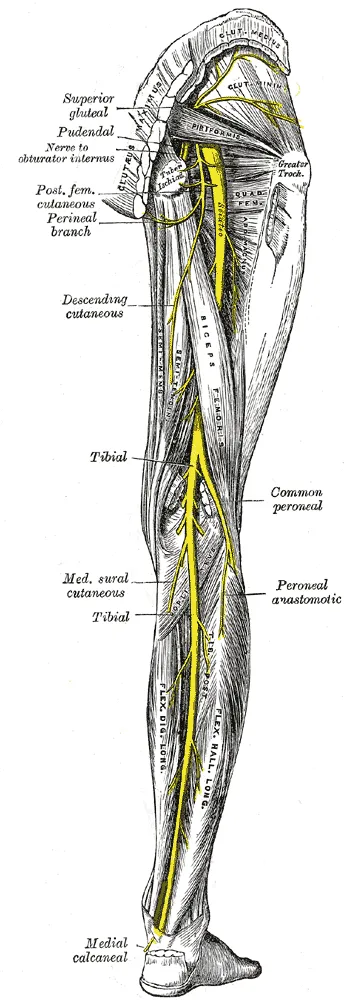
At the ankle it runs through the tarsal tunnel, a passage of bone and fibrous tissue1. The tunnel’s bony walls include the calcaneus (heel bone), the talus (ankle bone) and the medial malleolus, the bony bump on the inner ankle1. Its roof is the flexor retinaculum, a band stretched from that bump to the heel bone8. Three tendons and the nerve’s blood vessels share the tunnel, among them the tibialis posterior tendon1.
The medial calcaneal nerve leaves at the ankle, pierces the retinaculum and supplies skin on the back and underside of the heel7.
How it divides in the foot
Inside the tunnel the nerve typically divides into the medial and lateral plantar nerves2. These supply the small muscles of the foot and the skin of the sole2.
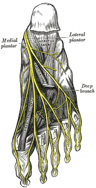
- The medial plantar nerve supplies four sole muscles, including the abductor hallucis, and the skin of the inner three and a half toes2.
- The lateral plantar nerve supplies the quadratus plantae, the abductor digiti minimi, the adductor hallucis, the interossei and one more sole muscle2.
- The inferior calcaneal nerve drives the abductor digiti minimi and sometimes the flexor digitorum brevis, and carries feeling from the heel bone’s covering1.
The inferior calcaneal nerve is often called Baxter’s nerve1. One paper describes the origin of the nerve to abductor digiti minimi as variable9. A review notes that in most feet each of these branches runs in its own channel of bone and fibrous tissue8. That, it says, is why one branch can be squeezed alone8.
How it slides and stretches
Walking asks the nerve to adapt as the ankle bends up and the foot rolls inward10. A review of studies in living people found joint movement slid the tibial nerve 0.7 to 5.2 mm11. Three studies in living people give mean slides10,12,13.
| Movement | Who was measured | Mean slide (mm) |
|---|---|---|
| Ankle from pointed down to pulled up, standing on the foot | 16 people without symptoms | 2.9910 |
| Active pulling up of the foot, measured behind the knee | Healthy individuals | 2.1812 |
| Forward bending, measured behind the knee | 24 healthy subjects | 12.213 |
With the hip bent first, the slide from the ankle was about three times smaller12.
Dorsiflexion is pulling the foot and toes up toward the shin. In 20 healthy adults, the nerve grew stiffer as the ankle moved further into it14. In ten cadavers, nerve strain (stretch as a share of resting length) at the ankle rose 3.9% on average with dorsiflexion3. It was higher with the knee straight or the hip bent first3.
Breakdown
In the tarsal tunnel
A 2024 scoping review, which maps the research on a topic, defines tarsal tunnel syndrome as a compressive neuropathy, a nerve problem caused by squeezing15. It affects the tibial nerve, or one of its heel or plantar divisions, inside the tunnel15. One narrative review describes it as uncommon next to other such problems, often missed, and sometimes mistaken for plantar fasciitis8.
Sources differ on how often a cause is found1,16. One review states that no exact cause is identified in 20% to 40% of cases1. A 2022 review reports that the syndrome most often has no known cause16. Causes the two reviews list include injury, cysts and other lumps, swollen veins, and whole-body illness1,16. Described risk factors include female sex, taking part in sport, diabetes and an underactive thyroid16.
In 10 cadaver specimens, mean tunnel pressure was 2 mmHg (millimetres of mercury) with the foot pointed slightly down and not turned17. It was 32 mmHg with the sole turned fully outward and 17 mmHg with it turned fully inward17.
One case series, a report on patients without a comparison group, covered 18 people operated on for sport-related tarsal tunnel syndrome18. The triggers were activities that load the ankle heavily, such as sprinting and jumping18. Their predisposing features included flatfoot, extra muscles and bony fragments around the tunnel18.
A narrative review states the syndrome has been reported as more frequent in avid runners, especially where the foot rolls in too far8. Another notes that little is published on how often sport causes foot and ankle nerve problems1. The cases reported most often involve running and dancing1.
Above the ankle
A review lists trapping sites above the ankle: at the inner gastrocnemius, under the arch of the soleus, and after a popliteus tear1. Squeezing under the soleus arch is called soleal sling syndrome, and a literature review calls it rare and under-recognised19.
Forward knee dislocations, usually from the knee being forced past straight, carry a high risk of pulling injury to the nerve1.
Branches in the heel and arch
A review lists places where Baxter’s nerve can be trapped, including between two sole muscles and at a ligament1. Bony heel spurs and tears of the plantar fascia can also press on it1. One paper states that the inner heel pain this produces may not be distinguishable from plantar fasciitis heel pain20.
Atrophy is wasting of a muscle. Among 602 consecutive patients having foot and ankle MRI (a scan showing soft tissue), 6.3% had fatty atrophy of the abductor digiti minimi alone21. That finding is a reported marker of this nerve being trapped21. In 69 heels that had this nerve surgically released, the pain had begun during sport in 3422.
A 1978 paper gave the name “jogger’s foot” to trapping of the medial plantar nerve in the arch23. A review names two common sites, one being the knot of Henry, where two long toe tendons cross1.
Between the toes
The authors of one meta-analysis define Morton’s neuroma as a compressive neuropathy24. The nerve involved is the common plantar digital nerve, most often in the third space between the toes24. In 253 UK general practices in 2000, new presentations ran at 50.2 per 100,000 men and 87.5 per 100,000 women yearly25. A narrative review describes the typical person as a woman in her late 40s or early 50s26. It adds that many are active, and that running and dancing can be associated with it26.
The same review states that high-heeled or narrow shoes usually make the pain worse26. In 68 people with the neuroma and 32 without, it was strongly associated with restricted ankle dorsiflexion27.
Thickened nerves are also found in feet without symptoms28,29.
- MRI of 70 people without symptoms showed a presumed Morton neuroma in 30%28.
- Ultrasound, a scan made with sound waves, of 48 volunteers with symptom-free forefeet showed a nerve thickening over 5 mm in 54%29.
- Under the microscope, one comparison found the nerve looked the same in people without symptoms as in patients30. Loss of myelin, the fibres’ fatty wrap, was more common in patients30.
Recovery
The general course
A review of nerve tissue states that most nerve compressions are the mildest grade of injury, neurapraxia31. Another describes neurapraxia as a temporary signal block that clears fully within 12 weeks once myelin is restored32. Where fibres themselves are damaged, regrowth is slow, about 1 mm a day, and often incomplete32.
What studies of trapped nerves report
| Group studied | What was reported |
|---|---|
| Tarsal tunnel syndrome, 32 observational studies in a scoping review | After care, excellent or good results in 75.3% of cases, fair or poor in 24.7%15 |
| 18 patients operated on for sport-related tarsal tunnel syndrome | Most could return to the same sport; mean follow-up 58.6 months18 |
| 15 runners who had 21 nerve releases in the foot and ankle, one surgeon | Good to excellent results in each of the 15, who returned to their earlier level of running33 |
| 69 heels with long-lasting heel pain that had Baxter’s nerve surgically released | 89% excellent or good; pain fully gone in 83%; average follow-up 49 months22 |
| 99 consecutive patients who had a Morton’s neuroma removed | 63% pain-free six months after surgery; 10 patients dissatisfied34 |
A narrative review gives a wider range for tarsal tunnel surgery, with reported success from 44% to 96%8. The scoping review judged its 32 studies low quality15. For Morton’s neuroma, a systematic review of 25 studies of non-surgical approaches found no high-quality evidence for any single one24.
After a tibial nerve injury
A systematic review of 19 studies covered 677 patients who had surgery after tibial nerve injury4. Those operated on within 7 months were more likely to regain useful muscle power than those operated on later4. Gunshot wounds were the most common cause, and people injured in sport had the highest share of good outcomes4.
In 119 patients with nerves fully divided by missile wounds, repair did significantly worse with gaps over 5 cm or surgery delays over 4 months35.
Connections
Muscles it drives
The nerve supplies the gastrocnemius and the soleus36,37. Through its plantar branches it reaches the small muscles of the foot2,38. In healthy people walking and running, numbing the tibial nerve stopped the small sole muscles from contracting39. The joints at the front of the foot could not then stiffen enough for a normal push-off39.
Stretches that load it
In cadavers with the knee straight, dorsiflexion moved and strained the nerve at hip angles from 0 to 90 degrees40. In 41 healthy adults, a stiffer tibial nerve went with a smaller maximum dorsiflexion angle41. Calf muscle stiffness was not significantly related to that angle41.
In six cadavers, bending the toe joints back raised strain by 0.4% in the tibial nerve and 2.0% in the medial plantar nerve42. Related stretch pages: calf stretch, soleus stretch, plantar fascia stretch3,41,42.
Tibial nerve supplies
Tibial nerve supplies, through its plantar branches,
Tibial nerve leaves the sciatic nerve alongside
Tibial nerve shares the tarsal tunnel with
Tibial nerve has a heel branch whose pain can resemble
Tibial nerve is loaded by
Sources
- Farag JI, McDougall AN, Catapano M (2025). Common sports-related nerve injuries seen by the electrodiagnostic medical consultant.. Muscle & nerve. doi:10.1002/mus.28298 PMID 39535438
- Dev V D, U S (2025). An Anatomical Study on the Tibial Nerve Bifurcation Level in Relation to the Malleolar-Calcaneal Axis and Its Clinical Significance.. Cureus. doi:10.7759/cureus.86511 PMID 40698204
- Alshami AM, Babri AS, Souvlis T, Coppieters MW (2008). Strain in the tibial and plantar nerves with foot and ankle movements and the influence of adjacent joint positions.. Journal of applied biomechanics. doi:10.1123/jab.24.4.368 PMID 19075306
- Garg SP, Hassan AM, Patel AA, Perez MM, Stoehr JR, Ketheeswaran S, Chappell AG, Galiano RD, Ko JH (2021). Outcomes of Tibial Nerve Repair and Transfer: A Structured Evidence-Based Systematic Review and Meta-Analysis.. The Journal of foot and ankle surgery : official publication of the American College of Foot and Ankle Surgeons. doi:10.1053/j.jfas.2021.07.001 PMID 34366221
- Liyew WA, Abebe S (2026). Anatomical variation of the sciatic nerve division in the cadaver studies: a systematic review and meta-analysis.. Anatomy & cell biology. doi:10.5115/acb.25.139 PMID 41851007
- Desai SS, La Rose J (2026). Anatomy, Bony Pelvis and Lower Limb: Tibial Nerve. StatPearls. PMID 30725713
- Banik S, Guria LR (2021). Variable Branching Pattern of Tibial Nerve in the Tarsal Tunnel: A Gross Anatomical Study With Clinical Implications.. Cureus. doi:10.7759/cureus.13729 PMID 33717767
- Rodríguez-Merchán EC, Moracia-Ochagavía I (2021). Tarsal tunnel syndrome: current rationale, indications and results.. EFORT open reviews. doi:10.1302/2058-5241.6.210031 PMID 35839088
- Rodrigues RN, Lopes AA, Torres JM, Mundim MF, Silva LL, Silva BR (2015). Compressive neuropathy of the first branch of the lateral plantar nerve: a study by magnetic resonance imaging.. Radiologia brasileira. doi:10.1590/0100-3984.2013.0028 PMID 26811554
- Carroll M, Yau J, Rome K, Hing W (2012). Measurement of tibial nerve excursion during ankle joint dorsiflexion in a weight-bearing position with ultrasound imaging.. Journal of foot and ankle research. doi:10.1186/1757-1146-5-5 PMID 22397397
- Silva A, Manso A, Andrade R, Domingues V, Brandão MP, Silva AG (2014). Quantitative in vivo longitudinal nerve excursion and strain in response to joint movement: A systematic literature review.. Clinical Biomechanics. doi:10.1016/j.clinbiomech.2014.07.006 PMID 25168082
- Boyd BS, Gray AT, Dilley A, Wanek L, Topp KS (2012). The pattern of tibial nerve excursion with active ankle dorsiflexion is different in older people with diabetes mellitus.. Clinical biomechanics (Bristol, Avon). doi:10.1016/j.clinbiomech.2012.06.013 PMID 22809735
- Shum GL, Attenborough AS, Marsden JF, Hough AD (2013). Tibial nerve excursion during lumbar spine and hip flexion measured with diagnostic ultrasound.. Ultrasound in medicine & biology. doi:10.1016/j.ultrasmedbio.2012.11.023 PMID 23465136
- Kawanishi K, Nariyama Y, Anegawa K, Tsutsumi M, Kudo S (2022). Changes in tibial nerve stiffness during ankle dorsiflexion according to in-vivo analysis with shear wave elastography.. Medicine. doi:10.1097/md.0000000000029840 PMID 35777040
- Haq II, Banerjee AA, Arshad Z, Iqbal AM, Bhatia M (2024). The management of tarsal tunnel syndrome: A scoping review.. Journal of clinical orthopaedics and trauma. doi:10.1016/j.jcot.2024.102489 PMID 39101044
- Vij N, Kaley HN, Robinson CL, Issa PP, Kaye AD, Viswanath O, Urits I (2022). Clinical Results Following Conservative Management of Tarsal Tunnel Syndrome Compared With Surgical Treatment: A Systematic Review.. Orthopedic reviews. doi:10.52965/001c.37539 PMID 36072502
- Trepman E, Kadel NJ, Chisholm K, Razzano L (1999). Effect of foot and ankle position on tarsal tunnel compartment pressure.. Foot & ankle international. doi:10.1177/107110079902001108 PMID 10582848
- Kinoshita M, Okuda R, Yasuda T, Abe M (2006). Tarsal tunnel syndrome in athletes.. The American journal of sports medicine. doi:10.1177/0363546506286344 PMID 16567455
- Hilbig-Vlatten L, Grauberger JN, Engmann TF, Stadelmeier LF, Dunn RM, Mandeville R, Ko JH, D'Hemecourt P, Dowlatshahi S (2025). Soleal Sling Syndrome: A Narrative Review.. Journal of the peripheral nervous system : JPNS. doi:10.1111/jns.70032 PMID 40391847
- Chundru U, Liebeskind A, Seidelmann F, Fogel J, Franklin P, Beltran J (2008). Plantar fasciitis and calcaneal spur formation are associated with abductor digiti minimi atrophy on MRI of the foot.. Skeletal radiology. doi:10.1007/s00256-008-0455-2 PMID 18286281
- Recht MP, Grooff P, Ilaslan H, Recht HS, Sferra J, Donley BG (2007). Selective atrophy of the abductor digiti quinti: an MRI study.. AJR. American journal of roentgenology. doi:10.2214/ajr.07.2229 PMID 17715077
- Baxter DE, Pfeffer GB (1992). Treatment of chronic heel pain by surgical release of the first branch of the lateral plantar nerve.. Clinical orthopaedics and related research. PMID 1600660
- Rask MR (1978). Medial plantar neurapraxia (jogger's foot): report of 3 cases.. Clinical orthopaedics and related research. PMID 729244
- Matthews BG, Hurn SE, Harding MP, Henry RA, Ware RS (2019). The effectiveness of non-surgical interventions for common plantar digital compressive neuropathy (Morton's neuroma): a systematic review and meta-analysis.. Journal of foot and ankle research. doi:10.1186/s13047-019-0320-7 PMID 30809275
- Latinovic R, Gulliford MC, Hughes RA (2006). Incidence of common compressive neuropathies in primary care.. Journal of neurology, neurosurgery, and psychiatry. doi:10.1136/jnnp.2005.066696 PMID 16421136
- Gougoulias N, Lampridis V, Sakellariou A (2019). Morton's interdigital neuroma: instructional review.. EFORT open reviews. doi:10.1302/2058-5241.4.180025 PMID 30800476
- Naraghi R, Bremner A, Slack-Smith L, Bryant A (2016). The relationship between foot posture index, ankle equinus, body mass index and intermetatarsal neuroma.. Journal of foot and ankle research. doi:10.1186/s13047-016-0179-9 PMID 27980684
- Zanetti M, Strehle JK, Zollinger H, Hodler J (1997). Morton neuroma and fluid in the intermetatarsal bursae on MR images of 70 asymptomatic volunteers.. Radiology. doi:10.1148/radiology.203.2.9114115 PMID 9114115
- Symeonidis PD, Iselin LD, Simmons N, Fowler S, Dracopoulos G, Stavrou P (2012). Prevalence of interdigital nerve enlargements in an asymptomatic population.. Foot & ankle international. doi:10.3113/fai.2012.0543 PMID 22835390
- Bourke G, Owen J, Machet D (1994). Histological comparison of the third interdigital nerve in patients with Morton's metatarsalgia and control patients.. The Australian and New Zealand journal of surgery. doi:10.1111/j.1445-2197.1994.tb02243.x PMID 7516653
- Menorca RM, Fussell TS, Elfar JC (2013). Nerve physiology. Hand Clinics. doi:10.1016/j.hcl.2013.04.002 PMID 23895713
- Grinsell D, Keating CP (2014). Peripheral nerve reconstruction after injury: a review of clinical and experimental therapies.. BioMed Research International. doi:10.1155/2014/698256 PMID 25276813
- Murphy PC, Baxter DE (1985). Nerve entrapment of the foot and ankle in runners.. Clinics in sports medicine. PMID 4053199
- Bucknall V, Rutherford D, MacDonald D, Shalaby H, McKinley J, Breusch SJ (2016). Outcomes following excision of Morton's interdigital neuroma. The bone & joint journal. doi:10.1302/0301-620x.98b10.37610 PMID 27694592
- Roganović Z, Pavlićević G, Petković S (2005). Missile-induced complete lesions of the tibial nerve and tibial division of the sciatic nerve: results of 119 repairs.. Journal of neurosurgery. doi:10.3171/jns.2005.103.4.0622 PMID 16266043
- Bordoni B, Varacallo MA (2026). Anatomy, Bony Pelvis and Lower Limb: Gastrocnemius Muscle.. StatPearls. PMID 30422541
- Trybulski R, Gałęziok K, Matuszczyk F, Halski T, Muracki J (2025). Problems in Diagnosis and Treatment of Soleus Muscle Injuries-Narrative Review and Case Report.. Journal of Clinical Medicine. doi:10.3390/jcm14061955 PMID 40142763
- Ficke J, Fakoya AO, Byerly DW (2026). Anatomy, Bony Pelvis and Lower Limb: Foot and Ankle. StatPearls. PMID 31536304
- Farris DJ, Kelly LA, Cresswell AG, Lichtwark GA (2019). The functional importance of human foot muscles for bipedal locomotion.. Proceedings of the National Academy of Sciences of the United States of America. doi:10.1073/pnas.1812820116 PMID 30655349
- Bueno-Gracia E, Estébanez-de-Miguel E, López-de-Celis C, Shacklock M, Caudevilla-Polo S, González-Rueda V, Pérez-Bellmunt A (2020). Effect of ankle dorsiflexion on displacement and strain in the tibial nerve and biceps femoris muscle at the posterior knee during the straight leg raise: Investigation of specificity of nerve movement.. Clinical biomechanics (Bristol, Avon). doi:10.1016/j.clinbiomech.2020.105003 PMID 32335471
- Mukai H, Umehara J, Saeki J, Yanase K, Wang Z, Tateuchi H, Ichihashi N (2026). Tibial nerve stiffness is related to maximum angle of ankle dorsiflexion.. Journal of biomechanics. doi:10.1016/j.jbiomech.2025.113050 PMID 41187609
- Alshami AM, Babri AS, Souvlis T, Coppieters MW (2007). Biomechanical evaluation of two clinical tests for plantar heel pain: the dorsiflexion-eversion test for tarsal tunnel syndrome and the windlass test for plantar fasciitis.. Foot & ankle international. doi:10.3113/FAI.2007.0499 PMID 17475147
Changes
- First published.