Hip joint
Also called hip, coxal joint, coxal articulation
The ball-and-socket joint between the thigh bone and the hip bone. How it moves, what holds it together, the forces it carries, how it wears or dislocates, and what crosses it.
- Region
- Hip
- Muscles
- gluteus maximus, gluteus medius, gluteus minimus, hamstrings, hip adductors, hip flexors, deep hip rotators, tensor fasciae latae
- Updated
- October 7, 2026
- Reviewed
- October 7, 2026 · how entries are checked
Function
A ball in a deep socket
The hip is a ball-and-socket joint that moves in many directions1. The rounded head at the top of the femur (thigh bone) fits into the acetabulum (the hip socket)1. Three bones of the pelvis, the ilium, ischium and pubis, meet on the outer side of the hip bone to form that socket1.
The socket is deep and has a large contact area for the head of the femur1. That gives the hip its stability and its ability to bear weight1. The hip’s built-in stability comes mainly from its bones and the way they fit together5.
Articular cartilage (the thin layer of cartilage covering bone ends in a joint) covers the whole head except one small pit, the fovea6. In the socket, the cartilage forms an incomplete ring around a central hollow that holds a pad of fat6.
The hip labrum is a rim of fibrocartilage (a tough, fibrous kind of cartilage) that runs around the edge of the socket7. One review article reports that some studies show it deepens the socket by 21%7.
What holds it together
A joint capsule (the sleeve of tough tissue that encloses a joint) wraps the hip, thickened into three ligaments1. They run from the rim of the socket to the base of the femoral neck, the angled part of the femur below the ball1. On the way they wind in a spiral around the femur’s head and neck1.
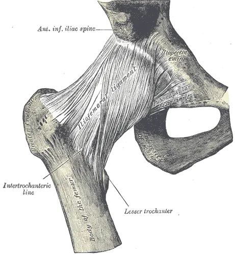
| Ligament | Where it lies | What it checks, in a 1918 anatomy text |
|---|---|---|
| Iliofemoral | In front of the joint, dividing into two bands that fix to the femur6 | Extension (moving the thigh backward); its outer band checks outward rotation6 |
| Pubofemoral | From the pubis, blending below with the capsule6 | Abduction (moving the thigh out to the side), together with the inner band of the iliofemoral ligament6 |
| Ischiofemoral | From the ischium, below and behind the socket6 | Inward rotation, together with the back of the capsule6 |
The ligaments tighten when the hip extends1. In upright standing this pulls the head of the femur firmly into the socket and allows very little extension beyond that position1. The hip ligaments steady the joint, so a person can stand upright with only minimal muscle work1.
Laboratory studies have tested cadaveric hips, from bodies donated for study8. In nine of them, each capsular ligament was the main restraint to hip rotation somewhere in the range of movement8. In another, of 12 matched pairs of cadaveric hips, the ischiofemoral ligament controlled internal rotation (turning the limb inward) in both flexion and extension9.
How far it moves
The hip moves in flexion (bending), extension, abduction, adduction (toward the midline), rotation and circumduction (circling)6. Because it carries body weight and needs stability for standing and walking, it has less range of motion than the shoulder1. In 200 healthy people, hip flexion ranged from 80 to 140 degrees and extension from 5 to 40 degrees10.
With the knee bent, flexion stops when the soft tissue of the thigh and the abdomen press together6. With the knee straight, the hamstrings stop it, which is the position of a hamstring stretch6.
The forces it carries
In four patients with instrumented hip implants, the measured hip contact force was as follows2:
| Activity | Hip contact force, % of body weight |
|---|---|
| Walking at about 4 km/h, average | 2382 |
| Going upstairs | 2512 |
| Going downstairs | 2602 |
A later study measured ten people with such implants11. Its standardised high-load peak force for slow jogging at 7 km/h was 68% higher than for walking11. That force reached nearly five times body weight11.
Modelling studies estimate forces inside the body with a model in place of direct measurement. Two give estimates for healthy people12,13:
- In 20 young healthy adults, modelled peak hip contact force in running rose with speed12. It went from about 7.5 to 10 times body weight between 6 and 12 km/h12.
- In 29 top-ranked Austrian powerlifters squatting at 90% of their one-repetition maximum, the estimated peak averaged 15.5 times body weight13. A one-repetition maximum is the heaviest load a person can lift once.
- In those powerlifters, the estimated force peaked in the deepest positions of the back squat13.
Breakdown
Osteoarthritis
An anatomy textbook describes the hip as prone to osteoarthritis1. A systematic review and meta-analysis pooled 31 studies of 326,463 people3. It put the worldwide prevalence of hip osteoarthritis on X-ray at 8.55%3. Prevalence is the share of a group with a condition. The difference between men and women was not statistically significant3.
Reviews have linked several factors with hip osteoarthritis14,15,16:
- Body weight. A meta-analysis of 14 studies found each 5-unit rise in body mass index (a measure of weight for height) associated with an 11% higher risk14.
- Heavy work. A meta-analysis of 24 articles found higher odds with lifting or carrying loads at work, with a pooled odds ratio of 1.615. An odds ratio is how many times higher the odds are in one group than in another.
- Elite impact sport. A systematic review of 11 studies of mostly male elite athletes found a higher risk than in matched controls16. Its odds ratios ran from 1.8 to 8.716.
That review found inconsistent results across five studies of competitive long-distance running16. It concluded that such runners do not have a clearly raised risk16. A meta-analysis of 17 studies counted hip and knee osteoarthritis in runners and non-runners17. It found it in 3.5% of recreational runners, 13.3% of competitive runners and 10.2% of non-running controls17.
The shape of the bones
An international consensus defines primary cam morphology as a cartilage or bony bump where the head of the femur meets its neck18. The bump changes the head from round to out-of-round18. The same consensus calls it mostly benign and highly prevalent in many groups of athletes18.
A 2021 meta-analysis pooled prospective studies, which follow people forward in time19. Hips with a cam shape were about two and a half times as likely to develop hip osteoarthritis19. A 2026 pooled analysis followed 23,886 hips for 4 to 8 years20. Hips with cam morphology had 1.87 times the odds of new osteoarthritis on X-ray20. The page on femoroacetabular impingement covers these shapes in detail.
Dislocation
A dislocation is when a bone is forced fully out of its normal place at a joint. At the hip, it occurs with high-energy trauma or after a total hip replacement21. Backward (posterior) dislocations are the most common type21. Hip dislocation is rare in sport22. A sample of US emergency department visits from 2010 to 2019 recorded 102 sports-related hip dislocations, 91.2% of them in contact sports23.
A systematic review gathered 145 patients with a hip dislocation or subluxation (a partial slip of the joint) in sport22. Some of its studies looked inside the joint by arthroscopy (keyhole surgery using a small camera)22. They found labral tears in 100% of hips and cartilage injuries in 92%22. In traumatic hip dislocation, the sciatic nerve is the nerve most often injured24.
Recovery
After a dislocation in sport
The same systematic review reported what followed22:
- Of the 39 patients with return-to-play data, 87% returned successfully22.
- Of the 38 with imaging follow-up, 11% had X-ray evidence of osteonecrosis, the death of bone in the head of the femur22.
- Recurrence was reported in 3% of cases22.
How hip cartilage responds to load
A systematic review covered studies using MRI (a scan that shows soft tissue in detail) of knee and hip cartilage before and after joint loading25. It found too little hip cartilage data to pool25. It found two hip studies of cartilage straight after activity25. After simulated standing, cartilage thickness and volume were unchanged, and after 50 knee bends in healthy adults its composition was unchanged25.
What exercise trials found in hip osteoarthritis
A Cochrane review (a systematic review done to the Cochrane Collaboration’s published standards) pooled 18 randomised trials of 1,368 adults with hip osteoarthritis4. It compared exercise with no exercise programme, usual care or limited education4. Its findings:
- Exercise probably reduces pain slightly, by about 7 points on a 0 to 100 scale4.
- Exercise probably improves physical function slightly, by about 9 points on a 0 to 100 scale4.
- Both improvements are unlikely to be clinically meaningful4.
- For quality of life, exercise probably makes little to no difference4.
Connections
The bones and muscles
The joint is formed by the femur and the hip bone of the pelvis1. A 1918 anatomy text groups them by the movement they produce6:
| Movement | Muscles |
|---|---|
| Flexion | The psoas major and iliacus (the hip flexors), with the rectus femoris, sartorius, pectineus and two adductors6 |
| Extension | Mainly the gluteus maximus, helped by the hamstrings and part of the adductor magnus6 |
| Abduction | The gluteus medius and gluteus minimus, with the upper part of the gluteus maximus6 |
| Adduction | The three adductors, pectineus and gracilis (the hip adductors), with the lower part of the gluteus maximus6 |
Six deep hip rotators lie beneath the gluteus maximus and rotate the thigh outward1. The tensor fasciae latae works with the gluteus medius and minimus to abduct and internally rotate the hip26. The inguinal ligament is a band of tough tissue running across the groin. Below it, only the iliopsoas (a deep hip muscle that bends the hip) lies between the front of the hip capsule and the femoral nerve27.
Exercises and stretches that load it
Related exercise pages: hip thrust, lunge, split squat and step-up28,29,30,31. Each has been studied at the hip:
- Nineteen resistance trained males lifted 70% of one-repetition maximum28. The barbell hip thrust placed significantly greater extensor demand on the hip than on the knee28.
- After a study of 16 recreationally active, college-aged adults, the authors concluded that the lunge is hip-extensor-dominant in its joint kinetics29.
- Eight male college rugby players lifted their 10-repetition-maximum load in two exercises30. The peak hip extension moment (turning effect) was 47% greater in the rear-foot-elevated split squat than in the back squat30.
- In 20 healthy adults, the step-up had the lowest gluteus medius, quadriceps and hamstring activity of four stepping tasks31.
Related stretch pages: hip flexor stretch, adductor stretch and figure-4 stretch1,32,33. In eight cadaver specimens, the pubofemoral ligament was strained by about 3% at maximal hip abduction32. In a cross-over trial of 31 physically active people, two 30-second holds of a gluteal stretch were followed by improved passive hip range of motion33.
Hip joint is formed by
Hip joint is rimmed by
Hip joint is lined by
Hip joint is moved by
Hip joint lies next to
Hip joint is related to
Hip joint is compared with
Hip joint is loaded by
Hip joint is stretched by
Sources
- Betts JG, Young KA, Wise JA, Johnson E, Poe B, Kruse DH, Korol O, Johnson JE, Womble M, DeSaix P (2022). Anatomy and Physiology 2e. OpenStax. Link
- Bergmann G, Deuretzbacher G, Heller M, Graichen F, Rohlmann A, Strauss J, Duda GN (2001). Hip contact forces and gait patterns from routine activities.. Journal of Biomechanics. doi:10.1016/s0021-9290(01)00040-9 PMID 11410170
- Fan Z, Yan L, Liu H, Li X, Fan K, Liu Q, Li JJ, Wang B (2023). The prevalence of hip osteoarthritis: a systematic review and meta-analysis.. Arthritis research & therapy. doi:10.1186/s13075-023-03033-7 PMID 36991481
- Hall M, Lawford BJ, Hinman RS, Dobson F, Spiers L, Kimp A, French HP, Reichenbach S, Hernandez-Molina G, Bennell KL (2026). Exercise for osteoarthritis of the hip.. The Cochrane database of systematic reviews. doi:10.1002/14651858.CD007912.pub3 PMID 42486497
- Gold M, Munjal A, Varacallo MA (2023). Anatomy, Bony Pelvis and Lower Limb, Hip Joint. StatPearls. PMID 29262200
- Gray H; Lewis WH (editor) (1918). Anatomy of the Human Body. Lea & Febiger, Philadelphia (20th edition). Link
- Groh MM, Herrera J (2009). A comprehensive review of hip labral tears.. Current Reviews in Musculoskeletal Medicine. doi:10.1007/s12178-009-9052-9 PMID 19468871
- van Arkel RJ, Amis AA, Cobb JP, Jeffers JR (2015). The capsular ligaments provide more hip rotational restraint than the acetabular labrum and the ligamentum teres : an experimental study.. The bone & joint journal. doi:10.1302/0301-620X.97B4.34638 PMID 25820886
- Martin HD, Savage A, Braly BA, Palmer IJ, Beall DP, Kelly B (2008). The function of the hip capsular ligaments: a quantitative report.. Arthroscopy. doi:10.1016/j.arthro.2007.08.024 PMID 18237703
- Elson RA, Aspinall GR (2008). Measurement of hip range of flexion-extension and straight-leg raising.. Clinical orthopaedics and related research. doi:10.1007/s11999-007-0073-7 PMID 18196407
- Bergmann G, Bender A, Dymke J, Duda G, Damm P (2016). Standardized Loads Acting in Hip Implants.. PloS one. doi:10.1371/journal.pone.0155612 PMID 27195789
- Giarmatzis G, Jonkers I, Wesseling M, Van Rossom S, Verschueren S (2015). Loading of Hip Measured by Hip Contact Forces at Different Speeds of Walking and Running.. Journal of Bone and Mineral Research. doi:10.1002/jbmr.2483 PMID 25704538
- Pürzel A, Kaufmann P, Koller W, Wallnöfer EK, Baca A, Kainz H (2025). Biomechanical analysis of hip, knee, and ankle joint contact forces during squats in elite powerlifters.. PloS one. doi:10.1371/journal.pone.0327973 PMID 40705768
- Jiang L, Rong J, Wang Y, Hu F, Bao C, Li X, Zhao Y (2011). The relationship between body mass index and hip osteoarthritis: a systematic review and meta-analysis.. Joint bone spine. doi:10.1016/j.jbspin.2010.04.011 PMID 20580591
- Jahn A, Andersen JH, Seidler A, Christiansen DH, Dalbøge A (2024). Hip osteoarthritis and occupational mechanical exposures: a systematic review and meta-analysis.. Scandinavian journal of work, environment & health. doi:10.5271/sjweh.4152 PMID 38483209
- Vigdorchik JM, Nepple JJ, Eftekhary N, Leunig M, Clohisy JC (2017). What Is the Association of Elite Sporting Activities With the Development of Hip Osteoarthritis?. The American journal of sports medicine. doi:10.1177/0363546516656359 PMID 27474380
- Alentorn-Geli E, Samuelsson K, Musahl V, Green CL, Bhandari M, Karlsson J (2017). The Association of Recreational and Competitive Running With Hip and Knee Osteoarthritis: A Systematic Review and Meta-analysis.. The Journal of orthopaedic and sports physical therapy. doi:10.2519/jospt.2017.7137 PMID 28504066
- Dijkstra HP, Mc Auliffe S, Ardern CL, Kemp JL, Mosler AB, Price A, Blazey P, Richards D, Farooq A, Serner A, McNally E, Mascarenhas V, Willy RW, Oke JL, Khan KM, Glyn-Jones S, Clarke M, Greenhalgh T, Young Athlete’s Hip Research (YAHiR) Collaborative (2022). Oxford consensus on primary cam morphology and femoroacetabular impingement syndrome: part 1-definitions, terminology, taxonomy and imaging outcomes.. British journal of sports medicine. doi:10.1136/bjsports-2022-106085 PMID 36588401
- Casartelli NC, Maffiuletti NA, Valenzuela PL, Grassi A, Ferrari E, van Buuren MMA, Nevitt MC, Leunig M, Agricola R (2021). Is hip morphology a risk factor for developing hip osteoarthritis? A systematic review with meta-analysis.. Osteoarthritis and cartilage. doi:10.1016/j.joca.2021.06.007 PMID 34171473
- Tang J, Boel F, van Buuren MM, Riedstra NS, van den Berg MA, Ahedi H, Arden NK, Bierma-Zeinstra SMA, Boer CG, Cicutini F, Cootes T, Crossley KM, Felson DT, Gielis WP, Heerey JJ, Jones G, Kluzek S, Lane NE, Lindner C, Lynch JA, van Meurs J, Mosler AB, Nelson AE, Nevitt M, Oei EHG, Runhaar J, Weinans H, Agricola R (2026). Cam morphology and the risk of developing radiographic hip osteoarthritis within 8 years: an individual participant data meta-analysis of 23 886 hips from the world COACH consortium.. British journal of sports medicine. doi:10.1136/bjsports-2025-110144 PMID 41314784
- Dawson-Amoah K, Raszewski J, Duplantier N, Waddell BS (2018). Dislocation of the Hip: A Review of Types, Causes, and Treatment.. Ochsner journal. doi:10.31486/toj.17.0079 PMID 30275789
- Chona DV, Minetos PD, LaPrade CM, Cinque ME, Abrams GD, Sherman SL, Safran MR (2022). Hip Dislocation and Subluxation in Athletes: A Systematic Review.. The American journal of sports medicine. doi:10.1177/03635465211036104 PMID 34623933
- Moran J, Cheng R, Schneble CA, Mathew JI, Kahan JB, Li D, Gardner EC (2022). Epidemiology of Sports-Related Traumatic Hip Dislocations Reported in United States Emergency Departments, 2010-2019.. Orthopaedic journal of sports medicine. doi:10.1177/23259671221088009 PMID 35547614
- Cornwall R, Radomisli TE (2000). Nerve injury in traumatic dislocation of the hip.. Clinical orthopaedics and related research. doi:10.1097/00003086-200008000-00012 PMID 10943188
- Coburn SL, Crossley KM, Kemp JL, Warden SJ, West TJ, Bruder AM, Mentiplay BF, Culvenor AG (2023). Immediate and Delayed Effects of Joint Loading Activities on Knee and Hip Cartilage: A Systematic Review and Meta-analysis.. Sports medicine - open. doi:10.1186/s40798-023-00602-7 PMID 37450202
- Sunil Kumar KH, Rawal J, Nakano N, Sarmento A, Khanduja V (2021). Pathogenesis and contemporary diagnoses for lateral hip pain: a scoping review.. Knee Surgery, Sports Traumatology, Arthroscopy. doi:10.1007/s00167-020-06354-1 PMID 33341914
- Patton RS, Runner RP, Lazarus D, Bradbury TL (2018). Femoral neuropathy following direct anterior total hip arthroplasty: an anatomic review and case series†.. Journal of surgical case reports. doi:10.1093/jscr/rjy171 PMID 30206472
- Brazil A, Needham L, Palmer JL, Bezodis IN (2021). A comprehensive biomechanical analysis of the barbell hip thrust.. PloS one. doi:10.1371/journal.pone.0249307 PMID 33780488
- Riemann BL, Lapinski S, Smith L, Davies G (2012). Biomechanical analysis of the anterior lunge during 4 external-load conditions.. Journal of athletic training. doi:10.4085/1062-6050-47.4.16 PMID 22889652
- Arakawa H, Mori M, Tanimoto M (2023). Greater Hip Moments in Rear-Foot-Elevated Split Squats Than in Conventional Back Squats With the Same Relative Intensity of Loads.. Journal of strength and conditioning research. doi:10.1519/JSC.0000000000004351 PMID 36542838
- Hatfield GL, Charlton JM, Cochrane CK, Hammond CA, Napier C, Takacs J, Krowchuk NM, Hunt MA (2017). The Biomechanical Demands on the Hip During Progressive Stepping Tasks.. Journal of Strength and Conditioning Research. doi:10.1519/JSC.0000000000001756 PMID 27984498
- Hidaka E, Aoki M, Izumi T, Suzuki D, Fujimiya M (2014). Ligament strain on the iliofemoral, pubofemoral, and ischiofemoral ligaments in cadaver specimens: biomechanical measurement and anatomical observation.. Clinical anatomy (New York, N.Y.). doi:10.1002/ca.22425 PMID 24913440
- Cotteret C, Almazán-Polo J, Guérineau F, González de-la-Flor Á (2025). The effects of gluteal stretching vs. Lightback® on hip rotation range of motion and posterior chain flexibility in healthy subjects: a cross-over clinical trial.. PeerJ. doi:10.7717/peerj.18523 PMID 39926033
Changes
- First published.