Deep hip rotators
Also called short external rotators, short hip rotators
Six small muscles under the gluteus maximus that turn the thigh outward. Where they attach, how their job changes as the hip bends, how they strain, and how they sit around the sciatic nerve.
- Region
- Hip
- Starts on (piriformis)
- the front of the sacrum, the triangle-shaped bone at the base of the spine (Pelvis)1,7
- Starts on (obturator internus)
- inside the pelvis, on the pubic bone, the ischium (the hip bone's lower rear) and the hip bone's inner surface1,7
- Starts on (superior gemellus)
- the outer surface of the spine of the ischium, the lower rear part of the hip bone (Pelvis)1,7
- Starts on (inferior gemellus)
- the upper part of the ischial tuberosity, the sitting bone (Pelvis)1,7
- Starts on (quadratus femoris)
- the upper part of the outer border of the ischial tuberosity, the sitting bone (Pelvis)1,7
- Starts on (obturator externus)
- the outside of the front wall of the pelvis, on the pubic bone and ischium (the hip bone's lower rear)1,7
- Ends on (piriformis)
- the upper border of the greater trochanter, the bony bump at the top of the thigh bone7
- Ends on (obturator internus)
- the front of the inner surface of the greater trochanter, the bony bump at the top of the thigh bone1,7
- Ends on (superior gemellus)
- the inner surface of the greater trochanter of the thigh bone, with the obturator internus tendon1,7
- Ends on (inferior gemellus)
- the inner surface of the greater trochanter of the thigh bone, with the obturator internus tendon1,7
- Ends on (quadratus femoris)
- the upper part of the linea quadrata, a line running straight down the back of the thigh bone7
- Ends on (obturator externus)
- the trochanteric fossa, a shallow hollow on the thigh bone7
- Muscles
- piriformis, obturator internus, obturator externus, superior gemellus, inferior gemellus, quadratus femoris
- Updated
- October 7, 2026
- Reviewed
- October 7, 2026 · how entries are checked
Function
Six small muscles under the gluteus maximus
Six muscles lie deep to the gluteus maximus (the large muscle of the buttock) and rotate the thigh outward at the hip1. They are the piriformis, the obturator internus, the obturator externus, the superior gemellus, the inferior gemellus and the quadratus femoris1. The two gemelli are small muscle bundles that act as accessories to the obturator internus tendon7. That tendon lies in a groove between them7.
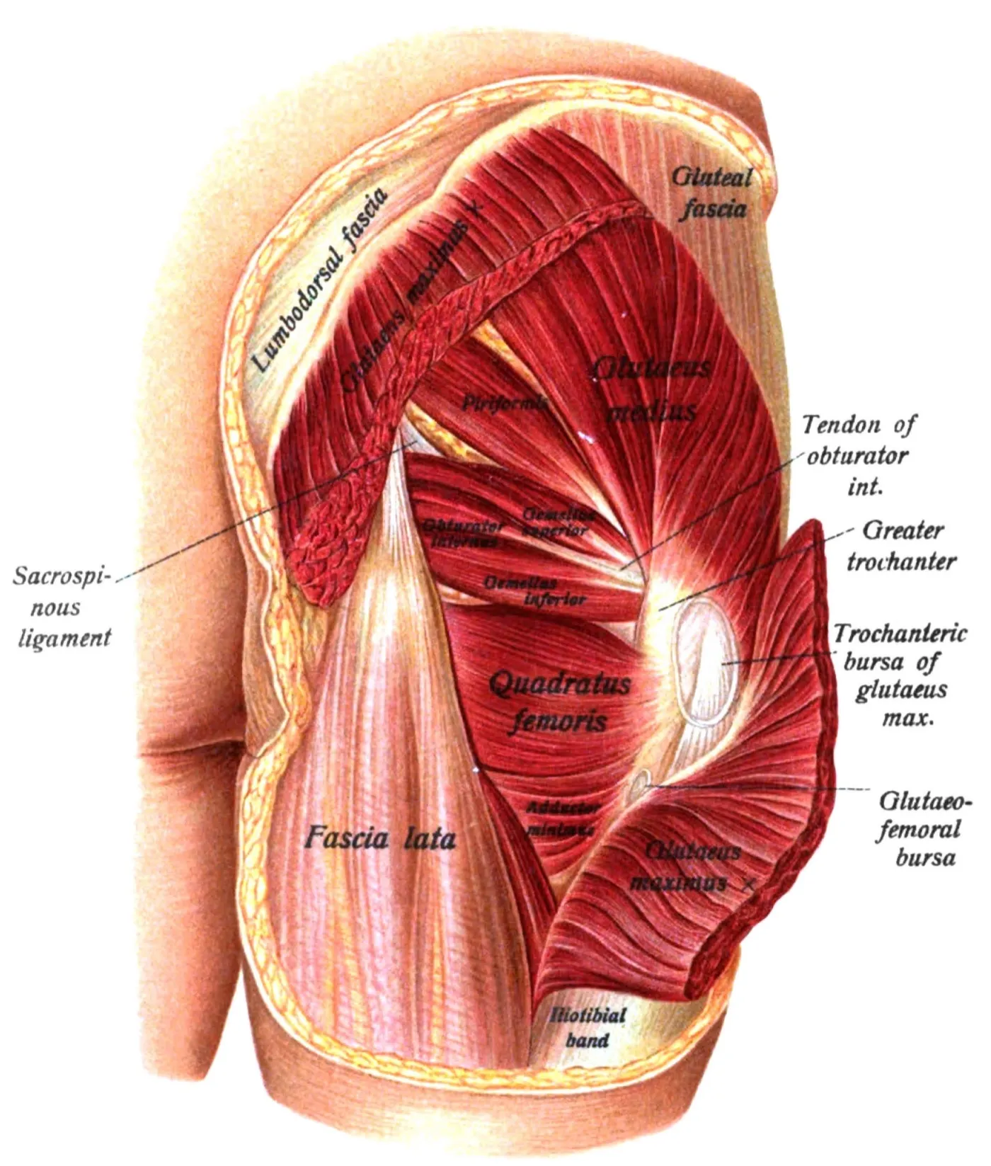
Each muscle starts on the pelvis and ends on the femur, the thigh bone7. Four end on the greater trochanter, the bony bump at the outer hip, at the top of the thigh bone7.
| Muscle | Starts on | Ends on |
|---|---|---|
| Piriformis | The front of the sacrum, the triangle-shaped bone at the base of the spine7 | The upper border of the greater trochanter7 |
| Obturator internus | Inside the pelvis, on the pubis, the ischium and the inner surface of the hip bone7 | The inner surface of the greater trochanter7 |
| Superior gemellus | The ischial spine, a point of bone on the ischium7 | The inner surface of the greater trochanter, with the obturator internus tendon7 |
| Inferior gemellus | The upper part of the ischial tuberosity, the “sitting bone”7 | The inner surface of the greater trochanter, with the obturator internus tendon7 |
| Quadratus femoris | The outer border of the ischial tuberosity7 | A line on the back of the femur7 |
| Obturator externus | The outer surface of the front wall of the pelvis, on the pubis and the ischium7 | The trochanteric fossa, a hollow on the femur7 |
The pubis is the bone at the lower front of the pelvis. The ischium is the lower rear part of the hip bone, which carries the sitting bone. The two gemelli join the obturator internus to form one conjoined tendon8. The piriformis tendon is often partly blended with that shared tendon7.
How their job changes as the hip bends
A classic anatomy text describes the deep muscles as powerful outward rotators of the thigh7. It says they stop rotating and become abductors when the thigh is flexed (bent) in sitting7. Abductors move a limb away from the midline of the body. The text names one exception: the obturator externus keeps turning the thigh bone outward7.
One study measured outward rotation torque (turning force) in 15 fresh-frozen cadaver hips2. For the piriformis, the obturator internus and their conjoined tendon, it peaked at 15° of hip flexion and fell as the hip flexed further2. The torque of the obturator externus rose with flexion instead2. At 75° of flexion or more, the obturator externus had the highest outward rotation torque of the short rotators2. On average it was 2.18 times that of the conjoined tendon at 90° and 3.80 times at 105°2.
What recordings from the working muscles show
The table shows recordings of three of these muscles made with fine-wire electrodes, very thin wire sensors placed into a muscle to record its activity9,10,11.
| Muscle | Who was recorded | When it was most active |
|---|---|---|
| Obturator internus | Ten participants | In isometric (held, with no joint movement) hip extension, then outward rotation, then abduction9 |
| Quadratus femoris | 10 healthy participants | In maximal isometric outward rotation and the clam movement10 |
| Piriformis | Eleven healthy men | In prone (face-down) hip extension with the hip rotated outward, of seven hip movements11 |
Whether they steady the joint
In 10 cadaver hips, each deep rotator had a relatively small force-producing cross-section and short fibres12. Some sources suggest these muscles help hold the femoral head (the ball at the top of the thigh bone) in its socket3,12,13:
- The authors of the 10-hip study pointed to the large combined cross-section and short fibres of the gemelli and obturators12. They concluded that these suggest the muscles mainly stabilise the hip joint more than they rotate it12.
- A narrative review is a summary of research written without a set, checkable search method. One proposed that the quadratus femoris, gemelli and both obturators, with three other deep muscles, may be the hip’s primary stabilisers3. It also noted that research on hip muscles is limited3.
- The obturator externus tendon is described as passing like a sling under the neck of the femur13. A string-model study of 18 fresh cadaver hips concluded that it helps hold the femoral head in the socket13.
Two modelling studies of 12 participants each, which estimate forces with a model rather than measuring them, tested the idea4,14. In one, of walking and squatting, simulated maximal activation of the six muscles pointed the hip contact force more towards the centre of the socket14. The other modelled walking4. Hip stiffness in the forward-backward plane was 0.8% lower with the muscles switched off and 3.2% higher with them fully on4. Those authors said this casts doubt on the muscles’ assumed role as hip stabilisers4.
The authors of the obturator internus recordings raised a doubt of their own9. They said the muscle’s direction-specific activity questions a general hip-stabilising role9.
Breakdown
Strains in kicking sports
Tears of the obturator externus and internus are uncommon injuries, known mostly from a few case reports in high-level athletes5. Three reports describe them5,15,16:
- One series covered four seasons in four teams of the Spanish first division5. In it, 16 professional soccer players had an obturator muscle tear confirmed by MRI, a scan that shows soft tissue5. Twelve tears were of the obturator externus and four of the obturator internus5. The main complaint was pain at the front of the hip5.
- In a case series of three professional soccer players with obturator externus injuries, two were hurt by long-distance shooting and one by long-distance passing15.
- A report of two cases described obturator internus strains in young male athletes in kicking sports16. One was a sudden strain alongside a strain of the adductor longus, one of the hip adductors16. The other was an overuse injury of both obturator internus muscles16.
The authors of the three-player series wrote that few studies describe injuries to the deep hip rotators15. They gave two reasons: such injuries are either under-reported, or mistaken for hamstring, adductor or quadriceps injuries15.
The quadratus femoris and the ischiofemoral space
The ischiofemoral space is the gap between the ischium and the femur17. One MRI study measured it in 12 hips of nine women, aged 30 to 71, with hip pain and abnormal quadratus femoris signal17. Their gap averaged 13 mm, against 23 mm in 11 control hips17.
In those 12 hips, the quadratus femoris showed oedema (swelling caused by fluid) in every one17. It showed a partial tear in 33%17. The nearby hamstring tendons showed oedema in 50%17.
A meta-analysis, which pools earlier studies, compared 217 hip MRIs with ischiofemoral impingement (pinching of soft tissue in that gap) and 140 control hips18. The space was about 15 mm in the impingement cases, against 26 mm18. In the five studies it pooled, 133 of the 154 people with the condition were female18.
In people with hip joint conditions
Two small studies looked at these muscles in people with a hip joint condition20,21.
The first was an MRI study of 12 people with a one-sided tear of the hip labrum and 12 matched healthy people20. A labrum is a rim of tough tissue around the edge of a joint socket. Piriformis and quadratus femoris cross-sectional area did not differ between groups20.
The second was a fine-wire study of squatting21. It compared 15 people with femoroacetabular impingement syndrome, a hip condition, with 14 healthy controls21. Unlike the controls, the impingement group activated the obturator internus on the way down about as much as on the way up21.
Recovery
Return to play after a strain
In the series of 16 professional soccer players with obturator muscle tears, the mean time back to play was 11.5 days5. In the case series of three players with obturator externus injuries, all three were playing again after 10 days15.
How their size changes with sport and training
One study scanned 46 elite Australian Football League players by MRI three times in a season22. Piriformis cross-sectional area, the size of a slice cut straight across the muscle, increased significantly between each scan22. Players whose piriformis grew less had slightly higher odds of sustaining an injury22.
The same study tested motor control training, exercise that trains how the deep trunk muscles are switched on and coordinated22. Two 30-minute sessions a week for 7 to 8 weeks were reported to increase piriformis size in players with low back pain22.
An MRI study compared 33 professional ballet dancers with 33 athletes matched for age and sex, at a single point in time23. Piriformis, gemelli and quadratus femoris cross-sectional areas were similar in the two groups, and unrelated to hip pain23.
Connections
The sciatic nerve
The sciatic nerve is the large nerve that runs from the lower back down the back of the thigh. It leaves the pelvis below the piriformis7. In the upper part of its course it rests on the obturator internus, the gemelli and the quadratus femoris, covered by the gluteus maximus7.
Common peroneal nerve is another name for the common fibular nerve. Two meta-analyses pooled how often the nerve takes this usual path, and how often one part of it pierces the muscle6,24:
| Meta-analysis | Nerve passes as one trunk below the piriformis | Common peroneal part passes through the piriformis |
|---|---|---|
| 44 cadaver studies | 90%6 | 8%6 |
| 45 studies, 7,068 lower limbs | 85.2%24 | 9.8%24 |
A 2010 systematic review described piriformis syndrome as sciatica (pain along the sciatic nerve) caused by the piriformis compressing the nerve25. It noted that the condition remained controversial after more than 70 years25.
The nerves that supply them
The obturator externus is supplied through the obturator nerve, a nerve that runs to the inner-thigh muscles7. One nerve supplies the obturator internus and the superior gemellus7. Another supplies the quadratus femoris and the inferior gemellus7. The piriformis has a nerve that may be double7.
Exercises and stretches
Related exercise pages: side-lying hip abduction and running10,26. A muscle MRI study of 10 healthy, active people compared exercises26. The clamshell, and side-lying hip abduction with outward rotation, activated the obturator internus more than a single-leg bridge or reverse clamshell26. The clamshell is a side-lying exercise that opens the knees apart with the hips and knees bent. In 10 healthy participants, quadratus femoris activity was very high in the stance phase of running, when the foot is on the ground10.
Related stretch page: the figure-4 stretch27. In 17 hips from nine embalmed cadavers, strings run along the short outward rotators were displaced significantly in each of four stretch positions tested27. The authors wrote that there was limited research showing how well common stretches work on these muscles27.
Deep hip rotators lies beneath
Deep hip rotators lies next to
Deep hip rotators starts on
Deep hip rotators attaches to
Deep hip rotators is crossed by
Deep hip rotators crosses
Deep hip rotators has an entry on
Deep hip rotators is loaded by
Deep hip rotators is stretched by
Sources
- Betts JG, Young KA, Wise JA, Johnson E, Poe B, Kruse DH, Korol O, Johnson JE, Womble M, DeSaix P (2022). Anatomy and Physiology 2e. OpenStax. Link
- Ito Y, Suzuki D, Kizawa F, Kanaizumi A, Hiraiwa T, Tamura T, Kawaguchi Y, Nagoya S (2025). Comparison of External Rotation Torque of Short External Rotator Muscles in Hip Flexion Position. Journal of Orthopaedic Research. doi:10.1002/jor.70005 PMID 40524428
- Retchford TH, Crossley KM, Grimaldi A, Kemp JL, Cowan SM (2013). Can local muscles augment stability in the hip? A narrative literature review. Journal of Musculoskeletal & Neuronal Interactions. PMID 23445909
- Meinders E, Pizzolato C, Goncalves BAM, Lloyd DG, Saxby DJ, Diamond LE (2022). The Deep Hip Muscles are Unlikely to Stabilize the Hip in the Sagittal Plane During Walking: A Joint Stiffness Approach. IEEE Transactions on Biomedical Engineering. doi:10.1109/tbme.2021.3114717 PMID 34559628
- Wong-On M, Turmo-Garuz A, Arriaza R, Gonzalez de Suso JM, Til-Perez L, Yanguas-Leite X, Diaz-Cueli D, Gasol-Santa X (2018). Injuries of the obturator muscles in professional soccer players. Knee Surgery, Sports Traumatology, Arthroscopy. doi:10.1007/s00167-017-4453-6 PMID 28188336
- Poutoglidou F, Piagkou M, Totlis T, Tzika M, Natsis K (2020). Sciatic Nerve Variants and the Piriformis Muscle: A Systematic Review and Meta-Analysis. Cureus. doi:10.7759/cureus.11531 PMID 33354475
- Gray H; Lewis WH (editor) (1918). Anatomy of the Human Body. Lea & Febiger, Philadelphia (20th edition). Link
- Lezak B, Massel DH (2026). Anatomy, Bony Pelvis and Lower Limb, Gemelli Muscles. StatPearls. PMID 32491352
- Hodges PW, McLean L, Hodder J (2014). Insight into the function of the obturator internus muscle in humans: observations with development and validation of an electromyography recording technique. Journal of Electromyography and Kinesiology. doi:10.1016/j.jelekin.2014.03.011 PMID 24788026
- Semciw AI, Freeman M, Kunstler BE, Mendis MD, Pizzari T (2015). Quadratus femoris: An EMG investigation during walking and running. Journal of Biomechanics. doi:10.1016/j.jbiomech.2015.05.029 PMID 26116043
- Morimoto Y, Oshikawa T, Imai A, Okubo Y, Kaneoka K (2018). Piriformis electromyography activity during prone and side-lying hip joint movement. Journal of Physical Therapy Science. doi:10.1589/jpts.30.154 PMID 29410588
- Parvaresh KC, Chang C, Patel A, Lieber RL, Ball ST, Ward SR (2019). Architecture of the Short External Rotator Muscles of the Hip. BMC Musculoskeletal Disorders. doi:10.1186/s12891-019-2995-0 PMID 31862009
- Gudena R, Alzahrani A, Railton P, Powell J, Ganz R (2015). The anatomy and function of the obturator externus. Hip International. doi:10.5301/hipint.5000249 PMID 25952918
- Meinders E, Pizzolato C, Gonçalves B, Lloyd DG, Saxby DJ, Diamond LE (2022). Activation of the deep hip muscles can change the direction of loading at the hip. Journal of Biomechanics. doi:10.1016/j.jbiomech.2022.111019 PMID 35248802
- Rhim HC, Gureck AE, Jang KM (2022). Acute Obturator Externus Injury in Professional Soccer Players: A Case Series. Medicina (Kaunas). doi:10.3390/medicina58091145 PMID 36143822
- Byrne C, Alkhayat A, O'Neill P, Eustace S, Kavanagh E (2017). Obturator internus muscle strains. Radiology Case Reports. doi:10.1016/j.radcr.2016.10.020 PMID 28228895
- Torriani M, Souto SC, Thomas BJ, Ouellette H, Bredella MA (2009). Ischiofemoral impingement syndrome: an entity with hip pain and abnormalities of the quadratus femoris muscle. American Journal of Roentgenology. doi:10.2214/ajr.08.2090 PMID 19542413
- Singer AD, Subhawong TK, Jose J, Tresley J, Clifford PD (2015). Ischiofemoral impingement syndrome: a meta-analysis. Skeletal Radiology. doi:10.1007/s00256-015-2111-y PMID 25672947
- Maraş Özdemir Z, Aydıngöz Ü, Görmeli CA, Sağır Kahraman A (2015). Ischiofemoral Space on MRI in an Asymptomatic Population: Normative Width Measurements and Soft Tissue Signal Variations. European Radiology. doi:10.1007/s00330-015-3625-3 PMID 25680726
- Mendis MD, Wilson SJ, Hayes DA, Hides JA (2020). Hip muscle atrophy in patients with acetabular labral joint pathology. Clinical Anatomy. doi:10.1002/ca.23429 PMID 31301249
- Diamond LE, van den Hoorn W, Bennell KL, Wrigley TV, Hinman RS, O'Donnell J, Hodges PW (2019). Deep hip muscle activation during squatting in femoroacetabular impingement syndrome. Clinical Biomechanics. doi:10.1016/j.clinbiomech.2019.07.017 PMID 31351343
- Leung FT, Mendis MD, Stanton WR, Hides JA (2015). The relationship between the piriformis muscle, low back pain, lower limb injuries and motor control training among elite football players. Journal of Science and Medicine in Sport. doi:10.1016/j.jsams.2014.06.011 PMID 25027772
- Emery S, Cook J, Ferrar K, Mayes S (2021). Deep hip external rotator muscle size in ballet dancers compared to non-dancing athletes, and associations to pain. Physical Therapy in Sport. doi:10.1016/j.ptsp.2021.06.010 PMID 34237542
- Tomaszewski KA, Graves MJ, Henry BM, Popieluszko P, Roy J, Pękala PA, Hsieh WC, Vikse J, Walocha JA (2016). Surgical anatomy of the sciatic nerve: A meta-analysis. Journal of Orthopaedic Research. doi:10.1002/jor.23186 PMID 26856540
- Hopayian K, Song F, Riera R, Sambandan S (2010). The clinical features of the piriformis syndrome: a systematic review.. European spine journal : official publication of the European Spine Society, the European Spinal Deformity Society, and the European Section of the Cervical Spine Research Society. doi:10.1007/s00586-010-1504-9 PMID 20596735
- Kim S, Burland JP, Morgan KD, DiStefano LJ, Boling MC, Glaviano NR (2026). Superficial and Deep Gluteal Muscle Activation During Common Therapeutic Exercises: A Muscle Functional Magnetic Resonance Imaging Analysis.. Sports Health. doi:10.1177/19417381251372979 PMID 40977436
- McGovern RP, Kivlan BR, Martin RL (2017). LENGTH CHANGE OF THE SHORT EXTERNAL ROTATORS OF THE HIP IN COMMON STRETCH POSITIONS: A CADAVERIC STUDY.. International journal of sports physical therapy. doi:10.26603/ijspt20171068 PMID 29234558
Changes
- First published.