Hamstrings
Also called hamstring muscles, hamstring group
The three long muscles at the back of the thigh. How they swing the thigh back and bend the knee, why they strain so often in sprinting sports, how they change with training, and what they work with.
- Region
- Thigh
- Starts on (all but the short head of biceps femoris)
- the ischial tuberosity, at the lower back of the hip bone; semitendinosus and biceps long head share one tendon there (Pelvis) (Proximal hamstring tendon)7,8,36
- Starts on (short head of biceps femoris)
- the linea aspera, a rough ridge down the back of the thigh bone2,7
- Ends on (biceps femoris)
- the outer side of the knee, at the top of the fibula and the outer shinbone7
- Ends on (semimembranosus and semitendinosus)
- the inner side of the shinbone7
- Tendons
- Proximal hamstring tendon
- Muscles
- biceps femoris, semitendinosus, semimembranosus
- Updated
- September 29, 2026
- Reviewed
- September 29, 2026 · how entries are checked
Function
Which muscles make up the group
The hamstring group is three long muscles at the back of the thigh and knee1,2. They are the biceps femoris, the semitendinosus and the semimembranosus2. The biceps femoris stands out from the other two because it has two parts, called heads: a long head and a short head7. Of the three, the semimembranosus has the largest cross-sectional area, and the semitendinosus has the smallest7. Cross-sectional area is the size of a slice cut straight across a muscle.
Where they attach
Most of the group starts at the ischial tuberosity, the rounded knob of bone at the lower back of the hip bone7,8. The short head of the biceps femoris is the exception7,8. It starts instead from the linea aspera, a long ridge down the back of the femur (thigh bone)7.
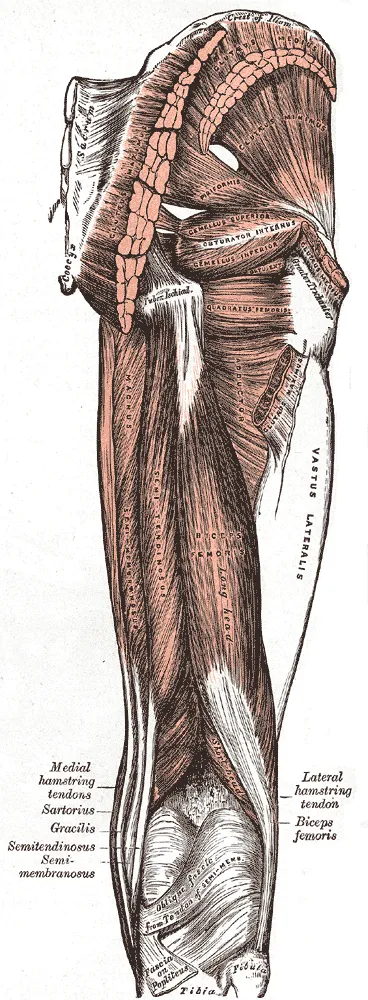
In a study of 97 half-pelvises from donors, the semimembranosus shared fibres at its start with the biceps femoris long head8. It also spread into the adductor magnus, a large muscle on the inner side of the thigh8.
At the knee, the group splits to either side, onto the tibia (shinbone) and the fibula (the thin outer bone of the lower leg)7:
| Muscle | Where it attaches below the knee |
|---|---|
| Biceps femoris (both heads) | Outer side: top of the fibula and outer tibia7 |
| Semitendinosus | Inner side of the tibia7 |
| Semimembranosus | Inner side of the tibia7 |
What they move
The hamstrings extend the hip, which swings the thigh backward, and flex the knee, which bends it2,3. Three of the four parts cross both joints: the semitendinosus, the semimembranosus and the long head of the biceps femoris7. The short head of the biceps femoris crosses the knee alone7. Muscles at the front of the thigh, including the quadriceps, do the reverse: they straighten the knee and help bend the hip2.
Which nerves supply them
The semitendinosus, the semimembranosus and the long head of the biceps femoris take their nerve supply from the tibial part of the sciatic nerve7. The sciatic nerve is the large nerve that runs from the lower back down the back of the thigh. The short head of the biceps femoris gets its supply from a different nerve, the common peroneal nerve7.
Breakdown
How common hamstring injuries are
Hamstring injuries are common in football4,9. In a cohort study of 2,299 professional male footballers, the hamstrings accounted for 37% of muscle injuries9. That was more than the adductors (the inner thigh muscles), at 23%, or the quadriceps, at 19%9. Another study followed 3,909 elite male players from 54 teams across 21 seasons4. In elite men’s football, hamstring injuries made up 19% of injuries of any kind over 21 seasons4.
That share did not stay level4. It was 12% of injuries in the first season studied, and 24% by the latest season4.
Where and how the muscle strains
A review reports that most hamstring injuries in athletes are strains where muscle meets tendon7. That meeting point is called the myotendinous junction, and the upper one, near the hip, is the most common site7. In the elite football study, running or sprinting caused most injuries4. It accounted for 62% of injuries with a visible tear and 51% without one4.
In elite football from 2011/12 on, 80% of injuries placed in one muscle were in the biceps femoris, not the two inner hamstrings4. A review of the few studies on deadlift injuries lists hamstring ruptures among the injuries reported10.
What raises the odds of a strain
A meta-analysis of 78 studies in 71,324 athletes named two strongest risk factors: older age and an earlier hamstring strain5. For earlier strains, it gave its figures as risk ratios, which say how many times as likely a strain was5. Any earlier strain went with a risk ratio of 2.7, and a recent strain with 4.85. A separate review of 16 studies found a recent hamstring strain was associated with quadriceps injury11.
A systematic review of 1982–2007 studies found other factors less clear12. It found evidence lacking that tight hamstrings raise the risk, though it could not rule this out12. In one group of amateur Australian Rules footballers, more flexible quadriceps went with fewer hamstring strains12.
The same review found mixed results for strength balance between the front and back of the thigh12. In one study, footballers with relatively stronger quadriceps than hamstrings went on to strain more12. A second study supported this, but a third found the imbalance did not predict a future strain12.
How often a strain comes back
A strain often returns4,13. A systematic review of five studies found repeat-injury rates from 13.9% to 63.3%13. Those rates were measured from the same season up to two years after the first injury13. In elite male footballers, 18% of hamstring injuries were repeats, and 69% of those came back within two months4. The review of 1982–2007 studies reports that footballers stayed about three times as likely to strain again even after a year12.
Recovery
How long players are out
A systematic review of adult athletes in the football codes reports 2 to 6 weeks away from sport for each hamstring strain12. In the elite football study, players with a visible tear on MRI (a scan that shows muscle in detail) were out longer4. The median time out, in days, was4:
| Type of hamstring injury | Median days out |
|---|---|
| Any hamstring injury | 134 |
| Visible fibre tear on MRI | 174 |
| No visible fibre tear | 64 |
In professional football, time out after a repeat hamstring injury was significantly longer on average than after the first14.
How training changes the muscle
Training that works the hamstrings while they lengthen may change the shape of one of them15. This is called eccentric work15. A meta-analysis of ten randomised trials looked at eccentric programmes of at least four weeks in healthy adults15. It found limited to moderate evidence of three changes in the long head of the biceps femoris15:
- Longer fascicles, the bundles of muscle fibres15.
- A thicker muscle15.
- A smaller pennation angle, the slant of fibres against the line of pull15.
In a study of 28 men, the type of work set the direction of change6. Six weeks of purely eccentric knee flexor training made the long head’s fascicles longer6. Six weeks of purely concentric work, done while the muscle shortens, made them shorter6. After 28 days without training, the longer fascicles had gone back to where they started6.
What the Nordic hamstring exercise shows
Many studies have looked at one exercise, the Nordic hamstring exercise16,17. Across 13 Nordic studies, programmes of six weeks or more showed very large before-to-after effects on eccentric strength and biceps femoris fascicle length16. They appeared with both high and low training volumes16. A meta-analysis pooled 15 studies of athletes across sports17. Injury-reduction programmes that included the exercise had a hamstring injury risk ratio of 0.49 compared with usual training or programmes without it17.
What happens with other training and disuse
In one trial, squat training grew other hip muscles but not the hamstrings18. In 17 men who trained full or half squats for 10 weeks, hamstring volume did not change18. Over the same time, gluteus maximus and adductor volume increased18. In 13 young men with one knee braced for 7 days, the hamstrings lost 3.5% of their muscle19. The quadriceps lost more, at 6.7%19.
Muscle soreness and strength loss after hard lowering work also differ between muscles20. In 15 inactive young men, the knee flexors lost more strength than the knee extensors, the plantar flexors (calf muscles) or the back muscles20. When the same session was repeated two weeks later, each measure of damage changed less20.
Connections
The muscles they work with
The gluteus maximus works with the hamstrings to extend the hip21. Its bulk lies over the hamstrings’ starting point22. When the body rises from a full forward bend, these two do most of the work in the first half23. In 38 women, half of them with long-term low back pain, the biceps femoris and low-back muscles switched on together early in a forward bend24. The gluteus maximus followed24.
The quadriceps, at the front of the thigh, are their opposite at the knee2. The quadriceps straighten the knee, while the hamstrings bend it2. One quadriceps muscle, the rectus femoris, opposes the hamstrings at the hip as well2,25.
Part of the adductor magnus is sometimes counted as a fourth hamstring3. This part also starts from the ischial tuberosity3. It shares the hamstrings’ nerve supply and action3.
The exercises that load them
Several exercises load the hamstrings, and studies compare how active they are26,27. The deadlift studies measured activity with surface electromyography, which records muscle activity through sensors on the skin26.
| Exercise | What studies found for the hamstrings |
|---|---|
| Deadlift | Among the muscles it is often done to strengthen26 |
| Deadlift | Biceps femoris less active than the back muscles and quadriceps, in trained lifters26 |
| Romanian deadlift | Biceps femoris and semitendinosus more active than the back muscles26 |
| Deadlift | More active in the parts of the lift where the knees were straighter28 |
| Glute bridge hold | On an unstable surface, biceps femoris activity rose as the knee straightened29 |
| Back squat | Moderate to high activity, generally rising as the knees bend more27 |
| Back squat | Stance width did not appear to change activity30 |
| Wall squats and one-leg squat | Modelled force of about 15 to 190 newtons, against 30 to 720 for the quadriceps31 |
| Farmer’s walk | Biceps femoris at about 54% of its maximum, in one study32 |
Within the group, the semitendinosus was slightly more active than the biceps femoris during deadlifts26. In a second bridge study, of 16 men, gluteus maximus activity did not differ across knee angles, while back-muscle activity changed like hamstring activity33. In 22 futsal players doing a single-leg bridge on the heel, the biceps femoris worked harder with the ankle also turned out34.
In 23 healthy adults, the one-leg wall squat produced the most biceps femoris activity of five one-leg exercises35. Even so, it stayed below the level the authors judged enough for strengthening35. Studies of how squat depth changes hamstring activity disagree30.
Hamstrings works with to extend the hip
Hamstrings opposes at the knee
Hamstrings is loaded by
Sources
- Hu Y, Raja A (2026). Anatomy, Bony Pelvis and Lower Limb, Hamstring Muscle. StatPearls. PMID 31536294
- Betts JG, Young KA, Wise JA, Johnson E, Poe B, Kruse DH, Korol O, Johnson JE, Womble M, DeSaix P (2022). Anatomy and Physiology 2e. OpenStax. Link
- Obey MR, Broski SM, Spinner RJ, Collins MS, Krych AJ (2016). Anatomy of the Adductor Magnus Origin. Orthopaedic Journal of Sports Medicine. doi:10.1177/2325967115625055 PMID 26798764
- Ekstrand J, Bengtsson H, Waldén M, Davison M, Khan KM, Hägglund M (2023). Hamstring injury rates have increased during recent seasons and now constitute 24% of all injuries in men's professional football: the UEFA Elite Club Injury Study from 2001/02 to 2021/22.. British Journal of Sports Medicine. doi:10.1136/bjsports-2021-105407 PMID 36588400
- Green B, Bourne MN, van Dyk N, Pizzari T (2020). Recalibrating the risk of hamstring strain injury (HSI): A 2020 systematic review and meta-analysis of risk factors for index and recurrent hamstring strain injury in sport.. British Journal of Sports Medicine. doi:10.1136/bjsports-2019-100983 PMID 32299793
- Timmins RG, Ruddy JD, Presland J, Maniar N, Shield AJ, Williams MD, Opar DA (2016). Architectural Changes of the Biceps Femoris Long Head after Concentric or Eccentric Training.. Medicine and Science in Sports and Exercise. doi:10.1249/MSS.0000000000000795 PMID 26460634
- Koolmees DS, Klott JD, Poppe TR, Bernholt DL (2025). Distal Hamstring Injuries and Disorders.. Journal of the American Academy of Orthopaedic Surgeons: Global Research & Reviews. doi:10.5435/JAAOSGlobal-D-25-00248 PMID 40972536
- Miguel-Pérez M, Iglesias-Chamorro P, Ortiz-Miguel S, Ortiz-Sagristà JC, Möller I, Blasi J, Agullò J, Martinoli C, Pérez-Bellmunt A (2024). Anatomical Relationships of the Proximal Attachment of the Hamstring Muscles with Neighboring Structures: From Ultrasound, Anatomical and Histological Findings to Clinical Implications.. Diagnostics (Basel). doi:10.3390/diagnostics14161725 PMID 39202213
- Ekstrand J, Hägglund M, Waldén M (2011). Epidemiology of muscle injuries in professional football (soccer).. The American Journal of Sports Medicine. doi:10.1177/0363546510395879 PMID 21335353
- Bengtsson V, Berglund L, Aasa U (2018). Narrative review of injuries in powerlifting with special reference to their association to the squat, bench press and deadlift.. BMJ Open Sport & Exercise Medicine. doi:10.1136/bmjsem-2018-000382 PMID 30057777
- Pietsch S, Pizzari T (2022). Risk Factors for Quadriceps Muscle Strain Injuries in Sport: A Systematic Review.. Journal of Orthopaedic & Sports Physical Therapy. doi:10.2519/jospt.2022.10870 PMID 35647880
- Prior M, Guerin M, Grimmer K (2009). An evidence-based approach to hamstring strain injury: a systematic review of the literature.. Sports Health. doi:10.1177/1941738108324962 PMID 23015867
- de Visser HM, Reijman M, Heijboer MP, Bos PK (2012). Risk factors of recurrent hamstring injuries: a systematic review.. British Journal of Sports Medicine. doi:10.1136/bjsports-2011-090317 PMID 22011915
- Ekstrand J, Krutsch W, Spreco A, van Zoest W, Roberts C, Meyer T, Bengtsson H (2020). Time before return to play for the most common injuries in professional football: a 16-year follow-up of the UEFA Elite Club Injury Study.. British Journal of Sports Medicine. doi:10.1136/bjsports-2019-100666 PMID 31182429
- Gérard R, Gojon L, Decleve P, Van Cant J (2020). The Effects of Eccentric Training on Biceps Femoris Architecture and Strength: A Systematic Review With Meta-Analysis.. Journal of Athletic Training. doi:10.4085/1062-6050-194-19 PMID 32216654
- Cuthbert M, Ripley N, McMahon JJ, Evans M, Haff GG, Comfort P (2020). The Effect of Nordic Hamstring Exercise Intervention Volume on Eccentric Strength and Muscle Architecture Adaptations: A Systematic Review and Meta-analyses.. Sports Medicine. doi:10.1007/s40279-019-01178-7 PMID 31502142
- van Dyk N, Behan FP, Whiteley R (2019). Including the Nordic hamstring exercise in injury prevention programmes halves the rate of hamstring injuries: a systematic review and meta-analysis of 8459 athletes.. British Journal of Sports Medicine. doi:10.1136/bjsports-2018-100045 PMID 30808663
- Kubo K, Ikebukuro T, Yata H (2019). Effects of squat training with different depths on lower limb muscle volumes.. European Journal of Applied Physiology. doi:10.1007/s00421-019-04181-y PMID 31230110
- Kilroe SP, Fulford J, Jackman SR, van Loon LJC, Wall BT (2020). Temporal Muscle-specific Disuse Atrophy during One Week of Leg Immobilization.. Medicine & Science in Sports & Exercise. doi:10.1249/MSS.0000000000002200 PMID 31688656
- Chen TC, Yang TJ, Huang MJ, Wang HS, Tseng KW, Chen HL, Nosaka K (2019). Damage and the repeated bout effect of arm, leg, and trunk muscles induced by eccentric resistance exercises.. Scandinavian Journal of Medicine & Science in Sports. doi:10.1111/sms.13388 PMID 30663816
- Lee DK, Oh JS (2018). Relationship between hamstring length and gluteus maximus strength with and without normalization.. Journal of Physical Therapy Science. doi:10.1589/jpts.30.116 PMID 29410579
- Grimaldi A, Mellor R, Nasser A, Vicenzino B, Hunter DJ (2024). Current and future advances in practice: tendinopathies of the hip.. Rheumatology Advances in Practice. doi:10.1093/rap/rkae022 PMID 38601140
- Pourahmadi M, Asadi M, Dommerholt J, Yeganeh A (2020). Changes in the macroscopic morphology of hip muscles in low back pain.. Journal of Anatomy. doi:10.1111/joa.13086 PMID 31475359
- Leinonen V, Kankaanpää M, Airaksinen O, Hänninen O (2000). Back and hip extensor activities during trunk flexion/extension: effects of low back pain and rehabilitation.. Archives of Physical Medicine and Rehabilitation. doi:10.1016/s0003-9993(00)90218-1 PMID 10638873
- Begum FA, Kayani B, Chang JS, Tansey RJ, Haddad FS (2020). The management of proximal rectus femoris avulsion injuries.. EFORT Open Reviews. doi:10.1302/2058-5241.5.200055 PMID 33312709
- Martín-Fuentes I, Oliva-Lozano JM, Muyor JM (2020). Electromyographic activity in deadlift exercise and its variants. A systematic review.. PLoS One. doi:10.1371/journal.pone.0229507 PMID 32107499
- Escamilla RF (2001). Knee biomechanics of the dynamic squat exercise.. Medicine and Science in Sports and Exercise. doi:10.1097/00005768-200101000-00020 PMID 11194098
- Escamilla RF, Francisco AC, Kayes AV, Speer KP, Moorman CT 3rd (2002). An electromyographic analysis of sumo and conventional style deadlifts.. Medicine and Science in Sports and Exercise. doi:10.1097/00005768-200204000-00019 PMID 11932579
- Kim S, Park J, Her J (2023). Comparison of Muscle Activity and Muscle Thickness According to Knee Flexion Angle during Supine Bridge Exercises using the Abdominal Drawing-in Maneuver on an Unstable Surface.. Journal of Sports Science and Medicine. doi:10.52082/jssm.2023.431 PMID 37711708
- Straub RK, Powers CM (2024). A Biomechanical Review of the Squat Exercise: Implications for Clinical Practice.. International Journal of Sports Physical Therapy. doi:10.26603/001c.94600 PMID 38576836
- Escamilla RF, Zheng N, Imamura R, Macleod TD, Edwards WB, Hreljac A, Fleisig GS, Wilk KE, Moorman CT 3rd, Andrews JR (2009). Cruciate ligament force during the wall squat and the one-leg squat.. Medicine and Science in Sports and Exercise. doi:10.1249/MSS.0b013e3181882c6d PMID 19127183
- Hindle BR, Lorimer A, Winwood P, Keogh JWL (2019). The Biomechanics and Applications of Strongman Exercises: a Systematic Review.. Sports Medicine - Open. doi:10.1186/s40798-019-0222-z PMID 31820223
- Hirose N, Tsuruike M (2018). Differences in the Electromyographic Activity of the Hamstring, Gluteus Maximus, and Erector Spinae Muscles in a Variety of Kinetic Changes.. Journal of Strength and Conditioning Research. doi:10.1519/JSC.0000000000002747 PMID 30102684
- Elias G, Jerez-Mayorga D, Intelangelo L (2024). Does the Position of the Ankle Matter During the Single Gluteal Bridge in Futsal Players? An Electromyographic Analysis.. Journal of Sport Rehabilitation. doi:10.1123/jsr.2022-0425 PMID 37917981
- Ayotte NW, Stetts DM, Keenan G, Greenway EH (2007). Electromyographical analysis of selected lower extremity muscles during 5 unilateral weight-bearing exercises.. Journal of Orthopaedic and Sports Physical Therapy. doi:10.2519/jospt.2007.2354 PMID 17366959
- Philippon MJ, Ferro FP, Campbell KJ, Michalski MP, Goldsmith MT, Devitt BM, Wijdicks CA, LaPrade RF (2015). A qualitative and quantitative analysis of the attachment sites of the proximal hamstrings.. Knee Surgery, Sports Traumatology, Arthroscopy. doi:10.1007/s00167-014-3074-6 PMID 24859732
Changes
- Added a figure showing where it sits, links to the exercise pages that load it, and tighter wording in a few places.
- First published.