Pelvis
Also called pelvic bones
The ring of bone at the base of the spine that carries the upper body's weight to the legs. How it is built, how it fractures in teenagers, athletes and older adults, how bone responds to load, and what attaches to it.
- Region
- Hip
- Muscles
- gluteus maximus, gluteus medius, gluteus minimus, hamstrings, hip adductors, quadriceps, rectus abdominis, external oblique, internal oblique, transversus abdominis, erector spinae, latissimus dorsi, Hip flexors, Tensor fasciae latae, Deep hip rotators, Quadratus lumborum
- Updated
- September 30, 2026
- Reviewed
- September 30, 2026 · how entries are checked
Function
Four bones locked together
Four bones make up the pelvis: a hip bone on each side, plus the sacrum and the coccyx (tailbone)1. The sacrum forms from five vertebrae (bones of the spine) that fuse together, a process that does not start until after age 201.
Each adult hip bone starts as three bones: the ilium, the ischium (the lower rear part) and the pubis1. These fuse together in the late teenage years1. On the outer side of the hip bone, the three meet to form the acetabulum (hip socket), the deep cup of the hip joint1.
The ilium (the wide upper part of the hip bone) is the largest part1. It is firmly joined to the sacrum at the sacroiliac joint, which barely moves1. At the front, the pubis (the bone at the lower front of the pelvis) of each side meets its partner at the pubic symphysis1. Each hip bone is where one leg attaches, and each is firmly joined to the sacrum at the base of the spine1.
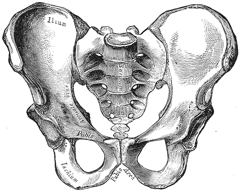
How it carries the body’s weight
The pelvis’s main job is to carry the upper body’s weight1:
It also gives muscles of the trunk and legs a place to attach, and it shields the organs inside the pelvis1.
The body’s centre of gravity sits in the region of the pelvis1. The shoulder girdle (the shoulder blades and collarbones) moves freely, but the pelvic bones are locked together into a firm, weight-bearing structure1. Because it is so stable, weight can pass from the spine through the hip joints into one leg while the other leg is lifted1.
Strong ligaments (tough bands of tissue that hold bones together at a joint) hold the sacrum still as it carries the body’s weight1. These include the sacroiliac ligaments and the sacrospinous ligament (a strong band at the back of the pelvis)1. The sacrotuberous ligament, running from the base of the spine to the sitting bone, helps too1.
A systematic review (a study that finds and sums up the research on a question in a set, checkable way) brought together seven 3D studies3. It found that the sacroiliac joint turned 8 degrees or less and shifted 8 mm or less about any axis3. A review of pelvic anatomy describes these joints as key to passing load between the spine and the legs4.
Landmarks where muscles attach
The iliac crest is the curved top rim of the hip bone1. Its rounded front end is the anterior superior iliac spine, and just below it sits the anterior inferior iliac spine1. Both are places where thigh muscles attach1. The ischial tuberosity (the “sitting bone”) lies at the bottom of the ischium1. It anchors the muscles at the back of the thigh and takes the body’s weight during sitting1.
Most muscles that attach to the femur (thigh bone) and move it start on the pelvis1. The pelvic floor is a sheet of muscle that closes the bottom of the pelvis1. Part of it stretches from the pubis to the coccyx1.
How male and female pelves differ
Male pelvic bones are generally thicker and heavier1. The female pelvis is wider, a shape suited to childbirth1. The angle below the pubic symphysis is over 80 degrees in females and under 70 degrees in males1.
Breakdown
Avulsion fractures in teenagers
An avulsion fracture is a break in which a muscle pulls off a piece of the bone it attaches to2. In the pelvis, these mostly happen in teenage athletes, from a sudden, strong muscle pull2. This happens while the growth plates (layers of cartilage where a growing bone lengthens) are still open2. In teenagers, the growth plates at the pelvic apophyses (bony bumps where muscles attach) give way more easily than the muscles and tendons2. So under a sudden, large pull, from a hard contraction or a forced stretch, the growth plate can fail first2.
A 2017 meta-analysis (a study that pools the results of several earlier studies) lists where these pieces can come away2:
- the anterior inferior iliac spine, where the rectus femoris (a front thigh muscle that also bends the hip) attaches2;
- the ischial tuberosity, where the hamstrings (the three long muscles at the back of the thigh) attach2;
- the anterior superior iliac spine, where the sartorius attaches, a muscle that runs from there to the inner upper shinbone1,2;
- the iliac crest, where the tensor fasciae latae (a small muscle at the upper outer thigh) attaches2;
- the upper corner of the pubic symphysis, where the rectus abdominis (a long, flat muscle down the front of the abdomen) attaches2.
The most common site differed between studies2,5,6,7:
| Study | Most common site | Next sites |
|---|---|---|
| 2017 meta-analysis, 596 teenagers2 | Anterior inferior iliac spine, 33.2%2 | Ischial tuberosity 29.7%; next, anterior superior iliac spine 27.9%2 |
| 2026 pooled data, 852 fractures5 | Anterior superior iliac spine, 33.1%5 | Anterior inferior iliac spine 30.4%, ischial tuberosity 15.5%5 |
| One children’s hospital, 719 fractures6 | Anterior inferior iliac spine, 33.4%6 | Anterior superior iliac spine 30.5%, ischial tuberosity 19.4%6 |
| 198 teenage competitive athletes, 203 fractures7 | Ischial tuberosity, 109 cases7 | Anterior inferior iliac spine, 45 cases7 |
These fractures happen in a narrow window of age and bone growth, often during sport6. In the series of 198 teenage athletes, they were most common in soccer and gymnastics7.
Stress injuries in athletes and recruits
An international consensus describes bone stress injuries as overuse injuries to bone that limit activity and can end high-level careers8. The term covers stress reactions (early bone stress injuries, before a crack forms) and stress fractures (small cracks in a bone from repeated loading)8.
A case-control study (one that compares people who have a condition with similar people who do not) looked at female Navy recruits9. The 25 with pelvic stress fractures were shorter and lighter than the 61 without9. Their self-rated fitness, past activity and a history of missed periods were not linked to the fractures9.
A sacral fatigue fracture is a stress fracture in normal bone from too much repeated load10. Across published imaging-confirmed cases, each one had a recent rise in training intensity and a poor diet10.
Fractures in older adults
In Finland, first pelvic fractures linked to osteoporosis (thin, weak bones) in people aged 60 or older numbered 128 in 1970 and 913 in 199711. That was a rise from 20 to 92 per 100,000 people aged 60 or older11. In 1997, the age-adjusted rate was 103 per 100,000 in women and 38 in men, both higher than in 197011.
A sacral insufficiency fracture is a stress fracture in bone too weak to bear normal loads10. In published imaging-confirmed cases, the most common risk factors were these10:
- osteoporosis10;
- radiation to the pelvis10;
- rheumatoid arthritis (a long-term disease in which the body’s immune system attacks the joints)10;
- long-term use of corticosteroids (drugs that copy a type of hormone the body makes)10;
- being postmenopausal (after menopause)10.
Recovery
How bone responds to load
Muscle contractions put the largest everyday forces on bone12. Bone adapts to the loads it usually carries12. It tunes its structure to how much it is strained, how fast and how often, and in what pattern12.
One theory, called the mechanostat, sets out a minimum level of strain12. Below it, bone is lost; above it, new bone forms, adding mass and thickness12. A 2017 review of bone mechanics adds that too little and too rare a load may lead to bone loss12. Too much and too often may lead to a stress reaction12.
Bone can also change its size, shape and inner structure, which makes it stronger and stiffer without adding mass12. Measures of bone mineral density (how much mineral is packed into a given amount of bone) leave out shape and inner structure12. Yet these add greatly to bone strength and resistance to repeated loading12.
How broken bone heals
Fracture repair rebuilds bone, repeating many of the steps by which the skeleton first forms before birth13. A review of fracture healing reports that repair usually returns bone to its earlier make-up, structure and function13. It puts the share of fractures that do not heal normally at about 10%13.
A meta-analysis pooled 16 studies of 560 bone stress injuries14. Injuries at sites rich in trabecular bone (light, spongy bone found inside bones), such as the pelvis, took longer to heal14. Sites rich in cortical bone (the dense, solid outer layer of a bone), such as the tibia (shinbone), healed sooner14.
Connections
Muscles of the hip and thigh
The hamstrings start from the ischial tuberosity, apart from the short head of the biceps femoris, one of the three1,15,16. In 5 of 6 cadaver pelvises, they began from one common tendon, covered on the proximal hamstring tendon page17. The adductor group, covered on the hip adductors page, generally starts on the pelvis and attaches to the femur18. The adductor longus tendon arises from the front of the pubic bone19.
In 11 cadaver limbs, the gluteus maximus (the large buttock muscle) arose partly from the ilium, sacrum, coccyx and nearby ligaments20. In 12 cadavers, the gluteus medius (a buttock muscle beneath the gluteus maximus) arose from three sites21. One was the back lower edge of the iliac crest21. In 16 cadaver hips, the gluteus minimus (a buttock muscle beneath the gluteus medius) arose from the outer surface of the ilium, among other sites22.
In the quadriceps (four muscles at the front of the thigh), the rectus femoris has one head from the anterior inferior iliac spine1,23. Its other head comes from the ridge of the hip socket23.
Muscles of the trunk
The rectus abdominis starts on the pubis1. The external oblique, the internal oblique and the transversus abdominis each attach to the ilium1. The transversus abdominis also attaches to the pubis1.
Farther up the back, the lumbar fibres of the erector spinae (long muscles beside the spine that straighten the back) attach to the ilium24. The latissimus dorsi (the broad muscle across the middle and lower back) has a broad origin that includes the iliac crest25,26.
Muscles that steady the pelvis
The hip adductors (inner thigh muscles) pull the thigh inward and keep the pelvis balanced during walking18. Weak gluteus medius and minimus muscles let the pelvis drop toward the other side during walking27. This pattern is called Trendelenburg gait27.
Bones, joints and exercises
Related exercise pages: glute bridge hold, side-lying hip abduction, deadlift and back squat28,29,30,31. In the glute bridge, the pelvis rises off the floor until the hip reaches a neutral position28. A review of side-lying hip abduction linked the spread in muscle-activity results partly to pelvis position29.
A narrative review (a summary of research written without a set search method) found few studies of injuries linked to the deadlift30. Those it found reported a stress fracture of the hip socket and avulsions at the anterior superior iliac spine, among others30.
Pelvis forms the hip socket for
Pelvis is attached to by
- MuscleHamstrings
- TendonProximal hamstring tendon
- MuscleGluteus maximus
- MuscleGluteus medius
- MuscleGluteus minimus
- MuscleHip adductors
- TendonAdductor longus tendon
- MuscleQuadriceps
- MuscleRectus abdominis
- MuscleExternal oblique
- MuscleInternal oblique
- MuscleTransversus abdominis
- MuscleErector spinae
- MuscleLatissimus dorsi
Pelvis is loaded by
Sources
- Betts JG, Young KA, Wise JA, Johnson E, Poe B, Kruse DH, Korol O, Johnson JE, Womble M, DeSaix P (2022). Anatomy and Physiology 2e. OpenStax. Link
- Eberbach H, Hohloch L, Feucht MJ, Konstantinidis L, Südkamp NP, Zwingmann J (2017). Operative versus conservative treatment of apophyseal avulsion fractures of the pelvis in the adolescents: a systematical review with meta-analysis of clinical outcome and return to sports.. BMC musculoskeletal disorders. doi:10.1186/s12891-017-1527-z PMID 28420360
- Goode A, Hegedus EJ, Sizer P, Brismee JM, Linberg A, Cook CE (2008). Three-dimensional movements of the sacroiliac joint: a systematic review of the literature and assessment of clinical utility.. The Journal of manual & manipulative therapy. doi:10.1179/106698108790818639 PMID 19119382
- Vleeming A, Schuenke MD, Masi AT, Carreiro JE, Danneels L, Willard FH (2012). The sacroiliac joint: an overview of its anatomy, function and potential clinical implications.. Journal of anatomy. doi:10.1111/j.1469-7580.2012.01564.x PMID 22994881
- Molina LL, Block AM, Carsen S, Crepeau AE, Stavinoha TJ, Chau MM, Nepple JJ (2026). Outcomes of Conservative Versus Surgical Treatment of Adolescent Pelvic and Hip Avulsion Fractures: A Systematic Review and Meta-Analysis.. Journal of pediatric orthopedics. doi:10.1097/BPO.0000000000003305 PMID 42084198
- Ferraro SL, Batty M, Heyworth BE, Cook DL, Miller PE, Novais EN (2023). Acute Pelvic and Hip Apophyseal Avulsion Fractures in Adolescents: A Summary of 719 Cases.. Journal of pediatric orthopedics. doi:10.1097/BPO.0000000000002355 PMID 36727766
- Rossi F, Dragoni S (2001). Acute avulsion fractures of the pelvis in adolescent competitive athletes: prevalence, location and sports distribution of 203 cases collected.. Skeletal radiology. doi:10.1007/s002560000319 PMID 11357449
- Hoenig T, Hollander K, Popp KL, Fredericson M, Kraus EA, Warden SJ, Tenforde AS, Bone Stress Injury Authorship Group (2025). International Delphi consensus on bone stress injuries in athletes. British journal of sports medicine. doi:10.1136/bjsports-2024-108616 PMID 39638438
- Kelly EW, Jonson SR, Cohen ME, Shaffer R (2000). Stress fractures of the pelvis in female navy recruits: an analysis of possible mechanisms of injury.. Military medicine. PMID 10709377
- Yoder K, Bartsokas J, Averell K, McBride E, Long C, Cook C (2013). Risk factors associated with sacral stress fractures: a systematic review.. The Journal of manual & manipulative therapy. doi:10.1179/2042618613Y.0000000055 PMID 26109829
- Kannus P, Palvanen M, Niemi S, Parkkari J, Järvinen M (2000). Epidemiology of osteoporotic pelvic fractures in elderly people in Finland: sharp increase in 1970-1997 and alarming projections for the new millennium.. Osteoporosis international : a journal established as result of cooperation between the European Foundation for Osteoporosis and the National Osteoporosis Foundation of the USA. doi:10.1007/s001980070112 PMID 10912847
- Hart NH, Nimphius S, Rantalainen T, Ireland A, Siafarikas A, Newton RU (2017). Mechanical basis of bone strength: influence of bone material, bone structure and muscle action.. Journal of musculoskeletal & neuronal interactions. PMID 28860414
- Einhorn TA, Gerstenfeld LC (2015). Fracture healing: mechanisms and interventions.. Nature reviews. Rheumatology. doi:10.1038/nrrheum.2014.164 PMID 25266456
- Hoenig T, Tenforde AS, Strahl A, Rolvien T, Hollander K (2022). Does Magnetic Resonance Imaging Grading Correlate With Return to Sports After Bone Stress Injuries? A Systematic Review and Meta-analysis.. The American journal of sports medicine. doi:10.1177/0363546521993807 PMID 33720786
- Koolmees DS, Klott JD, Poppe TR, Bernholt DL (2025). Distal Hamstring Injuries and Disorders.. Journal of the American Academy of Orthopaedic Surgeons: Global Research & Reviews. doi:10.5435/JAAOSGlobal-D-25-00248 PMID 40972536
- Miguel-Pérez M, Iglesias-Chamorro P, Ortiz-Miguel S, Ortiz-Sagristà JC, Möller I, Blasi J, Agullò J, Martinoli C, Pérez-Bellmunt A (2024). Anatomical Relationships of the Proximal Attachment of the Hamstring Muscles with Neighboring Structures: From Ultrasound, Anatomical and Histological Findings to Clinical Implications.. Diagnostics (Basel). doi:10.3390/diagnostics14161725 PMID 39202213
- Neuschwander TB, Benke MT, Gerhardt MB (2015). Anatomic Description of the Origin of the Proximal Hamstring.. Arthroscopy. doi:10.1016/j.arthro.2015.02.025 PMID 25896274
- Jeno SH, Launico MV, Schindler GS (2023). Anatomy, Bony Pelvis and Lower Limb: Thigh Adductor Magnus Muscle.. StatPearls. PMID 30521263
- Opara K, Pinkerman S, Kaiser K (2025). Adductor Strain.. StatPearls. PMID 29630218
- Barker PJ, Hapuarachchi KS, Ross JA, Sambaiew E, Ranger TA, Briggs CA (2014). Anatomy and biomechanics of gluteus maximus and the thoracolumbar fascia at the sacroiliac joint.. Clinical Anatomy. doi:10.1002/ca.22233 PMID 23959791
- Flack NA, Nicholson HD, Woodley SJ (2014). The anatomy of the hip abductor muscles.. Clinical Anatomy. doi:10.1002/ca.22248 PMID 23625344
- Beck M, Sledge JB, Gautier E, Dora CF, Ganz R (2000). The anatomy and function of the gluteus minimus muscle.. The Journal of Bone and Joint Surgery. British Volume. doi:10.1302/0301-620x.82b3.10356 PMID 10813169
- Begum FA, Kayani B, Chang JS, Tansey RJ, Haddad FS (2020). The management of proximal rectus femoris avulsion injuries.. EFORT Open Reviews. doi:10.1302/2058-5241.5.200055 PMID 33312709
- Macintosh JE, Bogduk N (1987). 1987 Volvo award in basic science. The morphology of the lumbar erector spinae.. Spine. doi:10.1097/00007632-198709000-00004 PMID 3686217
- Lehane KM, Faust T, Romeo AA, Erickson BJ (2025). Approach to diagnosing and treating tears of the latissimus dorsi and teres major.. JSES Reviews, Reports, and Techniques. doi:10.1016/j.xrrt.2025.05.015 PMID 41179423
- Hovsepian JM, Cartaya MA, Glaser J, Oehler N, Solovyov I, Abi Mansour C, Avila JC, Vogt S (2026). Surgical anatomy, biomechanics and clinical outcomes of latissimus dorsi transfer for irreparable rotator cuff tears: a review.. Journal of Orthopaedic Surgery and Research. doi:10.1186/s13018-026-07200-9 PMID 42661214
- Gandbhir VN, Lam JC, Lui F, Rayi A (2024). Trendelenburg Gait.. StatPearls. PMID 31082138
- Colonna S, D'Alessandro A, Tarozzi R, Casacci F (2025). Supine Bridge Exercise: A Narrative Review of the Literature (Part I).. Cureus. doi:10.7759/cureus.80349 PMID 40206899
- Macadam P, Cronin J, Contreras B (2015). AN EXAMINATION OF THE GLUTEAL MUSCLE ACTIVITY ASSOCIATED WITH DYNAMIC HIP ABDUCTION AND HIP EXTERNAL ROTATION EXERCISE: A SYSTEMATIC REVIEW.. International Journal of Sports Physical Therapy. PMID 26491608
- Bengtsson V, Berglund L, Aasa U (2018). Narrative review of injuries in powerlifting with special reference to their association to the squat, bench press and deadlift.. BMJ Open Sport & Exercise Medicine. doi:10.1136/bmjsem-2018-000382 PMID 30057777
- Straub RK, Powers CM (2024). A Biomechanical Review of the Squat Exercise: Implications for Clinical Practice.. International Journal of Sports Physical Therapy. doi:10.26603/001c.94600 PMID 38576836
Changes
- First published.