Rectus abdominis
The pair of long, straight muscles down the front of the belly. How they bend the trunk, how they strain in throwing and racket sports, what studies found about their size in trained people, and what they work with.
- Region
- Trunk
- Starts on
- the pubic crest and pubic symphysis, at the front of the pelvis1,2
- Ends on
- the fifth to seventh rib cartilages (costal cartilages) and the xiphoid process, the tip of the breastbone2,7
- Updated
- September 30, 2026
- Reviewed
- September 30, 2026 · how entries are checked
Revised September 30, 2026: Corrected the upper attachment to the fifth to seventh rib cartilages and the xiphoid process; the earlier textbook table was misaligned.
Function
Where the muscle sits
The rectus abdominis is a pair of long, straight muscles that run the length of the trunk1. They lie toward the middle of the front wall of the abdomen1. Each one starts low, at the pubic crest and the pubic symphysis1. These are the ridge along the top of the pubic bone and the joint where the two pubic bones meet. Each one ends at the xiphoid process, the lower tip of the breastbone, and at the cartilages of the fifth to seventh ribs2,7.
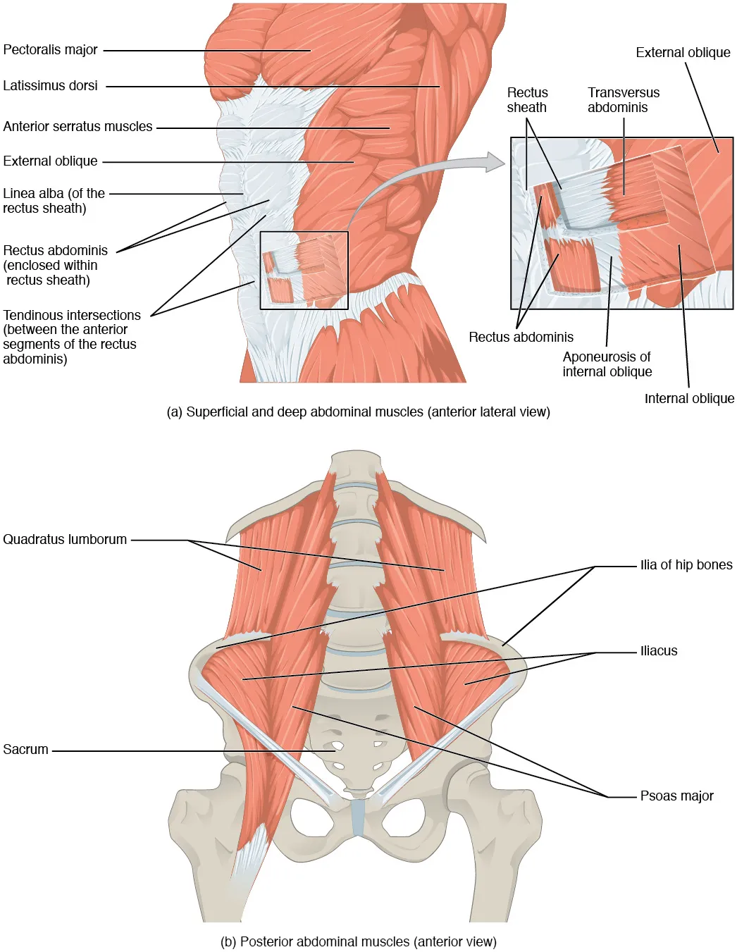
At the pubis, the attachment is shared with muscles of the inner thigh8. This was seen in 10 dissected halves of the pelvis8. The aponeuroses (flat, sheet-like tendons) of the rectus abdominis and the small pyramidalis muscle were attached to the pubic crest8. There they blended with the aponeurosis of two inner-thigh muscles, the gracilis and adductor brevis8. Together they formed one shared sheet, attached across the front and underside of the pubis8.
What wraps around it
Each muscle is wrapped in its own rectus sheath, a covering of connective tissue1. Where the left and right sheaths meet in the midline, they form the linea alba, a white band of tough fibre1. To each side, the belly wall has three flat muscle layers: the external oblique, the internal oblique and, deepest, the transversus abdominis1.
The sheath is built from the aponeuroses of those three flat muscles, and its make-up changes along the muscle’s length9. In the upper abdomen, the main part of the front layer is the external oblique’s aponeurosis, which attaches to the linea alba10. The pattern as it is usually described:
| Flat muscle’s aponeurosis | Above the navel | Some centimetres below the navel |
|---|---|---|
| External oblique | Passes in front of the muscle9 | Passes in front of the muscle9 |
| Internal oblique | Splits, with one layer in front of the muscle and one behind9 | Passes in front, without splitting9 |
| Transversus abdominis | Stays behind the muscle9 | Moves to the front, from a level that varies between people9 |
How cross bands divide it
Three bands of collagen, a strong rope-like protein, run across each muscle and split it into segments1. These bands are called tendinous intersections1. In one cadaver study, 30 of 32 bodies had three pairs of these bands, and 2 had four pairs11. Most bands did not pass through the whole thickness of the muscle11. Of 168 bands checked, 18% did11.
In four cadavers, the 8th, 9th, 10th and 12th intercostal nerves (nerves that run between the ribs) supplied the eight segments of the muscle12. Small nerve branches crossed the tendinous intersections to reach the segments next door12.
What it moves
The rectus abdominis is the prime mover for flexing the spine, which means bending it forward, as in sitting up1. A study of six men recorded the muscle with fine wires inside it13. During a hard push against a closed throat, called a Valsalva manoeuvre, the men also pushed the trunk forward or back13. Rectus abdominis activity rose sharply when the men strained to bend forward and fell when they strained to bend back13. Transversus abdominis activity stayed about the same13.
In these six men, during repeated Valsalva manoeuvres, peak pressure inside the belly tracked the activity of each abdominal muscle recorded except the rectus abdominis13. The authors concluded that the transversus abdominis appears to be the muscle whose activity most consistently follows that pressure13. In trunk rotation, a study’s background describes the rectus abdominis as working on the side opposite the turn, as the external oblique does14.
Breakdown
Strains in racket and throwing sports
Strains of the rectus abdominis are described as common injuries in competitive tennis players, and ones that hold players back3. A 2007 report gives the accepted mechanism3. First the muscle on the non-dominant side is overloaded while being stretched, during the cocking phase of the serve3. A forced contraction of that same side follows3.
Studies in tennis and handball point to one side and to the lower part of the muscle4,7:
- Tennis: ultrasound of 61 professional players found scar tissue in the muscle in 18, or 29.5%4. Each scar sat on the side of the non-dominant arm, and 16 of the 18 lay below the navel4.
- Handball: in five elite players with a torn rectus abdominis, four tears were on the side opposite the throwing arm7. Each of the five tears lay below the navel7.
Whether uneven muscle size plays a part is not settled4,15. The authors of the ultrasound study concluded that uneven growth of the muscle appears to be a risk factor for injury there4. An MRI study of eight professional male players found the two sides differed in size15. Its authors said it remains to be found out whether this imbalance raises injury risk15.
A gap between the two muscles after pregnancy
Diastasis recti is a condition in which the two rectus muscles sit an abnormal distance apart along their length9. It is acquired rather than present from birth, and it comes without a defect in the fascia, the connective tissue between them9. The studies below are of women during and after pregnancy, and their findings describe that group5,16.
A cohort study followed 300 first-time mothers from pregnancy to one year after birth5. A gap was counted when at least two fingerbreadths could be felt5. The share with a gap changed like this5:
| Time point | Share of women with diastasis recti |
|---|---|
| 21 weeks of pregnancy | 33.1%5 |
| 6 weeks after birth | 60.0%5 |
| 6 months after birth | 45.4%5 |
| 12 months after birth | 32.6%5 |
In that group, women with and without the gap did not differ significantly in the risk factors studied5. Women who reported heavy lifting at least 20 times a week had about twice the odds of the gap (odds ratio 2.18)5.
How the gap relates to pain
Findings on back pain are mixed5,16,17,18:
- In a cohort of first-time mothers, 30.3% had a gap over 30 mm at 12 months after birth17. They had worse back pain than women without it, but no significant difference in leaking urine17.
- In two other cohorts, of 300 and 84 first-time mothers, low back and pelvic pain did not differ significantly with or without the gap5,16.
- In a survey of 177 women within two years of giving birth, a self-reported gap went with belly discomfort and early urinary urgency19. It showed no significant link with low back pain or pelvic girdle pain19.
- A systematic review of 13 studies found that 5 linked the gap with low back pain and 8 did not18. It called for better studies18.
Recovery
What size studies found after training and in athletes
MRI studies measured the muscle’s volume, its whole size, after one Pilates programme and in professional players compared with inactive men6,15,20:
| Group measured | Rectus abdominis volume |
|---|---|
| 9 inactive women, after 36 weeks of Pilates | 21% more than before training6 |
| 15 professional male soccer players | 26% more than 6 inactive men20 |
| 8 professional male tennis players | 58% more than 6 inactive men15 |
In eight professional tennis players, the muscle was 35% bigger on the side of the non-dominant arm than on the dominant side15. The six inactive men showed no difference between sides15.
Coming back to sport after a tear
In the five elite handball players with torn rectus abdominis muscles, each followed the same recovery programme7. They took 16 to 22 days to return to play, 18.2 days on average7. At follow-up 15 months later, no player showed signs of a new tear or lasting discomfort7.
How the gap changes after birth
In 84 healthy first-time mothers measured by ultrasound, it was found in 100% at 35 weeks of pregnancy and in 39% at 6 months after birth16. A review notes that the gap often narrows on its own early after birth21. It puts this down to the effects of hormones21.
Connections
The muscles around it
The erector spinae, the group of long muscles beside the spine, makes up most of the muscle of the back1. It is the main muscle for straightening the spine, the opposite of what the rectus abdominis does1. In 10 healthy people bending slowly forward and back, the six trunk muscles recorded included the rectus abdominis22. Flexors and extensors worked together around a neutral spine posture, more so with a 32 kg mass added to the torso22. A model linked this to keeping the low back stable22.
What exercise studies show
Studies using EMG, a recording of the electrical activity in a muscle, show how hard the rectus abdominis works in different exercises23,24. Exercise pages that load it: plank, hollow hold, side plank, glute bridge hold, farmer’s carry, deadlift, overhead press. In the plank, position changes altered its activity25,26:
- Compared with crunches: in 18 adults, face-down exercises including the plank on toes gave rectus abdominis activity similar to the crunch and bent-knee sit-up23.
- Feet raised: in 24 young adults who did recreational gymnastics, raising the feet on wall bars gave higher activity than the usual plank25.
- Long lever and pelvis tucked: in 19 people, a longer plank with the pelvis tilted back raised upper rectus abdominis activity26.
- Unstable supports: unstable devices raised activity in the surface trunk muscles as a group in 12 adults27. Rectus abdominis activity peaked in 16 men with feet in rings and elbows on a half-ball28.
- Drawing the belly in: in 20 people, this did not significantly change rectus abdominis activity, though it raised activity in other trunk muscles29.
Studies of other exercises also recorded the muscle30,31:
| Exercise | What the study found for the rectus abdominis |
|---|---|
| Two-leg raise, held | More activity than the plank, in 18 active women24 |
| Leg lift held on the back | More activity with the pelvis tilted back than tilted forward, in 16 men32 |
| V-sit | No difference from the plank detected, in 30 adults33 |
| Side plank | More active on the lower, weight-bearing side, except in the rotational side-bridge, in 25 people34 |
| Rotational side-bridge | Highest of four side-bridge exercises, at 43.9% of its hardest possible effort, in 25 people34 |
| Glute bridge hold | Lower relative activity than the obliques, in 30 students35 |
| Single-leg glute bridge | On an unstable surface or with the hip moved out to the side, more activity than the standard bridge, in 15 adults36 |
| Farmer’s carry | More activity walking with two dumbbells than standing still with them37 |
| Suitcase carry | More activity on the side holding the weight than when standing still, but not on the other side37 |
| Deadlift | More activity with a lifting belt than without30 |
| Dumbbell shoulder press | About 81% less activity seated than standing, in 15 men31 |
| Row | No significant difference between one-arm and two-arm rows, in 15 trained men38 |
Rectus abdominis sheathed in part by
Rectus abdominis opposes at the spine
Rectus abdominis is loaded by
Sources
- Betts JG, Young KA, Wise JA, Johnson E, Poe B, Kruse DH, Korol O, Johnson JE, Womble M, DeSaix P (2022). Anatomy and Physiology 2e. OpenStax. Link
- Rai R, Azih LC, Iwanaga J, Loukas M, Mortazavi M, Oskouian RJ, Tubbs RS (2018). Tendinous Inscriptions of the Rectus Abdominis: A Comprehensive Review.. Cureus. doi:10.7759/cureus.3100 PMID 30338176
- Maquirriain J, Ghisi JP, Kokalj AM (2007). Rectus abdominis muscle strains in tennis players.. British Journal of Sports Medicine. doi:10.1136/bjsm.2007.036129 PMID 17957025
- Balius R, Pedret C, Galilea P, Idoate F, Ruiz-Cotorro A (2012). Ultrasound assessment of asymmetric hypertrophy of the rectus abdominis muscle and prevalence of associated injury in professional tennis players.. Skeletal Radiology. doi:10.1007/s00256-012-1429-y PMID 22618761
- Sperstad JB, Tennfjord MK, Hilde G, Ellström-Engh M, Bø K (2016). Diastasis recti abdominis during pregnancy and 12 months after childbirth: prevalence, risk factors and report of lumbopelvic pain.. British Journal of Sports Medicine. doi:10.1136/bjsports-2016-096065 PMID 27324871
- Dorado C, Calbet JA, Lopez-Gordillo A, Alayon S, Sanchis-Moysi J (2012). Marked effects of Pilates on the abdominal muscles. Medicine and Science in Sports and Exercise. doi:10.1249/MSS.0b013e31824fb6ae PMID 22357302
- Balius R, Pedret C, Pacheco L, Gutierrez JA, Vives J, Escoda J (2011). Rectus abdominis muscle injuries in elite handball players: management and rehabilitation.. Open Access Journal of Sports Medicine. doi:10.2147/OAJSM.S17504 PMID 24198573
- Tharnmanularp S, Muro S, Nimura A, Ibara T, Akita K (2024). Significant relationship between musculoaponeurotic attachment of the abdominal and thigh adductor muscles to the pubis: implications for the diagnosis of groin pain.. Anatomical Science International. doi:10.1007/s12565-023-00750-6 PMID 37985575
- Cavalli M, Aiolfi A, Bruni PG, Manfredini L, Lombardo F, Bonfanti MT, Bona D, Campanelli G (2021). Prevalence and risk factors for diastasis recti abdominis: a review and proposal of a new anatomical variation.. Hernia. doi:10.1007/s10029-021-02468-8 PMID 34363190
- Medina Pedrique M, Robin Valle de Lersundi Á, Avilés Oliveros A, Ruiz SM, López-Monclús J, Munoz-Rodriguez J, Blázquez Hernando LA, Martinez Caballero J, García-Urena MÁ (2023). Incisions in Hepatobiliopancreatic Surgery: Surgical Anatomy and its Influence to Open and Close the Abdomen.. Journal of Abdominal Wall Surgery. doi:10.3389/jaws.2023.11123 PMID 38312419
- Broyles JM, Schuenke MD, Patel SR, Vail CM, Broyles HV, Dellon AL (2018). Defining the Anatomy of the Tendinous Intersections of the Rectus Abdominis Muscle and Their Clinical Implications in Functional Muscle Neurotization.. Annals of Plastic Surgery. doi:10.1097/SAP.0000000000001193 PMID 28671887
- Grinsell D, Azizeddin A, Overland J (2020). Intramuscular pathway and fascicular characteristics of the segmental intercostal innervation to rectus abdominis.. ANZ Journal of Surgery. doi:10.1111/ans.15682 PMID 31957163
- Cresswell AG, Grundström H, Thorstensson A (1992). Observations on intra-abdominal pressure and patterns of abdominal intra-muscular activity in man.. Acta Physiologica Scandinavica. doi:10.1111/j.1748-1716.1992.tb09314.x PMID 1534959
- Sugaya T, Sakamoto M, Nakazawa R, Wada N (2016). Relationship between spinal range of motion and trunk muscle activity during trunk rotation.. Journal of Physical Therapy Science. doi:10.1589/jpts.28.589 PMID 27065549
- Sanchis-Moysi J, Idoate F, Dorado C, Alayón S, Calbet JA (2010). Large asymmetric hypertrophy of rectus abdominis muscle in professional tennis players.. PLoS ONE. doi:10.1371/journal.pone.0015858 PMID 21209832
- Fernandes da Mota PG, Pascoal AG, Carita AI, Bø K (2015). Prevalence and risk factors of diastasis recti abdominis from late pregnancy to 6 months postpartum, and relationship with lumbo-pelvic pain.. Manual Therapy. doi:10.1016/j.math.2014.09.002 PMID 25282439
- Fitzpatrick SE, Crittenden T, Watson DI, Dean NR (2026). Prevalence of Symptomatic Established Rectus Diastasis of Parity in Primiparous Women: A Prospective Cohort Study From Early Pregnancy to 1-Year Postpartum.. World Journal of Surgery. doi:10.1002/wjs.70227 PMID 41506782
- Sokunbi G, Camino-Willhuber G, Paschal PK, Olufade O, Hussain FS, Shue J, Abjornson C, Zelenty WD, Lebl DR, Cammisa FP, Girardi FP, Hughes AP, Sama AA (2023). Is Diastasis Recti Abdominis Associated With Low Back Pain? A Systematic Review.. World Neurosurgery. doi:10.1016/j.wneu.2023.03.014 PMID 36894002
- Simpson E, Hannington M, Bø K, Hahne A (2026). Association Between Diastasis of the Rectus Abdominis Muscles and Musculoskeletal Conditions in the First 2 Years Postpartum: A Cross-Sectional Study.. Musculoskeletal Care. doi:10.1002/msc.70209 PMID 41872055
- Idoate F, Calbet JA, Izquierdo M, Sanchis-Moysi J (2011). Soccer attenuates the asymmetry of rectus abdominis muscle observed in non-athletes.. PLoS ONE. doi:10.1371/journal.pone.0019022 PMID 21541351
- Du Y, Huang M, Wang S, Yang L, Lin Y, Yu W, Pan Z, Ye Z (2025). Diastasis recti abdominis: A comprehensive review.. Hernia. doi:10.1007/s10029-025-03417-5 PMID 40622579
- Cholewicki J, Panjabi MM, Khachatryan A (1997). Stabilizing function of trunk flexor-extensor muscles around a neutral spine posture.. Spine. doi:10.1097/00007632-199710010-00003 PMID 9346140
- Escamilla RF, Lewis C, Pecson A, Imamura R, Andrews JR (2016). Muscle Activation Among Supine, Prone, and Side Position Exercises With and Without a Swiss Ball.. Sports Health. doi:10.1177/1941738116653931 PMID 27302152
- Park DJ, Park SY (2019). Which trunk exercise most effectively activates abdominal muscles? A comparative study of plank and isometric bilateral leg raise exercises.. Journal of Back and Musculoskeletal Rehabilitation. doi:10.3233/BMR-181122 PMID 30856100
- Moreno-Navarro P, Sabido R, Barbado D, Prat-Luri A, Vera-Garcia FJ (2024). Trunk muscle activation in prone plank exercises with different body tilts.. Journal of Back and Musculoskeletal Rehabilitation. doi:10.3233/BMR-230209 PMID 38217576
- Schoenfeld BJ, Contreras B, Tiryaki-Sonmez G, Willardson JM, Fontana F (2014). An electromyographic comparison of a modified version of the plank with a long lever and posterior tilt versus the traditional plank exercise.. Sports Biomechanics. doi:10.1080/14763141.2014.942355 PMID 25325773
- Snarr RL, Esco MR (2014). Electromyographical comparison of plank variations performed with and without instability devices.. Journal of Strength and Conditioning Research. doi:10.1519/JSC.0000000000000521 PMID 25264667
- Sağlam AF, Aydın EM, Demirkan E, Gündoğan B, Favre M (2025). Effects of Surface Stability on Muscle Activation during Plank Exercise.. Journal of Human Kinetics. doi:10.5114/jhk/203417 PMID 42211810
- García-Jaén M, Cortell-Tormo JM, Hernández-Sánchez S, Tortosa-Martínez J (2020). Influence of Abdominal Hollowing Maneuver on the Core Musculature Activation during the Prone Plank Exercise.. International Journal of Environmental Research and Public Health. doi:10.3390/ijerph17207410 PMID 33053717
- Escamilla RF, Francisco AC, Kayes AV, Speer KP, Moorman CT 3rd (2002). An electromyographic analysis of sumo and conventional style deadlifts.. Medicine and Science in Sports and Exercise. doi:10.1097/00005768-200204000-00019 PMID 11932579
- Saeterbakken AH, Fimland MS (2012). Muscle activity of the core during bilateral, unilateral, seated and standing resistance exercise.. European Journal of Applied Physiology. doi:10.1007/s00421-011-2141-7 PMID 21877146
- Workman JC, Docherty D, Parfrey KC, Behm DG (2008). Influence of pelvis position on the activation of abdominal and hip flexor muscles.. Journal of Strength and Conditioning Research. doi:10.1519/JSC.0b013e3181739981 PMID 18714231
- Musalem LL, Stankovic T, Glisic D, Cook GE, Beach TA (2015). Biomechanical and Electromyographic Comparisons of Isometric Trunk Flexor Endurance Test Postures: Prone Plank Versus V-Sit.. Journal of Applied Biomechanics. doi:10.1123/jab.2014-0197 PMID 26252077
- Youdas JW, Boor MM, Darfler AL, Koenig MK, Mills KM, Hollman JH (2014). Surface electromyographic analysis of core trunk and hip muscles during selected rehabilitation exercises in the side-bridge to neutral spine position.. Sports Health. doi:10.1177/1941738114539266 PMID 25177418
- Stevens VK, Bouche KG, Mahieu NN, Coorevits PL, Vanderstraeten GG, Danneels LA (2006). Trunk muscle activity in healthy subjects during bridging stabilization exercises.. BMC Musculoskeletal Disorders. doi:10.1186/1471-2474-7-75 PMID 16987410
- Yoon JO, Kang MH, Kim JS, Oh JS (2018). Effect of modified bridge exercise on trunk muscle activity in healthy adults: a cross sectional study.. Brazilian Journal of Physical Therapy. doi:10.1016/j.bjpt.2017.09.005 PMID 28943402
- Ellestad SH, Holcomb TP, Swiergol AM, Holmstrup ME, Dicus JR (2024). The Quantification of Muscle Activation During the Loaded Carry Movement Pattern.. International Journal of Exercise Science. doi:10.70252/NWUE9985 PMID 38665162
- Saeterbakken A, Andersen V, Brudeseth A, Lund H, Fimland MS (2015). The Effect of Performing Bi- and Unilateral Row Exercises on Core Muscle Activation.. International Journal of Sports Medicine. doi:10.1055/s-0034-1398646 PMID 26134664
Changes
- Revised. Corrected the upper attachment to the fifth to seventh rib cartilages and the xiphoid process; the earlier textbook table was misaligned.
- Added a figure showing where it sits, links to the exercise pages that load it, and tighter wording in a few places.
- First published.