Erector spinae
The long muscle group that runs up the back and straightens the spine. How it is built, how it tires and shrinks, how it builds back, and what it works with.
- Region
- Spine
- Starts on (lumbar part)
- the accessory processes of the lower back (lumbar) vertebrae, and side spurs (transverse processes) of the top four of them6
- Starts on (thoracic part)
- the thorax, in a series of small segments6
- Ends on (lumbar part)
- the ilium, the wide upper part of the hip bone (Pelvis)1,6
- Ends on (thoracic part)
- the back of the iliac crest, the top rim of the hip bone, through long tendons forming a flat sheet (Pelvis)1,6,22
- Muscles
- iliocostalis, longissimus, spinalis
- Updated
- September 29, 2026
- Reviewed
- September 29, 2026 · how entries are checked
Function
Three columns along the spine
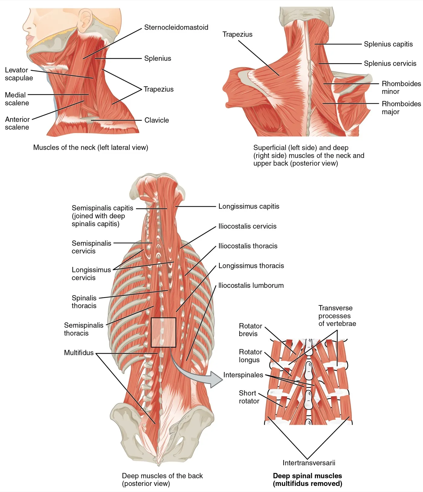
The erector spinae is a muscle group that makes up most of the back’s muscle mass1. It is the main extensor of the vertebral column, the stack of bones that forms the spine1. An extensor straightens a joint, or here, the spine. The group sits in three columns side by side1. Each column is split again into parts named for the body region they belong to1:
| Group | Where it lies | Parts by region |
|---|---|---|
| Iliocostalis | Outermost | Neck, chest and lower back1 |
| Longissimus | Middle | Head, neck and chest1 |
| Spinalis | Nearest the midline | Head, neck and chest1 |
The figure below labels the three columns of the erector spinae1.
How it attaches in the lower back
In the lumbar region, the lower back, the erector spinae is made of two muscles6. They are iliocostalis lumborum and longissimus thoracis, and each has its own thoracic (chest) part and lumbar part6. The thoracic parts have tiny muscle bellies that start level by level on the chest6. Their long tendons run downward and together form the erector spinae aponeurosis, a broad flat sheet of tendon6.
The lumbar parts start on small bony points of the lumbar vertebrae6. These include the transverse processes, the side spurs, of the first four lumbar vertebrae6. The fibres end on the ilium, the upper bone of the pelvis, apart from the aponeurosis6.
At the first four lumbar levels, each dorsal ramus, a spinal nerve branch to the back, tends to split in three7. The outer branch goes to iliocostalis, the middle one to longissimus, and the inner one to multifidus, a deep muscle along the spine7.
What it does in the trunk
Besides straightening the spine, the erector spinae controls its flexion (bending forward), lateral flexion (bending to the side) and rotation (twisting)1. It also holds the lumbar curve, the curve of the lower back1.
In 10 healthy people, the back extensors and the trunk flexors in front worked together around an upright spine8. This shared effort rose when a 32 kg mass was added to the torso8.
At full forward bend, the activity of the muscles beside the spine can drop, which is called flexion relaxation9. In four experienced powerlifters doing deadlifts, the lower spine stayed 1.5 to 13 degrees short of full bend, bar one trial10. The authors concluded that the ligaments took little of the forward-bending load, leaving it to the muscles10.
Breakdown
Soreness after lowering work
One study tested nine muscle groups in 15 inactive young men11. Each group did 5 sets of 10 eccentric contractions, with a load of 80% of maximum strength11. In an eccentric contraction, the muscle works while it lengthens, as when lowering a weight. Strength in the nine groups was down 1 to 4 days later11.
Soreness that came on later was greater in the erector spinae than in six of the other groups11. Yet the erector spinae lost less strength than the knee flexors, the muscles that bend the knee11.
Shrinking with disuse
In the studies below, the back muscles mostly shrank when load was taken away2,12,13. In head-down-tilt bed rest, a research set-up, people stay in bed with the head tilted slightly below the feet. Paraspinal means beside the spine; the paraspinal muscles include the erector spinae13.
| Setting | People | What happened to the back muscles |
|---|---|---|
| 60 days of bed rest | 24 healthy adults | Lumbar erector spinae volume fell 11.2% on average, but grew at the lowest level2 |
| 60 days of bed rest | Non-exercising group of a 16-woman study | Lumbar paraspinal area fell 10.9%; the erector spinae made up 76% of the paraspinal loss12 |
| About six months in space | 17 astronauts | Total lumbar paraspinal area fell about 1.0% a month13 |
A rare rise in pressure
Acute lumbar paraspinal compartment syndrome is a rare condition14. Pressure rises inside a closed space bounded by tough tissue and bone14. A 2018 review of published reports counted 23 cases14. Most followed weight-lifting; others followed other exercise, a direct injury, or surgery elsewhere14.
Findings in people with low back pain
- Size and make-up. A meta-analysis pooling 20 imaging studies identified no differences in lumbar erector spinae size or make-up linked to low back pain4. The multifidus, by contrast, was somewhat smaller and held more fat4. A systematic review of 25 studies found the evidence on erector spinae size conflicting5.
- Flexion relaxation. A meta-analysis estimated that in 55% of people with long-term nonspecific low back pain, back-muscle activity kept going at full bend9. The pooled studies differed a lot from one another9.
- Endurance. A systematic review of 26 prospective cohort studies found strong evidence of no link between trunk muscle endurance and later low back pain15. Its authors warned that the studies differed, so the results need caution15. In another systematic review of 12 prospective cohort articles (5,459 participants), the back-muscle endurance meta-analysis for developing low back pain was non-significant16. A single Danish study of 928 adults differed: its author wrote that good back-muscle endurance “may prevent first-time occurrence” of low back trouble in men17.
Recovery
How strength builds with training
In a two-year randomised controlled trial, healthy postmenopausal women did progressive back-strengthening exercise3. Back extensor strength was measured in kilograms3:
| When measured | Exercise group (27 women) | Control group (23 women) |
|---|---|---|
| Start | 39.4 kg3 | 36.9 kg3 |
| After two years | 66.8 kg3 | 49.0 kg3 |
| Eight years after it stopped | 32.9 kg3 | 26.9 kg3 |
In this trial, strength fell a long way once the set exercise stopped3. Even so, the exercise group was still significantly stronger than the controls ten years after the start3.
Training studies in people with low back pain
Other studies measured the back muscles before and after exercise in people with low back pain18,19.
- A meta-analysis of 8 randomised trials with 508 people found that exercise gave better lumbar extensor endurance than passive or active control groups18. It also gave more strength than passive controls and more electrical activity than active controls18.
- A trial randomly split 50 people with long-term low back pain into two 12-week exercise groups20. Movement-control work plus lower-back extension exercise grew erector spinae and multifidus area more than general exercise20. The authors call these results preliminary20.
- In a 16-week free-weight programme with no comparison group, fat within the erector spinae and multifidus fell at two lumbar levels19. Working muscle area rose at those levels, while fat did not change significantly at the lowest level19.
Coming back after disuse
In one bed-rest study, exercise limited the losses12. Women who did treadmill and flywheel resistance exercise lost 4.3% of paraspinal area, against 10.9% in those who did no exercise12. Their spine-straightening strength fell 4.3%, against 16.6%12.
After 60 days of bed rest, 13 days of reconditioning did not bring back erector spinae volume at the top of the lower back2. The authors judged a two-week programme too short to restore the lower back and pelvic muscles fully2. Astronauts back on Earth regained paraspinal area at about 0.72% a month over a year13.
Less damage the second time
A systematic review and meta-analysis of 20 studies in healthy adults compared two bouts of multi-joint muscle-damaging exercise21. A multi-joint exercise moves more than one joint at once. Soreness and creatine kinase were significantly higher 24 and 48 hours after the first bout than after the second21. Creatine kinase is a protein found in muscle; blood tests measure its level as a marker of muscle damage. The authors suggest that a first bout of common eccentric exercises may protect against damage in later bouts21.
In one study of 15 inactive men aged 20 to 25, damage measures changed less when the same eccentric exercise was repeated two weeks later11. This protective effect was similar across the nine muscle groups, erector spinae included11.
Connections
The sheath and sheet it shares
The thoracolumbar fascia is a set of tough tissue layers in the back22. It keeps the paraspinal muscles apart from the muscles at the rear of the abdominal wall22. The deeper sheet of its back layer wraps the paraspinal muscles in a sheath22. At the outer edge of that sheath, a seam forms where it meets the sheet of the transversus abdominis, a deep belly muscle22. A review describes this seam as placed to pass tension from nearby trunk and limb muscles into the fascia22.
Low in the lumbar region, the multifidus and both erector spinae muscles attach firmly to the inside of the aponeurosis, making it a shared sheet22. From about the fifth lumbar vertebra down, the aponeurosis merges with the layers that lie over it into one thick sheet22. This sheet attaches to the top edge of the pelvis and spreads to join the gluteus maximus, the large buttock muscle22. In 11 donor limbs, gluteus maximus fibres arose in part from the erector spinae aponeurosis23.
In 10 donor bodies, a simulated muscle pull moved the back layer of the fascia24. The authors inferred that in life, contraction of the gluteus maximus, erector muscle and latissimus dorsi would tense the outer sheet of that layer24. The latissimus dorsi is the broad muscle of the lower back.
Its neighbour, the multifidus
In the lower back, the multifidus lies closer to the midline than the erector spinae22. It belongs to a different group, the transversospinalis22. In eight donor spines, it weighed about the same as published figures for longissimus thoracis25. Yet its physiological cross-sectional area, a measure of size taken straight across all of its fibres, was more than twice that of any other lumbar muscle25.
In 40 active adults holding a back bridge, the glute bridge hold, the ratio of multifidus to erector spinae activity was 2.7626.
Working with the hips
One study compared 19 women with long-term low back pain and 19 without27. Early in a forward bend, the lumbar back muscles and the biceps femoris, a hamstring muscle, switched on together27. The gluteus maximus switched on after them27. The back muscles and biceps femoris followed a similar order in both groups27.
What exercise studies show
Several of these studies measured muscle activity with electromyography, which picks up a muscle’s electrical signals28,29,30. Some figures are a share of maximum voluntary contraction, the hardest effort a person can make29,31.
| Exercise | What the studies found |
|---|---|
| Deadlift | A review of 19 studies found the erector spinae and quadriceps, the front thigh muscles, more active than the gluteus maximus and biceps femoris28 |
| Romanian deadlift | In the same review, the Romanian deadlift was linked to lower erector spinae activity than hamstring activity, the reverse of other deadlift styles28 |
| Barbell row | In 15 resistance-trained men, the free-weight row drew more erector spinae activity than a machine row30. The two-arm free-weight row drew 12% more than the one-arm free-weight row30 |
| Back squat | The low-bar style can show more erector spinae activity than the high-bar style32 |
| Farmer’s carry | In one study, lower erector spinae activity averaged 106% and 144% of maximum on the left and right31 |
| Plank | Prone (face-down) exercises, including the plank on toes, drew lumbar paraspinal activity similar to a crunch33. It was higher on the toes than on the knees33 |
| Side plank | Longissimus thoracis activity stayed below half of maximum29 |
| Overhead press | Erector spinae activity was similar seated and standing34 |
Erector spinae gives origin to part of
Erector spinae tenses the back fascia with
Erector spinae switches on with in a forward bend
Erector spinae works with in the deadlift
Erector spinae is loaded by
Sources
- Betts JG, Young KA, Wise JA, Johnson E, Poe B, Kruse DH, Korol O, Johnson JE, Womble M, DeSaix P (2022). Anatomy and Physiology 2e. OpenStax. Link
- De Martino E, Hides J, Elliott JM, Hoggarth MA, Zange J, Lindsay K, Debuse D, Winnard A, Beard D, Cook JA, Salomoni SE, Weber T, Scott J, Hodges PW, Caplan N (2022). The Effects of Reconditioning Exercises Following Prolonged Bed Rest on Lumbopelvic Muscle Volume and Accumulation of Paraspinal Muscle Fat.. Frontiers in Physiology. doi:10.3389/fphys.2022.862793 PMID 35774286
- Sinaki M, Itoi E, Wahner HW, Wollan P, Gelzcer R, Mullan BP, Collins DA, Hodgson SF (2002). Stronger back muscles reduce the incidence of vertebral fractures: a prospective 10 year follow-up of postmenopausal women.. Bone. doi:10.1016/s8756-3282(02)00739-1 PMID 12052450
- Seyedhoseinpoor T, Taghipour M, Dadgoo M, Sanjari MA, Takamjani IE, Kazemnejad A, Khoshamooz Y, Hides J (2022). Alteration of lumbar muscle morphology and composition in relation to low back pain: a systematic review and meta-analysis.. The Spine Journal. doi:10.1016/j.spinee.2021.10.018 PMID 34718177
- Ranger TA, Cicuttini FM, Jensen TS, Peiris WL, Hussain SM, Fairley J, Urquhart DM (2017). Are the size and composition of the paraspinal muscles associated with low back pain? A systematic review.. The Spine Journal. doi:10.1016/j.spinee.2017.07.002 PMID 28756299
- Macintosh JE, Bogduk N (1987). 1987 Volvo award in basic science. The morphology of the lumbar erector spinae.. Spine. doi:10.1097/00007632-198709000-00004 PMID 3686217
- Bogduk N, Wilson AS, Tynan W (1982). The human lumbar dorsal rami.. Journal of Anatomy. PMID 7076562
- Cholewicki J, Panjabi MM, Khachatryan A (1997). Stabilizing function of trunk flexor-extensor muscles around a neutral spine posture.. Spine. doi:10.1097/00007632-199710010-00003 PMID 9346140
- Gouteron A, Tabard-Fougère A, Bourredjem A, Casillas JM, Armand S, Genevay S (2022). The flexion relaxation phenomenon in nonspecific chronic low back pain: prevalence, reproducibility and flexion-extension ratios. A systematic review and meta-analysis.. European Spine Journal. doi:10.1007/s00586-021-06992-0 PMID 34553264
- Cholewicki J, McGill SM (1992). Lumbar posterior ligament involvement during extremely heavy lifts estimated from fluoroscopic measurements.. Journal of Biomechanics. doi:10.1016/0021-9290(92)90242-s PMID 1733981
- Chen TC, Yang TJ, Huang MJ, Wang HS, Tseng KW, Chen HL, Nosaka K (2019). Damage and the repeated bout effect of arm, leg, and trunk muscles induced by eccentric resistance exercises.. Scandinavian Journal of Medicine & Science in Sports. doi:10.1111/sms.13388 PMID 30663816
- Holt JA, Macias BR, Schneider SM, Watenpaugh DE, Lee SM, Chang DG, Hargens AR (2016). WISE 2005: Aerobic and resistive countermeasures prevent paraspinal muscle deconditioning during 60-day bed rest in women.. Journal of Applied Physiology. doi:10.1152/japplphysiol.00532.2015 PMID 26893030
- Coulombe JC, Johannesdottir F, Burkhart KA, Brummer H, Allaire BT, Bouxsein ML (2023). Changes in Vertebral Bone Density and Paraspinal Muscle Morphology Following Spaceflight and 1 Year Readaptation on Earth.. JBMR Plus. doi:10.1002/jbm4.10810 PMID 38130751
- Alexander W, Low N, Pratt G (2018). Acute lumbar paraspinal compartment syndrome: a systematic review.. ANZ Journal of Surgery. doi:10.1111/ans.14342 PMID 29316189
- Hamberg-van Reenen HH, Ariëns GA, Blatter BM, van Mechelen W, Bongers PM (2007). A systematic review of the relation between physical capacity and future low back and neck/shoulder pain.. Pain. doi:10.1016/j.pain.2006.11.004 PMID 17222512
- Sadler SG, Spink MJ, Ho A, De Jonge XJ, Chuter VH (2017). Restriction in lateral bending range of motion, lumbar lordosis, and hamstring flexibility predicts the development of low back pain: a systematic review of prospective cohort studies.. BMC Musculoskeletal Disorders. doi:10.1186/s12891-017-1534-0 PMID 28476110
- Biering-Sørensen F (1984). Physical measurements as risk indicators for low-back trouble over a one-year period.. Spine. doi:10.1097/00007632-198403000-00002 PMID 6233709
- Clael S, Campos LF, Correia KL, de Lucena JMS, Gentil P, Durigan JL, Ribeiro ALA, Martins WR (2021). Exercise interventions can improve muscle strength, endurance, and electrical activity of lumbar extensors in individuals with non-specific low back pain: a systematic review with meta-analysis.. Scientific Reports. doi:10.1038/s41598-021-96403-7 PMID 34413400
- Welch N, Moran K, Antony J, Richter C, Marshall B, Coyle J, Falvey E, Franklyn-Miller A (2015). The effects of a free-weight-based resistance training intervention on pain, squat biomechanics and MRI-defined lumbar fat infiltration and functional cross-sectional area in those with chronic low back.. BMJ Open Sport & Exercise Medicine. doi:10.1136/bmjsem-2015-000050 PMID 27900136
- Fortin M, Rye M, Roussac A, Montpetit C, Burdick J, Naghdi N, Rosenstein B, Bertrand C, Macedo LG, Elliott JM, Dover G, DeMont R, Weber MH, Pepin V (2023). The Effects of Combined Motor Control and Isolated Extensor Strengthening versus General Exercise on Paraspinal Muscle Morphology, Composition, and Function in Patients with Chronic Low Back Pain: A Randomized Controlled Trial.. Journal of Clinical Medicine. doi:10.3390/jcm12185920 PMID 37762861
- Doma K, Matoso B, Protzen G, Singh U, Boullosa D (2023). The Repeated Bout Effect of Multiarticular Exercises on Muscle Damage Markers and Physical Performances: A Systematic Review and Meta-Analyses.. Journal of Strength and Conditioning Research. doi:10.1519/JSC.0000000000004628 PMID 38015738
- Willard FH, Vleeming A, Schuenke MD, Danneels L, Schleip R (2012). The thoracolumbar fascia: anatomy, function and clinical considerations.. Journal of Anatomy. doi:10.1111/j.1469-7580.2012.01511.x PMID 22630613
- Barker PJ, Hapuarachchi KS, Ross JA, Sambaiew E, Ranger TA, Briggs CA (2014). Anatomy and biomechanics of gluteus maximus and the thoracolumbar fascia at the sacroiliac joint.. Clinical Anatomy. doi:10.1002/ca.22233 PMID 23959791
- Vleeming A, Pool-Goudzwaard AL, Stoeckart R, van Wingerden JP, Snijders CJ (1995). The posterior layer of the thoracolumbar fascia. Its function in load transfer from spine to legs.. Spine. PMID 7701385
- Ward SR, Kim CW, Eng CM, Gottschalk LJ 4th, Tomiya A, Garfin SR, Lieber RL (2009). Architectural analysis and intraoperative measurements demonstrate the unique design of the multifidus muscle for lumbar spine stability.. The Journal of Bone and Joint Surgery. American Volume. doi:10.2106/JBJS.G.01311 PMID 19122093
- Khosrokiani Z, Letafatkar A, Sheikhi B, Thomas AC, Aghaie-Ataabadi P, Hedayati MT (2022). Hip and Core Muscle Activation During High-Load Core Stabilization Exercises.. Sports Health. doi:10.1177/19417381211015225 PMID 34060953
- Leinonen V, Kankaanpää M, Airaksinen O, Hänninen O (2000). Back and hip extensor activities during trunk flexion/extension: effects of low back pain and rehabilitation.. Archives of Physical Medicine and Rehabilitation. doi:10.1016/s0003-9993(00)90218-1 PMID 10638873
- Martín-Fuentes I, Oliva-Lozano JM, Muyor JM (2020). Electromyographic activity in deadlift exercise and its variants. A systematic review.. PLoS One. doi:10.1371/journal.pone.0229507 PMID 32107499
- Ekstrom RA, Osborn RW, Hauer PL (2008). Surface electromyographic analysis of the low back muscles during rehabilitation exercises.. Journal of Orthopaedic and Sports Physical Therapy. doi:10.2519/jospt.2008.2865 PMID 19195137
- Saeterbakken A, Andersen V, Brudeseth A, Lund H, Fimland MS (2015). The Effect of Performing Bi- and Unilateral Row Exercises on Core Muscle Activation.. International Journal of Sports Medicine. doi:10.1055/s-0034-1398646 PMID 26134664
- Hindle BR, Lorimer A, Winwood P, Keogh JWL (2019). The Biomechanics and Applications of Strongman Exercises: a Systematic Review.. Sports Medicine - Open. doi:10.1186/s40798-019-0222-z PMID 31820223
- Glassbrook DJ, Helms ER, Brown SR, Storey AG (2017). A Review of the Biomechanical Differences Between the High-Bar and Low-Bar Back-Squat.. Journal of Strength and Conditioning Research. doi:10.1519/JSC.0000000000002007 PMID 28570490
- Escamilla RF, Lewis C, Pecson A, Imamura R, Andrews JR (2016). Muscle Activation Among Supine, Prone, and Side Position Exercises With and Without a Swiss Ball.. Sports Health. doi:10.1177/1941738116653931 PMID 27302152
- Saeterbakken AH, Fimland MS (2012). Muscle activity of the core during bilateral, unilateral, seated and standing resistance exercise.. European Journal of Applied Physiology. doi:10.1007/s00421-011-2141-7 PMID 21877146
Changes
- Tidy pass: minor review findings fixed, glosses aligned with the reviewed glossary.
- Added a figure showing where it sits, links to the exercise pages that load it, and tighter wording in a few places.
- First published.