Lumbar vertebrae
Also called lumbar spine bones, L1 to L5, lower back vertebrae
The five bones of the lower back. How each is built, how the body, facet joints and pars share load, how and in whom the pars cracks, and how lumbar bone responds to loading.
- Region
- Spine
- Muscles
- hip flexors, quadratus lumborum, erector spinae, multifidus
- Updated
- October 9, 2026
- Reviewed
- October 9, 2026 · how entries are checked
Function
Five bones built to carry weight
The lumbar region of the spine has five vertebrae (the bones stacked to form the spine), named L1 to L51. They carry more body weight than any other vertebrae, and have large, thick bodies as a result2. Together they form a lordotic curve, the inward curve of the lower back1. That curve develops as a child begins to stand and then walk2.
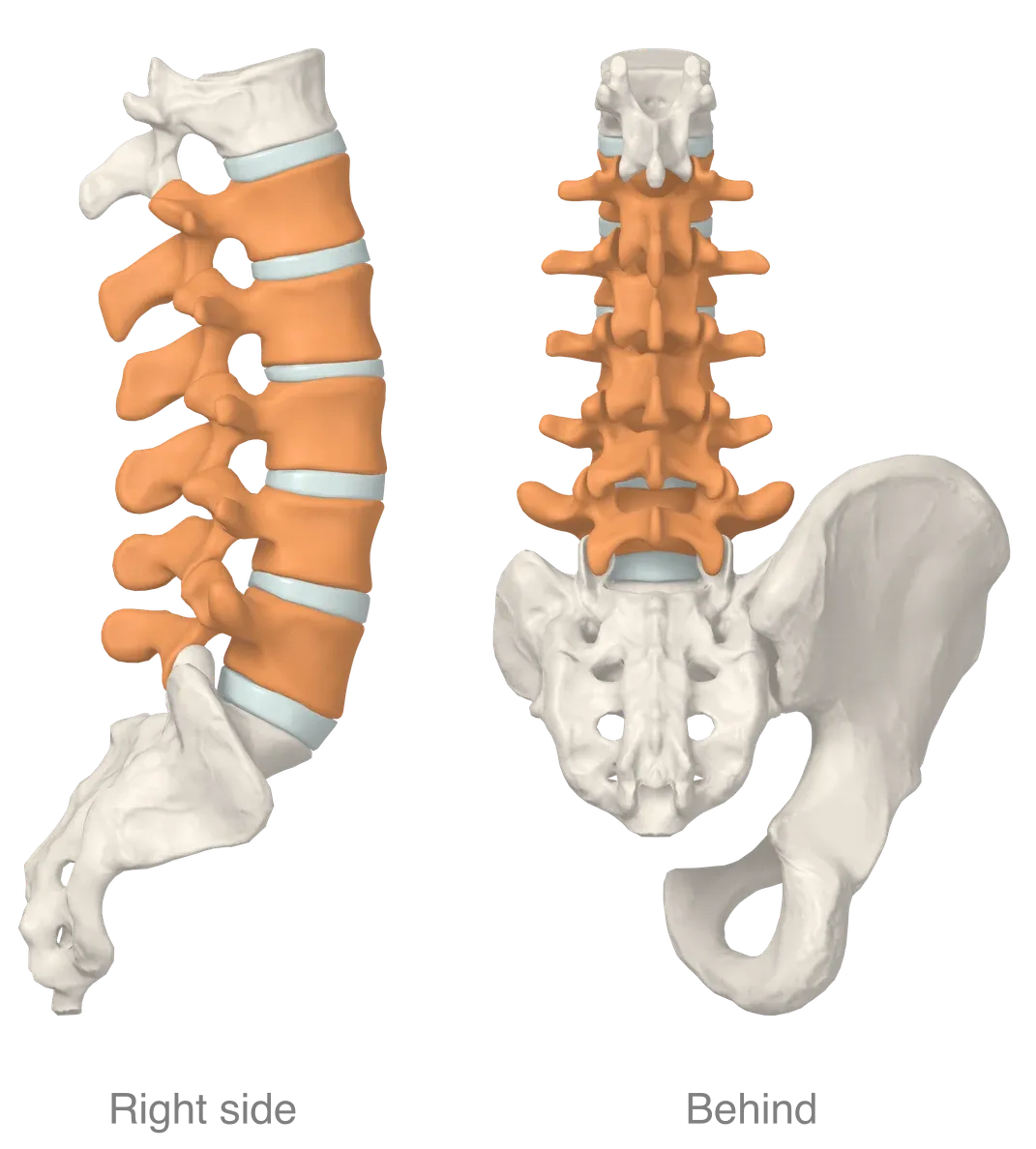
The parts of one vertebra
A typical vertebra has a body, an arch and seven processes (parts of a bone that stick out)2.
- The body is the front part, and it supports body weight2. It is spongy bone holding red marrow, inside a thin outer layer of compact bone1. It bears most of the load passed down the length of the spine1.
- The arch forms the back of the vertebra2. It has a pedicle on each side, anchored to the back of the body, and a lamina on each side forming the roof2. In lumbar vertebrae the pedicles are very strong, and the laminae are broad, short and strong7.
- The transverse processes and the spinous process are short in lumbar vertebrae, and the blunt spinous process points backward2.
The openings between arch and body line up to form the vertebral canal, the bony passage that protects the spinal cord2.
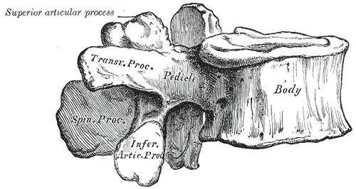
The facet joints and the pars
Each vertebra has an upward-facing and a downward-facing articular process on each side2. The upper pair of one vertebra joins the lower pair of the vertebra above, forming slightly movable joints2. In a lumbar vertebra these processes are large, and they rise and hang from the points where pedicles and laminae meet2,7. The joints they form are the facet joints1. Facet joints keep the vertebrae aligned, limit how far they move and bear weight in certain positions1.
The pars interarticularis is a small segment of bone between the upper and lower articular processes3. The lamina and the pedicle border it too8. This page calls it the pars.
L5 has the largest body and transverse processes of all the vertebrae1. Its body is taller in front than behind1. That shape helps form the angle where the lumbar spine meets the sacrum (the triangle-shaped bone at the base of the spine)1. The lower articular processes of L5 join the upper articular processes of the sacrum2.
How the parts share load
Laboratory tests, some on bones from cadavers (bodies donated for study), and one account of spinal mechanics describe how the parts share load9,10:
- Compression. In cadaver lumbar joints, the facet joints resisted about 16 per cent of the compressive force in a posture simulating upright standing9. They resisted none in a posture simulating upright sitting9. Both figures were measured once sustained loading had reduced disc height9.
- Forward shear. Shear is a force that pushes two surfaces to slide across each other. A 1976 account of spinal mechanics names three things that normally resist forward shear on a lumbar vertebra10. They are the outer ring of the disc, compression of the facet joints, and tension in the cortical bone (dense outer bone) of the pars10.
- Strength of the pars. In tests of single vertebrae, a backward force was applied to the lower articular processes10. Two reports tested the pars of L4 and L5 vertebrae this way10. The mean force at failure was 1800 newtons (a unit of force) in one report and 3200 newtons in the other10.
Two studies used a finite element model to look at stress inside the pars4,11. This is a computer model that splits a structure into many small pieces to work out the forces within it. In the model of an L4-L5 segment, pars stress was lowest under compression alone and high under extension (bending backward) and rotation4. In the model of the L3 to sacrum segment, the highest stresses in the L5 pars were at its lower front, in every loading mode11.
Breakdown
A fatigue crack in the pars
A crack in the pars is called a pars defect or spondylolysis12,13. A structured review found good evidence that, in children, it is an acquired fracture, on one side or on both14. A study of cricket fast bowlers describes lumbar stress fracture as a fatigue injury15. In that account, microdamage builds up and spreads across the arch faster than the bone can repair it15. A review describes repetitive loading as the cause, especially in activities needing spinal rotation and hyperextension (straightening a joint past its normal limit)3.
No pars defect was found in X-rays of 143 people who had never walked16. The authors took this to support the idea that the defect is a fatigue fracture arising from activities associated with walking16.
Three studies looked at where and why the bone gives way11,17,18:
- Where the crack starts. Ten adolescents aged 11 to 17 had incomplete stress fractures of the pars11. CT showed the fracture line only at the lower front of the pars in every case11.
- How thick the bone is. Vertebrae taken after death that had withstood repeated force without fracturing had a greater share of cortical bone in the pars than randomly selected ones17. The authors read this as suggesting that a thin pars may predispose to fatigue failure17.
- What a one-sided crack does. In a finite element model with a left-sided L5 pars defect, stresses on the opposite side rose in every loading mode18. In the same study, of thirteen athletes under 20 with a one-sided defect, three had a stress fracture on the opposite side18.
Which vertebra, and at what age
CT scans of 2000 Japanese adults aged 20 to 92 were taken for reasons unrelated to low back pain5. A pars defect was found in 5.9%: 7.9% of males and 3.9% of females5. Of the 124 affected vertebrae, 112 were L5, and 26 had a defect on one side only5. A review also reports L5 as the level most often affected3. It attributes this to L5 lying where the mobile lumbar spine meets the relatively fixed sacrum3.
In an X-ray study of 500 unselected first-grade children, 4.4% had a pars defect at age six, rising to 6% in adulthood12.
In CT scans of 188 adults aged 40 to 80 from the Framingham Heart Study, 11.5% had a pars defect19. Having one was not significantly associated with low back pain in the preceding 12 months19. The spondylolysis page covers who has symptoms and what follow-up studies found19.
Which sports
A literature review of pars defects in athletes of specific sports found the highest figures in these eight8:
| Sport | Athletes with a pars defect in one review |
|---|---|
| Diving | 35.38%8 |
| Cricket | 31.97%8 |
| Baseball and softball | 26.91%8 |
| Rugby | 22.22%8 |
| Weightlifting | 19.49%8 |
| Sailing | 17.18%8 |
| Table tennis | 15.63%8 |
| Wrestling | 14.74%8 |
One study followed 368 professional English cricket fast bowlers from 2010 to 2016, and 57 lumbar stress fractures were reported15. Of those fractures:
- They were most common at L4 and L5, at the pars (77.2%) or the pedicle (22.8%)15.
- 93% were on the side opposite the bowling arm15.
- 74% were in bowlers under 2515.
In the same study, a peak week above 234 deliveries was associated with 11 times the odds of fracture, against fewer than 19715.
The vertebral body with age
In L3 vertebral bodies from 101 people aged 18 to 96, compressive strength fell with age twice as much as bone density did20. A review of 62 articles found vertebral fractures on imaging in 18% to 26% of European women, depending on region21. In White North American women aged 50 or over, the figure was 20% to 24%21.
Recovery
Lumbar bone and loading
In eight power lifters, the bone mineral content of the L3 vertebra was extremely high6. It was closely correlated with the amount of weight lifted in training over the last year6.
On bone density scans, 23 cricket fast bowlers had more bone mineral on one side of L4: the side opposite the bowling arm22. The authors concluded that the lumbar spine responds to this one-sided, high-load activity by adding bone at the site of most strain22. They also reported that this adaptation was less marked in bowlers with a past lumbar stress fracture22. The bone page covers how bone tissue adapts in general22.
How these fractures heal
An MRI review covered 31 lumbar stress fractures in 20 elite male fast bowlers23. Bone healing was seen in 25, a median of 203 days after the first scan23.
A CT study followed 239 pars defects in 134 patients under 18 managed without surgery24. The main factors affecting bony union were the level of the spine and how advanced the defect was24. Another study grouped the defects of 37 patients under 18, who wore a hard brace, as early, progressive or terminal25. Union was seen in 94% of early defects and none of the terminal ones25. The spondylolysis page sets out healing by stage and what happened afterwards25.
Connections
The five lumbar vertebrae make up the lumbar region of the spine1. Each intervertebral disc keeps neighbouring vertebral bodies from touching bone to bone26. Below, L5 joins the sacrum, which is part of the pelvis2. The sacroiliac joint page covers how the sacrum meets the hip bones2.
Several muscles attach to these bones2:
- The psoas major, one of the hip flexors, arises from the transverse processes, bodies and discs of all the lumbar vertebrae7.
- The quadratus lumborum attaches by four small tendons to the upper four lumbar vertebrae, at the tips of their transverse processes7,27.
- The lumbar fibres of the erector spinae arise from the L1 to L4 transverse processes and from other lumbar processes called the accessory processes28.
- The lumbar multifidus was found in dissection studies to divide into five bands, each arising from a lumbar spinous process29.
Related exercise pages: good morning and deadlift. One study had four male nationally ranked weightlifters do good mornings with a typical training load30. A model estimated mean peak compressive force between L5 and the sacrum at 6712.7 newtons30. Among powerlifters at a national championship, a model-based analysis associated the sumo deadlift style with 8% lower L4/L5 shear force than the conventional style31.
Related stretch pages: child’s pose and knee-to-chest. One published description of child’s pose has the spine rounded32. In volunteers X-rayed sitting, flexing the hips widened the spaces between the lumbar spinous processes33.
Lumbar vertebrae forms part of
Lumbar vertebrae lies next to
Lumbar vertebrae joins
Lumbar vertebrae has an entry on
Lumbar vertebrae is related to
Lumbar vertebrae is a form of
Lumbar vertebrae is attached to by
Lumbar vertebrae is loaded by
Sources
- Waxenbaum JA, Reddy V, Williams C, Futterman B (2026). Anatomy, Back, Lumbar Vertebrae.. StatPearls. PMID 29083618
- Betts JG, Young KA, Wise JA, Johnson E, Poe B, Kruse DH, Korol O, Johnson JE, Womble M, DeSaix P (2022). Anatomy and Physiology 2e. OpenStax. Link
- Madden V, Ayoub A, Thomas J, Thomas I (2026). Spondylolysis: A Narrative Review of Etiology, Diagnosis, and Management.. International journal of environmental research and public health. doi:10.3390/ijerph23020153 PMID 41752236
- Chosa E, Totoribe K, Tajima N (2004). A biomechanical study of lumbar spondylolysis based on a three-dimensional finite element method.. Journal of orthopaedic research : official publication of the Orthopaedic Research Society. doi:10.1016/S0736-0266(03)00160-8 PMID 14656675
- Sakai T, Sairyo K, Takao S, Nishitani H, Yasui N (2009). Incidence of lumbar spondylolysis in the general population in Japan based on multidetector computed tomography scans from two thousand subjects.. Spine. doi:10.1097/BRS.0b013e3181b4abbe PMID 19934813
- Granhed H, Jonson R, Hansson T (1987). The loads on the lumbar spine during extreme weight lifting.. Spine. doi:10.1097/00007632-198703000-00010 PMID 3589805
- Gray H; Lewis WH (editor) (1918). Anatomy of the Human Body. Lea & Febiger, Philadelphia (20th edition). Link
- Tawfik S, Phan K, Mobbs RJ, Rao PJ (2020). The Incidence of Pars Interarticularis Defects in Athletes.. Global spine journal. doi:10.1177/2192568218823695 PMID 32002353
- Adams MA, Hutton WC (1980). The effect of posture on the role of the apophysial joints in resisting intervertebral compressive forces.. The Journal of bone and joint surgery. British volume. doi:10.1302/0301-620X.62B3.6447702 PMID 6447702
- Troup JD (1976). Mechanical factors in spondylolisthesis and spondylolysis.. Clinical orthopaedics and related research. PMID 1277687
- Terai T, Sairyo K, Goel VK, Ebraheim N, Biyani A, Faizan A, Sakai T, Yasui N (2010). Spondylolysis originates in the ventral aspect of the pars interarticularis. The Journal of bone and joint surgery. British volume. doi:10.1302/0301-620X.92B8.22883 PMID 20675758
- Fredrickson BE, Baker D, McHolick WJ, Yuan HA, Lubicky JP (1984). The natural history of spondylolysis and spondylolisthesis.. The Journal of bone and joint surgery. American volume. PMID 6373773
- Sima S, Chen X, Sheldrick K, Lu A, Diwan AD (2024). Imaging predictors of progression of lumbar spondylolysis to spondylolisthesis: a systematic review.. The spine journal : official journal of the North American Spine Society. doi:10.1016/j.spinee.2024.03.010 PMID 38499064
- Crawford CH 3rd, Ledonio CG, Bess RS, Buchowski JM, Burton DC, Hu SS, Lonner BS, Polly DW, Smith JS, Sanders JO (2015). Current Evidence Regarding the Etiology, Prevalence, Natural History, and Prognosis of Pediatric Lumbar Spondylolysis: A Report from the Scoliosis Research Society Evidence-Based Medicine Committee.. Spine deformity. doi:10.1016/j.jspd.2014.06.005 PMID 27927448
- Alway P, Brooke-Wavell K, Langley B, King M, Peirce N (2019). Incidence and prevalence of lumbar stress fracture in English County Cricket fast bowlers, association with bowling workload and seasonal variation.. BMJ open sport & exercise medicine. doi:10.1136/bmjsem-2019-000529 PMID 31205746
- Rosenberg NJ, Bargar WL, Friedman B (1981). The incidence of spondylolysis and spondylolisthesis in nonambulatory patients.. Spine. doi:10.1097/00007632-198101000-00005 PMID 7209672
- Cyron BM, Hutton WC (1979). Variations in the amount and distribution of cortical bone across the partes interarticulares of L5. A predisposing factor in spondylolysis?. Spine. doi:10.1097/00007632-197903000-00013 PMID 264031
- Sairyo K, Katoh S, Sasa T, Yasui N, Goel VK, Vadapalli S, Masuda A, Biyani A, Ebraheim N (2005). Athletes with unilateral spondylolysis are at risk of stress fracture at the contralateral pedicle and pars interarticularis: a clinical and biomechanical study.. The American journal of sports medicine. doi:10.1177/0363546504269035 PMID 15722292
- Kalichman L, Kim DH, Li L, Guermazi A, Berkin V, Hunter DJ (2009). Spondylolysis and spondylolisthesis. Spine. doi:10.1097/BRS.0b013e31818edcfd PMID 19139672
- Ebbesen EN, Thomsen JS, Beck-Nielsen H, Nepper-Rasmussen HJ, Mosekilde L (1999). Age- and gender-related differences in vertebral bone mass, density, and strength.. Journal of bone and mineral research : the official journal of the American Society for Bone and Mineral Research. doi:10.1359/jbmr.1999.14.8.1394 PMID 10457272
- Ballane G, Cauley JA, Luckey MM, El-Hajj Fuleihan G (2017). Worldwide prevalence and incidence of osteoporotic vertebral fractures.. Osteoporosis international : a journal established as result of cooperation between the European Foundation for Osteoporosis and the National Osteoporosis Foundation of the USA. doi:10.1007/s00198-017-3909-3 PMID 28168409
- Alway P, Peirce N, King M, Jardine R, Brooke-Wavell K (2019). Lumbar bone mineral asymmetry in elite cricket fast bowlers.. Bone. doi:10.1016/j.bone.2019.07.030 PMID 31369915
- Singh SP, Rotstein AH, Saw AE, Saw R, Kountouris A, James T (2021). Radiological healing of lumbar spine stress fractures in elite cricket fast bowlers.. Journal of science and medicine in sport. doi:10.1016/j.jsams.2020.06.018 PMID 32680702
- Fujii K, Katoh S, Sairyo K, Ikata T, Yasui N (2004). Union of defects in the pars interarticularis of the lumbar spine in children and adolescents. The Journal of bone and joint surgery. British volume. doi:10.1302/0301-620x.86b2.14339 PMID 15046438
- Sairyo K, Sakai T, Yasui N, Dezawa A (2012). Conservative treatment for pediatric lumbar spondylolysis to achieve bone healing using a hard brace: what type and how long?: Clinical article.. Journal of neurosurgery. Spine. doi:10.3171/2012.2.SPINE10914 PMID 22519929
- Waxenbaum JA, Reddy V, Futterman B (2026). Anatomy, Back, Intervertebral Discs. StatPearls. PMID 29262063
- Bordoni B, Sina RE, Varacallo MA (2024). Anatomy, Abdomen and Pelvis, Quadratus Lumborum.. StatPearls. PMID 30571028
- Macintosh JE, Bogduk N (1987). 1987 Volvo award in basic science. The morphology of the lumbar erector spinae.. Spine. doi:10.1097/00007632-198709000-00004 PMID 3686217
- Macintosh JE, Valencia F, Bogduk N, Munro RR (1986). The morphology of the human lumbar multifidus.. Clinical Biomechanics. doi:10.1016/0268-0033(86)90146-4 PMID 23915550
- Burnett A, Beard A, Netto K (2002). Back stress and assistance exercises in weightlifting. ISBS - Conference Proceedings Archive (20th International Symposium on Biomechanics in Sports, Cáceres, Spain), pp. 421-424. Link
- Cholewicki J, McGill SM, Norman RW (1991). Lumbar spine loads during the lifting of extremely heavy weights.. Medicine and Science in Sports and Exercise. PMID 1758295
- Zuhair M, Seitler S, Kaza N, Lim PB, Keene D, Khiani R (2025). Yoga in the emergency room: termination of SVT with the Child's pose.. Oxford medical case reports. doi:10.1093/omcr/omaf049 PMID 40443873
- Fisher A, Lupu L, Gurevitz B, Brill S, Margolin E, Hertzanu Y (2001). Hip flexion and lumbar puncture: a radiological study.. Anaesthesia. doi:10.1046/j.1365-2044.2001.01717-4.x PMID 11251435
Changes
- First published.