Spondylolysis
Also called pars defect, pars defects, pars interarticularis defect, pars fracture, pars stress fracture, lumbar spondylolysis, isthmic spondylolysis
A fracture of the pars interarticularis, a small part of a lower-back vertebra. How reviews define it, who has it, how often the bone united in series by stage, and what trials and long follow-ups found.
- Region
- Spine
- Updated
- October 9, 2026
- Reviewed
- October 9, 2026 · how entries are checked
What it is
A fracture in one small part of a vertebra
A structured literature review for the Scoliosis Research Society’s evidence committee covered paediatric lumbar spondylolysis1. It found good evidence that the fracture is in the pars interarticularis, is acquired, and occurs on one side or on both1. The pars is a small segment of bone between the upper and lower articular processes of a vertebra2. Those processes join each vertebra to its neighbours7. In the figure, the pars is the unlabelled bone between the two labelled articular processes2.
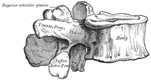
A critical review of over 125 articles concluded that the kind called isthmic spondylolysis is considered a fatigue fracture of the pars8. A fatigue fracture is a stress fracture in normal bone from too much repeated load. The lumbar vertebrae page covers the bone itself.
Which vertebra, and which stage
The lumbar spine has five vertebrae, named L1 to L59. In CT scans of 2,000 adults in Japan, 90.3% of the vertebrae with spondylolysis were L53. A CT scan builds detailed pictures of the inside of the body from X-rays.
A 1995 study of 185 people under 19 classed each pars defect as early, progressive or terminal10. Two series below also report a very early stage11,12.
A multicentre randomised trial in adolescents aged 10 to 19 used the term active spondylolysis6. It meant oedema at the pars of a lumbar vertebra on MRI, with or without a fracture6. A randomised trial assigns people to groups by chance. Oedema is swelling caused by fluid building up in body tissue, and MRI is a scan that shows soft tissue in detail.
When the vertebra slips
A 2024 systematic review (a study that searches for the research on a question in a set, checkable way) defines isthmic spondylolisthesis13. It is one lumbar vertebra shifting forward on the one below, as a result of a pars fracture on one or both sides13.
Who it affects
How common it is on scans
Four imaging studies report how common it is3,14,15,16:
| Who was imaged | Method | Share with spondylolysis |
|---|---|---|
| 500 unselected first-grade children, at age six | X-ray | 4.4%14 |
| The same group, by adulthood | X-ray | 6%14 |
| 532 children under eight, scanned for unrelated reasons | CT | 4.7%15 |
| 2,000 adults in Japan, scanned for reasons unrelated to back pain | CT | 5.9%3 |
| 188 adults aged 40 to 80 from the Framingham Heart Study | CT | 11.5%16 |
In the Japanese scans, 7.9% of the males and 3.9% of the females had it3.
Athletes with back pain
A meta-analysis (a study that pools the results of several earlier studies) covered 9 studies of 835 athletes with low back pain17. It estimated that 41.7% had lumbar spondylolysis, a figure that varied considerably with gender and age17.
Which sports the counts come from
- High-level athletes in Spain. A review of 3,152 case histories found spondylolysis in 8.02%18. Throwing sports had the highest figure, at 26.67%, ahead of artistic gymnastics and rowing, each near 17%18.
- Published reports. A literature review that used 114 articles found the highest reported incidence in diving (35.38%), cricket (31.97%) and baseball or softball (26.91%)19.
- Weightlifting. In a three-year MRI study of twelve competitive child and adolescent weightlifters, four had a lumbar pars defect by the final year20. Three of the twelve reported low back pain that year20.
With and without pain
A critical review concluded that most spondylolysis seen on X-ray in the general population probably comes without symptoms8. It added that pars lesions with symptoms appear to be a problem in adolescents in particular, especially adolescent athletes8.
A systematic review of 15 observational studies covered the general adult population4. It found no strong or consistent association between low back pain and spondylolysis, with or without isthmic spondylolisthesis4. An observational study watches what happens to people without the researchers choosing who gets what.
What studies found
What kind of evidence there is
A structured literature review of paediatric spondylolysis concluded that the evidence available to guide care is low quality21. Its reason was that uncontrolled case series dominate and comparative studies are lacking21. A case series is a report on a group of patients with the same condition, without a comparison group.
How often the bone united, by stage
Five reports give how often the bone healed, or united, at each stage5,10,11,12,22.
| Who was studied | What the patients did | Earliest stages | Progressive | Terminal |
|---|---|---|---|---|
| 346 defects in 185 people under 19, 199510 | Non-surgical management | Early: 73.0% | 38.5% | 0% |
| 63 defects in 37 patients under 185 | Wore a hard trunk brace | Early: 94% | 64% with high MRI signal in the bone beside the defect; 27% with normal signal | 0% |
| 60 patients aged 6 to 1712 | Rested, avoided sport and wore a trunk brace | Very early: 100%; early: 93.8% | 80.0% | Not given |
| The one-sided cases among 590 patients under 1811 | Managed without surgery | Very early: 98.2%; early: 96.0% | 64.3% | Not given |
| Pooled observational studies of children and young adults22 | Managed without surgery | Acute: 68.1% | 28.3% | 0 of 217 |
The 590-patient report is a case series, so it has no comparison group11. In the hard-brace study, defects that healed took a mean of 3.2 months when early and 5.4 to 5.7 months when progressive5.
The pooled observational studies of children and young adults managed without surgery also compared one side with both22. In ten studies of healing on imaging, 71% of one-sided defects healed and 18.1% of two-sided defects22.
What studies of braces found
A meta-analysis pooled 15 observational studies of 665 children and young adults managed without surgery22. A pooled 83.9% had a successful clinical outcome after at least a year22. The outcome did not differ significantly between those who wore a brace and those who did not22.
In one study, 57 children with acute spondylolysis wore a rigid brace or an elastic lumbar support for four months23. The first 14 were randomised and the other 43 chose their brace23. Bone union on CT was 69.0% with the rigid brace and 60.0% with the elastic support, a difference that was not significant23.
In a randomised trial of adolescent athletes with lumbar spondylolysis, no one wore a rigid brace24. Of 53 with MRI at the start and at 3 months, 81% showed healing and 11% worse findings24.
Whether healing goes with less pain
Three reports looked at this, over different lengths of time22,24,25.
- Pooled studies. In the same meta-analysis as the brace comparison above, most defects did not heal22. Across ten studies of healing on imaging, 28.0% healed22. Its authors took this to suggest that a good clinical outcome does not depend on healing22.
- Eight years on. A survey reached 61 patients a mean of 8 years after non-surgical management at ages 5 to 2125. Neither pain ratings nor return to sports correlated with whether the defect had shown healing on imaging25.
- Within one trial. A randomised trial of adolescent athletes with lumbar spondylolysis compared 53 of them by what their MRI showed, healing or not24. Of those whose MRI showed healing at 3 months, 97.6% were pain-free then24. The figure was 67% in those whose MRI was unchanged or worse24.
Starting a programme early or resting first
A multicentre randomised trial enrolled 64 adolescent athletes with active lumbar spondylolysis, at a median age of 14.26. One group started a therapist-led programme within 7 days, and the other rested until symptoms resolved before starting it6. Both groups rested from sport and neither wore a brace6.
- Pain and disability. The immediate group had less of both at 1 month6.
- Return to sport. The median time to meet the return-to-sport criteria was 74 days in the immediate group and 112 in the other6.
- Recurrence. Low back pain came back over 12 months in 3% of the immediate group and 29% of the other6.
- Limits. The authors listed a small sample, 14% dropout over a year, and participants from two US states6.
Exercise trials
A randomised trial assigned 44 patients with chronic low back pain and spondylolysis or spondylolisthesis on imaging26. One group did a 10-week programme training the deep abdominal muscles and the lumbar multifidus, a deep muscle running along the spine26. That group had a significant reduction in pain and disability, maintained at 30 months26. The control group, with care directed by their own practitioner, showed no significant change26.
A systematic review and meta-analysis pooled five randomised trials of lumbar segmental stabilisation exercise27. The 198 participants had spondylolysis or spondylolisthesis27. It found a trend toward less disability that fell short of significance, no significant effect on pain, and high inconsistency between trials27.
Return to sport and recurrence
| What was counted | Result |
|---|---|
| Return to sport at any level, systematic review of 14 studies | 92% after non-surgical care, 88% after surgery28 |
| Recurrence after union, 590 patients under 18 | 16.6%11 |
| Recurrence of significant symptoms, 121 athletes at 3.4 years | 45.5%29 |
The 14 studies in the systematic review covered 592 athletes with symptomatic spondylolysis and were level IV evidence28. Level IV evidence is a low rung on a scale that ranks study designs. A second systematic review compared surgery with non-surgical care for one-sided spondylolysis in athletes30. It concluded that the limited evidence allows no conclusion about which gets athletes back to sport faster30.
Slips and the long term
A structured literature review of paediatric spondylolysis looked at patients who develop chronic pars defects on both sides1. It found evidence that 43% to 74% go on to a grade 1 or 2 spondylolisthesis1. A slip grade is a score for how far a vertebra has shifted forward, and grade 1 is the smallest. In the Japanese CT scans, spondylolisthesis was present in 74.5% of adults with spondylolysis on both sides and 7.7% with it on one3.
A study began in 1955 with 500 first-grade children and followed the 30 with pars lesions for 45 years31. Slip progression slowed with each decade, and no one reached a 40% slip31. Their scores on the SF-36 health survey did not differ significantly from those of the general population of the same age31.
Connections
The bone and the spine
The lumbar vertebrae page covers the loads on the pars. One computer model covered an L4-L5 segment32. In the model, stress in the pars was lowest under compression alone and high under extension (bending backward) and rotation (twisting) in particular32.
A 1976 account of the mechanics of the spine lists what normally resists forward shear, a sliding force, on a lumbar vertebra33. It names the outer ring of the intervertebral disc, the facet joints (small joints between neighbouring vertebrae) and the pars33.
Related conditions and stretches
One critical review places isthmic spondylolysis among fatigue fractures, a kind of stress fracture8. The 15-study review above found no strong or consistent link with low back pain in adults4.
One randomised trial studied people with chronic low back pain, not spondylolysis in particular34. Its flexion-based (forward-bending) exercise programme included the knee-to-chest stretch34. A systematic review of non-surgical care for spondylolysis and spondylolisthesis found two studies, from one patient population, comparing flexion exercises with extension exercises35. The review concluded that the studies of non-surgical options lacked blinded assessors and control groups35.
Spondylolysis is a fracture in part of
Spondylolysis involves
Spondylolysis is described in a review as a fatigue fracture, one kind of
Spondylolysis has been studied for a link with
Sources
- Crawford CH 3rd, Ledonio CG, Bess RS, Buchowski JM, Burton DC, Hu SS, Lonner BS, Polly DW, Smith JS, Sanders JO (2015). Current Evidence Regarding the Etiology, Prevalence, Natural History, and Prognosis of Pediatric Lumbar Spondylolysis: A Report from the Scoliosis Research Society Evidence-Based Medicine Committee.. Spine deformity. doi:10.1016/j.jspd.2014.06.005 PMID 27927448
- Madden V, Ayoub A, Thomas J, Thomas I (2026). Spondylolysis: A Narrative Review of Etiology, Diagnosis, and Management.. International journal of environmental research and public health. doi:10.3390/ijerph23020153 PMID 41752236
- Sakai T, Sairyo K, Takao S, Nishitani H, Yasui N (2009). Incidence of lumbar spondylolysis in the general population in Japan based on multidetector computed tomography scans from two thousand subjects.. Spine. doi:10.1097/BRS.0b013e3181b4abbe PMID 19934813
- Andrade NS, Ashton CM, Wray NP, Brown C, Bartanusz V (2015). Systematic review of observational studies reveals no association between low back pain and lumbar spondylolysis with or without isthmic spondylolisthesis.. European spine journal : official publication of the European Spine Society, the European Spinal Deformity Society, and the European Section of the Cervical Spine Research Society. doi:10.1007/s00586-015-3910-5 PMID 25833204
- Sairyo K, Sakai T, Yasui N, Dezawa A (2012). Conservative treatment for pediatric lumbar spondylolysis to achieve bone healing using a hard brace: what type and how long?: Clinical article.. Journal of neurosurgery. Spine. doi:10.3171/2012.2.SPINE10914 PMID 22519929
- Selhorst M, Sweeney E, Martin LC, Yang J, Benedict J, Brna M, Spondylolysis Physician Group, Fischer AN (2026). Immediate physical therapy is beneficial for adolescent athletes with active lumbar spondylolysis: a multicentre randomised trial.. British journal of sports medicine. doi:10.1136/bjsports-2025-110606 PMID 41402030
- Betts JG, Young KA, Wise JA, Johnson E, Poe B, Kruse DH, Korol O, Johnson JE, Womble M, DeSaix P (2022). Anatomy and Physiology 2e. OpenStax. Link
- Standaert CJ, Herring SA (2000). Spondylolysis: a critical review.. British journal of sports medicine. doi:10.1136/bjsm.34.6.415 PMID 11131228
- Waxenbaum JA, Reddy V, Williams C, Futterman B (2026). Anatomy, Back, Lumbar Vertebrae.. StatPearls. PMID 29083618
- Morita T, Ikata T, Katoh S, Miyake R (1995). Lumbar spondylolysis in children and adolescents.. The Journal of bone and joint surgery. British volume. PMID 7615609
- Kuroshima K, Miyazaki S, Hiranaka Y, Ryu M, Inoue S, Yurube T, Kakutani K, Tadokoro K (2024). Rate and Duration of Bone Union for Conservative Treatment in Pediatric Lumbar Spondylolysis.. Spine. doi:10.1097/BRS.0000000000004849 PMID 37807612
- Sakai T, Tezuka F, Yamashita K, Takata Y, Higashino K, Nagamachi A, Sairyo K (2017). Conservative Treatment for Bony Healing in Pediatric Lumbar Spondylolysis.. Spine. doi:10.1097/BRS.0000000000001931 PMID 27755499
- Sima S, Chen X, Sheldrick K, Lu A, Diwan AD (2024). Imaging predictors of progression of lumbar spondylolysis to spondylolisthesis: a systematic review.. The spine journal : official journal of the North American Spine Society. doi:10.1016/j.spinee.2024.03.010 PMID 38499064
- Fredrickson BE, Baker D, McHolick WJ, Yuan HA, Lubicky JP (1984). The natural history of spondylolysis and spondylolisthesis.. The Journal of bone and joint surgery. American volume. PMID 6373773
- Lemoine T, Fournier J, Odent T, Sembély-Taveau C, Merenda P, Sirinelli D, Morel B (2018). The prevalence of lumbar spondylolysis in young children: a retrospective analysis using CT.. European spine journal : official publication of the European Spine Society, the European Spinal Deformity Society, and the European Section of the Cervical Spine Research Society. doi:10.1007/s00586-017-5339-5 PMID 29030702
- Kalichman L, Kim DH, Li L, Guermazi A, Berkin V, Hunter DJ (2009). Spondylolysis and spondylolisthesis. Spine. doi:10.1097/BRS.0b013e31818edcfd PMID 19139672
- Li J, Liang J, Xu Y, Du D, Feng F, Shen J, Cui Y (2023). Incidence of lumbar spondylolysis in athletes with low back pain: A systematic evaluation and single-arm meta-analysis.. Medicine. doi:10.1097/MD.0000000000034857 PMID 37747004
- Soler T, Calderón C (2000). The prevalence of spondylolysis in the Spanish elite athlete.. The American journal of sports medicine. doi:10.1177/03635465000280012101 PMID 10653544
- Tawfik S, Phan K, Mobbs RJ, Rao PJ (2020). The Incidence of Pars Interarticularis Defects in Athletes.. Global spine journal. doi:10.1177/2192568218823695 PMID 32002353
- Shimozaki K, Nakase J, Yoshioka K, Takata Y, Asai K, Kitaoka K, Tsuchiya H (2018). Incidence rates and characteristics of abnormal lumbar findings and low back pain in child and adolescent weightlifter: A prospective three-year cohort study.. PloS one. doi:10.1371/journal.pone.0206125 PMID 30372456
- Crawford CH 3rd, Ledonio CG, Bess RS, Buchowski JM, Burton DC, Hu SS, Lonner BS, Polly DW, Smith JS, Sanders JO (2015). Current Evidence Regarding the Surgical and Nonsurgical Treatment of Pediatric Lumbar Spondylolysis: A Report from the Scoliosis Research Society Evidence-Based Medicine Committee.. Spine deformity. doi:10.1016/j.jspd.2014.06.004 PMID 27927449
- Klein G, Mehlman CT, McCarty M (2009). Nonoperative treatment of spondylolysis and grade I spondylolisthesis in children and young adults. Journal of pediatric orthopedics. doi:10.1097/BPO.0b013e3181977fc5 PMID 19352240
- Virkki E, Holstila M, Kolari T, Lastikka M, Mattila K, Malmi S, Pajulo O, Helenius I (2023). Elastic Lumbar Support Versus Rigid Thoracolumbar Orthosis for Acute Pediatric Spondylolysis: A Prospective Controlled Study.. Spine. doi:10.1097/BRS.0000000000004424 PMID 35834380
- Sweeney EA, Fischer A, Brna M, Martin L, Yang J, Selhorst M (2026). Rest Before Physical Therapy Is Not Necessary to Achieve Bony Healing of Lumbar Spondylolysis in Adolescent Athletes.. Orthopaedic journal of sports medicine. doi:10.1177/23259671261421595 PMID 42099883
- Sousa T, Skaggs DL, Chan P, Yamaguchi KT Jr, Borgella J, Lee C, Sawyer J, Moisan A, Flynn JM, Gunderson M, Hresko MT, D'Hemecourt P, Andras LM (2017). Benign Natural History of Spondylolysis in Adolescence With Midterm Follow-Up.. Spine deformity. doi:10.1016/j.jspd.2016.10.005 PMID 28259265
- O'Sullivan PB, Phyty GD, Twomey LT, Allison GT (1997). Evaluation of specific stabilizing exercise in the treatment of chronic low back pain with radiologic diagnosis of spondylolysis or spondylolisthesis.. Spine. doi:10.1097/00007632-199712150-00020 PMID 9431633
- Lin LH, Lin TY, Chang KV, Wu WT, Özçakar L (2024). Effectiveness of Lumbar Segmental Stabilization Exercises in Managing Disability and Pain Intensity Among Patients With Lumbar Spondylolysis and Spondylolisthesis. Spine. doi:10.1097/BRS.0000000000004989 PMID 38514931
- Grazina R, Andrade R, Santos FL, Marinhas J, Pereira R, Bastos R, Espregueira-Mendes J (2019). Return to play after conservative and surgical treatment in athletes with spondylolysis: A systematic review.. Physical therapy in sport : official journal of the Association of Chartered Physiotherapists in Sports Medicine. doi:10.1016/j.ptsp.2019.02.005 PMID 30826586
- Selhorst M, Fischer A, Graft K, Ravindran R, Peters E, Rodenberg R, MacDonald J (2016). Long-Term Clinical Outcomes and Factors That Predict Poor Prognosis in Athletes After a Diagnosis of Acute Spondylolysis: A Retrospective Review With Telephone Follow-up.. The Journal of orthopaedic and sports physical therapy. doi:10.2519/jospt.2016.7028 PMID 27825292
- Scheepers MS, Streak Gomersall J, Munn Z (2015). The effectiveness of surgical versus conservative treatment for symptomatic unilateral spondylolysis of the lumbar spine in athletes: a systematic review.. JBI database of systematic reviews and implementation reports. doi:10.11124/jbisrir-2015-1926 PMID 26447055
- Beutler WJ, Fredrickson BE, Murtland A, Sweeney CA, Grant WD, Baker D (2003). The natural history of spondylolysis and spondylolisthesis. Spine. doi:10.1097/01.BRS.0000061992.98108.A0 PMID 12768144
- Chosa E, Totoribe K, Tajima N (2004). A biomechanical study of lumbar spondylolysis based on a three-dimensional finite element method.. Journal of orthopaedic research : official publication of the Orthopaedic Research Society. doi:10.1016/S0736-0266(03)00160-8 PMID 14656675
- Troup JD (1976). Mechanical factors in spondylolisthesis and spondylolysis.. Clinical orthopaedics and related research. PMID 1277687
- Park CH, Beom J, Chung CK, Kim CH, Lee MY, Park MW, Kim K, Chung SG (2024). Long-term effects of lumbar flexion versus extension exercises for chronic axial low back pain: a randomized controlled trial.. Scientific reports. doi:10.1038/s41598-024-51769-2 PMID 38302483
- Garet M, Reiman MP, Mathers J, Sylvain J (2013). Nonoperative treatment in lumbar spondylolysis and spondylolisthesis. Sports health. doi:10.1177/1941738113480936 PMID 24427393
Changes
- First published.