Multifidus
Also called lumbar multifidus
The deep back muscle that lies closest to the spine. How it is built, how it shrinks with disuse and in some people with low back pain, how it comes back, and what it works with.
- Region
- Spine
- Updated
- September 30, 2026
- Reviewed
- September 30, 2026 · how entries are checked
Function
Where it sits
The multifidus belongs to the transversospinales, a group that runs between bony points on the vertebrae, the bones of the spine1. It runs from the side spurs to the bony points at the back1.
In the lower back, three muscles sit side by side: iliocostalis on the outside, then longissimus, then multifidus nearest the middle7. The first two belong to the erector spinae, and the multifidus belongs to a different group7.
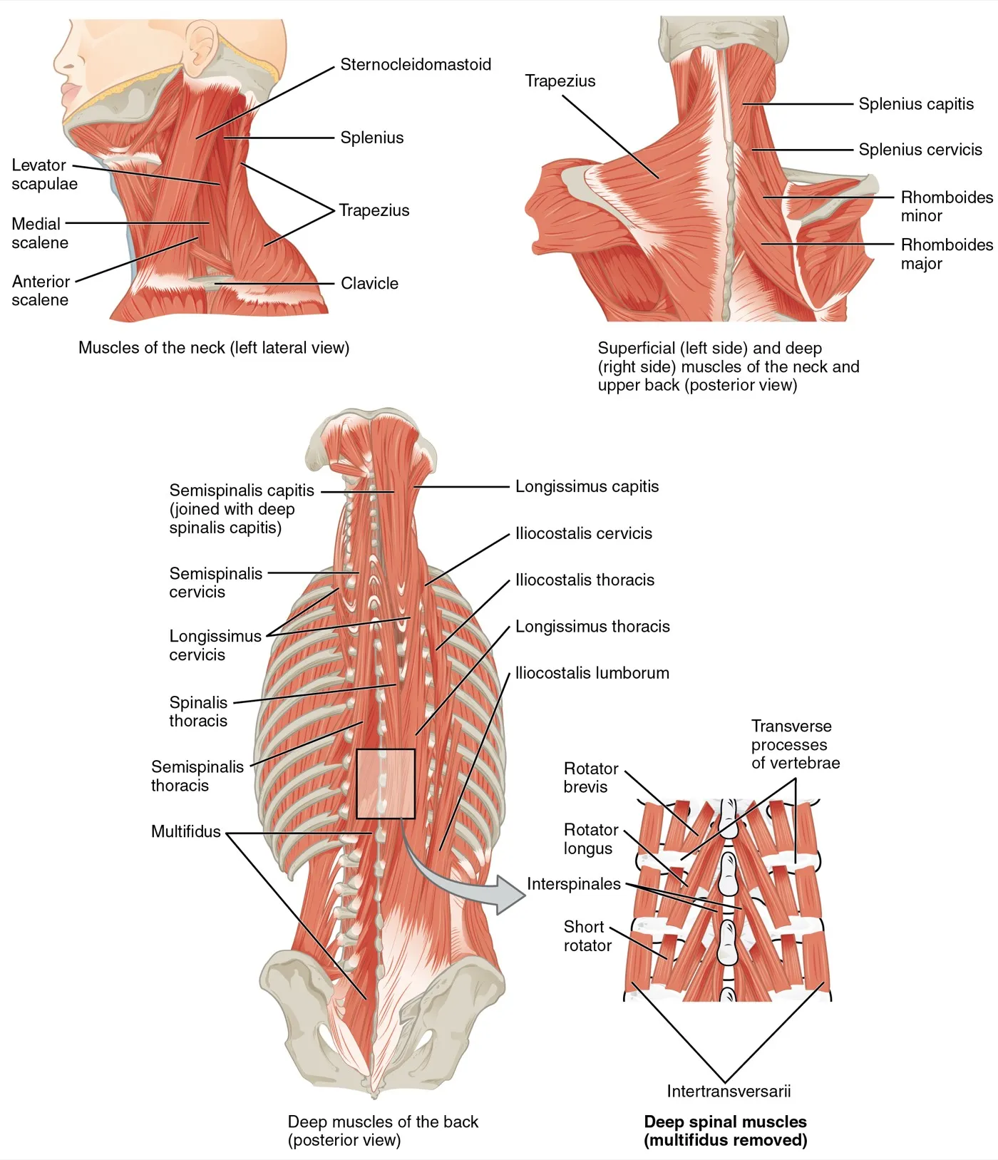
Dissection studies found natural dividing lines that split the lumbar multifidus into five bands, each starting from one lumbar spinous process8. Each band is supplied by a single spinal nerve8. So the fibres that move one segment of the spine get their nerve supply from that segment8.
Layers from surface to deep
L1 to L4 are the top four vertebrae of the lower back. In 10 embalmed cadavers, the lumbar multifidus from L1 to L4 had three regions with a distinct build: superficial, intermediate and deep9. The intermediate region was missing at L5, the lowest vertebra of the lower back, in every specimen9. The average fibre bundle angle grew from the surface region to the deep one9. The authors saw this as support for separate roles in movement and in stability9.
Built for large forces
In eight cadaver lumbar spines, the multifidus had short fibres for the muscle’s length2. Its physiological cross-sectional area, a size measure taken across all its fibres, was more than twice that of any other lumbar muscle2. From this build, the researchers concluded the muscle is designed as a stabiliser that produces large forces2. Tissue samples from 16 people having spine surgery suggest the muscle gets stronger as the spine leans forward2.
What it does
One analysis plotted where the muscle’s fibre bundles attach on X-rays of 5 cadavers and 21 living people10. Its main action was to tip the lumbar vertebrae backward, which is extension without backward sliding10. The same analysis concluded that any twisting it produces is minor, a side effect linked to extension10.
In a lab test on seven human lower spines with simulated muscle forces, the multifidus group had the strongest influence in reducing spinal motion11.
Deep and superficial fibres
One cross-sectional study recorded deep and superficial multifidus during arm movements, using electrodes placed inside the muscle12. When the superficial fibres switched on depended on which way the arm moved, but the deep fibres’ timing did not12. The authors took this to support superficial fibres controlling spine orientation, and deep fibres controlling motion between vertebrae12.
Breakdown
Shrinking with disuse and injury
The multifidus lost size in each of these settings3,13,14:
- Bed rest. In 10 young men on 10 days of flat bed rest, its cross-sectional area fell, with the drop detectable by day 43.
- Spaceflight. In one astronaut after 6 months in space, exercise in flight kept its size at L2 to L4, but its size at L5 fell13.
- Experimental injury in pigs. In 21 pigs, its area fell within 3 or 6 days of an experimental disc or nerve injury14. After a disc injury, the loss was on the same side at one level14. After a nerve injury, it spread across three of the lowest levels of the lower back14.
Fat, size and age
Fatty infiltration means fat building up inside a muscle. In 80 healthy volunteers aged 20 to 62, fat in the multifidus and erector spinae rose with age, but muscle volume did not change15.
Findings on size disagree. A systematic review of 27 studies found multifidus thickness and area did not correlate with age16. In one other study, smaller multifidus area went with greater age3. A review of adults aged 50 and over found limited evidence that ageing or spinal conditions harm the abdominal and multifidus muscles17.
Findings in people with low back pain
One review of the multifidus in low back pain describes shrinkage, fat and a build-up of connective tissue, mostly in the deep fibres18. It adds that results are not uniform and differ between acute, recurrent and chronic stages18,19.
Studies that found a smaller or fattier muscle20,21:
- A meta-analysis of imaging studies found a medium rise in multifidus fat4. The muscle was somewhat smaller too, though the likely range for size included no difference4.
- A systematic review of 25 studies found evidence that a smaller multifidus area goes with low back pain20.
- A meta-analysis in chronic low back pain found less cross-sectional area and more fat than in controls, mostly from L3/L4 down to L521. Of the muscles beside the spine that were compared, the multifidus showed the most consistent changes21.
- A systematic review found more significant left-right differences in people with chronic low back pain than in those without16.
Studies that found no clear difference, or mixed results5,19:
- A meta-analysis of 18 studies with 1,142 subjects looked at athletes with and without chronic non-specific low back pain5. It found no significant difference in multifidus size or make-up5. The quality of those studies was poor5.
- A systematic review of 15 studies found moderate evidence of shrinkage in chronic low back pain19. It also found moderate evidence of no shrinkage in any lumbar muscle in acute or recurrent low back pain19.
- On fat, the same review found results in chronic pain inconsistent, and moderate evidence of no fat build-up in recurrent pain19.
- A review of muscle-activity studies calls the results somewhat inconsistent18. They often report lower multifidus activity, with more use of the superficial multifidus and erector spinae18.
One side or both
In 26 patients with acute low back pain on one side, the multifidus was markedly smaller on the painful side, by 31%, at one level22. In 51 people without pain, side-to-side differences were under 5%22. In 24 of the 26 patients, the level of wasting matched the level of symptoms judged by clinicians22. How big the difference was did not track symptom severity22.
Chronic pain looked different in another study23. Fourteen active people with chronic pain on one side were matched with 14 healthy people23. Their multifidus volume was 18.1% smaller at L5-S1 only, with no difference between the painful and pain-free sides23. The authors concluded that, unlike acute pain on one side, chronic pain goes with a smaller muscle on both sides23.
A meta-analysis looked at people with a lumbar disc herniation on one side, a slipped disc in the lower back24. On the side of the herniation, type I (slow) and type II (fast) fibres were smaller, and type I fibres were more common24. Whole-muscle size did not differ significantly between sides24.
Which comes first
In 412 Danish 40-year-olds, severe fatty infiltration was strongly linked to ever having had low back pain, with an odds ratio of 9.225. An odds ratio says how many times higher the odds of an outcome are in one group than another. The ratio for pain in the past year was 4.1, and there was no such link in 13-year-olds25. The authors noted that studies over time are needed to tell whether the muscle change causes the pain or the reverse25.
Recovery
After a first bout of acute pain
One trial enrolled 39 patients with a first bout of acute low back pain on one side, whose multifidus was affected at one level6. They were randomly given either medical care only or care with added exercise6. In the medical-care group, multifidus size did not recover on its own as the pain went away6. At 10 weeks, that group still had smaller multifidus muscles, although they had gone back to normal activity6. In the exercise group, size came back faster and more fully, while other outcomes were similar at 4 weeks6.
In a randomised trial of 39 patients with first-time acute low back pain, one group exercised the multifidus with the transversus abdominis26. At one year, pain had come back in 30% of the exercise group and 84% of the control group26. At two to three years, the figures were 35% and 75%26. These figures come from this one small trial alone26.
Training studies in chronic pain
Training studies in chronic low back pain have found multifidus size gains, but the evidence is preliminary or of very low quality27,28. Motor control training is exercise that trains how the deep trunk muscles are switched on and coordinated.
- In a randomised trial of 50 people, 12 weeks of motor control training plus lumbar extension exercise was compared with general exercise27. It gave greater gains in multifidus and erector spinae area27. The authors call the results preliminary27.
- A review looked at 9 randomised trials with 451 people28. It found very low-quality evidence that 36 sessions of motor control training beat general physiotherapy at producing small but measurable gains in multifidus area28.
- A review of randomised trials found low-quality evidence that changes in multifidus size after this training bore no relation to pain or disability28.
In healthy people and after spaceflight
In a cluster-randomised trial of 64 healthy firefighters, whole groups were assigned by chance29. Against controls, 24 weeks of twice-weekly supervised back and core exercise made no significant difference to multifidus area or left-right balance29. In the astronaut whose L5 multifidus shrank in space, its size returned to the pre-mission level during a daily programme back on Earth13.
Connections
Its neighbour, the erector spinae
Low in the lumbar region, the multifidus and both lumbar erector spinae muscles attach firmly inside one aponeurosis, which they share7. An aponeurosis is a broad, flat sheet of tendon. At L1 to L4, each dorsal ramus, a nerve branch that runs to the back, tends to split in three30. The inner branch goes to the multifidus, and the other two to the erector spinae’s iliocostalis and longissimus30.
In 40 active adults holding a back bridge, the glute bridge hold, the ratio of multifidus to erector spinae activity was 2.7631.
The belly muscles
One muscle-activity study of trunk rotation describes, as background, three muscles acting on the opposite side: the external oblique, rectus abdominis and lumbar multifidus32.
What exercise studies show
Some figures below are shares of maximum voluntary contraction, the hardest effort a person can make33.
| Exercise | What the studies found |
|---|---|
| Bird dog | In 34 healthy adults, the deep multifidus did relatively more in the bird dog than in back extension, glute bridge or reverse hyperextension34 |
| Four-point kneeling | In 30 healthy volunteers doing three hands-and-knees exercises, the same-side lumbar multifidus and gluteus maximus were most active, above 20% of maximum35 |
| Back extension | In adults aged 21 to 35, resisted face-down extension to end range drew multifidus activity above 92% of maximum33 |
| Barbell row | One of three studies found the most multifidus activity in the bent-over row at 45% of body weight (about 58% of maximum)36. The deadlift came close behind36 |
| Farmer’s carry | Walking with two dumbbells drew more multifidus activity on both sides than holding them still37 |
| Side plank | In adults aged 21 to 35, multifidus activity in the side bridge stayed below half of maximum33 |
Multifidus shares an aponeurosis with
Multifidus was trained with in a low back pain trial
Multifidus works with in trunk rotation
Multifidus works with in the bird dog
Multifidus has been measured in studies of
Multifidus is loaded by
Sources
- Betts JG, Young KA, Wise JA, Johnson E, Poe B, Kruse DH, Korol O, Johnson JE, Womble M, DeSaix P (2022). Anatomy and Physiology 2e. OpenStax. Link
- Ward SR, Kim CW, Eng CM, Gottschalk LJ 4th, Tomiya A, Garfin SR, Lieber RL (2009). Architectural analysis and intraoperative measurements demonstrate the unique design of the multifidus muscle for lumbar spine stability.. The Journal of Bone and Joint Surgery. American Volume. doi:10.2106/JBJS.G.01311 PMID 19122093
- Sarto F, Monti E, Tagliaferri S, Simunič B, Salvi M, Jandova T, Franchi MV, Pišot R, Maggio M, Narici MV (2026). Changes in human multifidus muscle size with aging and short-term disuse.. Experimental Gerontology. doi:10.1016/j.exger.2026.113090 PMID 41802687
- Seyedhoseinpoor T, Taghipour M, Dadgoo M, Sanjari MA, Takamjani IE, Kazemnejad A, Khoshamooz Y, Hides J (2022). Alteration of lumbar muscle morphology and composition in relation to low back pain: a systematic review and meta-analysis.. The Spine Journal. doi:10.1016/j.spinee.2021.10.018 PMID 34718177
- Wong WK, Fu ACL, Tsang SMH (2026). Morphological and Compositional Features of Lumbar Multifidus in Athletes with Chronic Non-specific Low Back Pain: A Systematic Review and Meta-Analysis.. International Journal of Sports Physical Therapy. doi:10.26603/001c.162340 PMID 42245760
- Hides JA, Richardson CA, Jull GA (1996). Multifidus muscle recovery is not automatic after resolution of acute, first-episode low back pain.. Spine. doi:10.1097/00007632-199612010-00011 PMID 8979323
- Willard FH, Vleeming A, Schuenke MD, Danneels L, Schleip R (2012). The thoracolumbar fascia: anatomy, function and clinical considerations.. Journal of Anatomy. doi:10.1111/j.1469-7580.2012.01511.x PMID 22630613
- Macintosh JE, Valencia F, Bogduk N, Munro RR (1986). The morphology of the human lumbar multifidus.. Clinical Biomechanics. doi:10.1016/0268-0033(86)90146-4 PMID 23915550
- Rosatelli AL, Ravichandiran K, Agur AM (2008). Three-dimensional study of the musculotendinous architecture of lumbar multifidus and its functional implications.. Clinical Anatomy. doi:10.1002/ca.20659 PMID 18627104
- Macintosh JE, Bogduk N (1986). The biomechanics of the lumbar multifidus.. Clinical Biomechanics. doi:10.1016/0268-0033(86)90147-6 PMID 23915551
- Wilke HJ, Wolf S, Claes LE, Arand M, Wiesend A (1995). Stability increase of the lumbar spine with different muscle groups. Spine. doi:10.1097/00007632-199501150-00011 PMID 7716624
- Moseley GL, Hodges PW, Gandevia SC (2002). Deep and superficial fibers of the lumbar multifidus muscle are differentially active during voluntary arm movements.. Spine. doi:10.1097/00007632-200201150-00013 PMID 11805677
- Hides JA, Lambrecht G, Stanton WR, Damann V (2016). Changes in multifidus and abdominal muscle size in response to microgravity: possible implications for low back pain research.. European Spine Journal. doi:10.1007/s00586-015-4311-5 PMID 26582165
- Hodges P, Holm AK, Hansson T, Holm S (2006). Rapid atrophy of the lumbar multifidus follows experimental disc or nerve root injury.. Spine. doi:10.1097/01.brs.0000248453.51165.0b PMID 17139223
- Crawford RJ, Filli L, Elliott JM, Nanz D, Fischer MA, Marcon M, Ulbrich EJ (2016). Age- and Level-Dependence of Fatty Infiltration in Lumbar Paravertebral Muscles of Healthy Volunteers.. AJNR. American Journal of Neuroradiology. doi:10.3174/ajnr.A4596 PMID 26635285
- Rummens S, Robben E, De Groef A, Van Wambeke P, Janssens L, Brumagne S, Desloovere K, Peers K (2020). Factors Associated With the Ultrasound Characteristics of the Lumbar Multifidus: A Systematic Review.. PM&R. doi:10.1002/pmrj.12212 PMID 31264799
- Cuellar WA, Wilson A, Blizzard CL, Otahal P, Callisaya ML, Jones G, Hides JA, Winzenberg TM (2017). The assessment of abdominal and multifidus muscles and their role in physical function in older adults: a systematic review.. Physiotherapy. doi:10.1016/j.physio.2016.06.001 PMID 27667760
- Hodges PW, Danneels L (2019). Changes in Structure and Function of the Back Muscles in Low Back Pain: Different Time Points, Observations, and Mechanisms.. Journal of Orthopaedic & Sports Physical Therapy. doi:10.2519/jospt.2019.8827 PMID 31151377
- Goubert D, Oosterwijck JV, Meeus M, Danneels L (2016). Structural Changes of Lumbar Muscles in Non-specific Low Back Pain: A Systematic Review.. Pain Physician. PMID 27676689
- Ranger TA, Cicuttini FM, Jensen TS, Peiris WL, Hussain SM, Fairley J, Urquhart DM (2017). Are the size and composition of the paraspinal muscles associated with low back pain? A systematic review.. The Spine Journal. doi:10.1016/j.spinee.2017.07.002 PMID 28756299
- Kalli I, Niglas M, Naeini MK, Freidin M, Thomas L, Menni C, Williams F (2026). Paraspinal muscle quality in chronic low back pain: a systematic review and meta-analysis of muscle atrophy and fat infiltration.. European Spine Journal. doi:10.1007/s00586-025-09454-z PMID 41148279
- Hides JA, Stokes MJ, Saide M, Jull GA, Cooper DH (1994). Evidence of lumbar multifidus muscle wasting ipsilateral to symptoms in patients with acute/subacute low back pain.. Spine. doi:10.1097/00007632-199401001-00009 PMID 8153825
- Beneck GJ, Kulig K (2012). Multifidus atrophy is localized and bilateral in active persons with chronic unilateral low back pain.. Archives of Physical Medicine and Rehabilitation. doi:10.1016/j.apmr.2011.09.017 PMID 22289241
- Stevens S, Agten A, Timmermans A, Vandenabeele F (2020). Unilateral changes of the multifidus in persons with lumbar disc herniation: a systematic review and meta-analysis.. The Spine Journal. doi:10.1016/j.spinee.2020.04.007 PMID 32325246
- Kjaer P, Bendix T, Sorensen JS, Korsholm L, Leboeuf-Yde C (2007). Are MRI-defined fat infiltrations in the multifidus muscles associated with low back pain?. BMC Medicine. doi:10.1186/1741-7015-5-2 PMID 17254322
- Hides JA, Jull GA, Richardson CA (2001). Long-term effects of specific stabilizing exercises for first-episode low back pain.. Spine. doi:10.1097/00007632-200106010-00004 PMID 11389408
- Fortin M, Rye M, Roussac A, Montpetit C, Burdick J, Naghdi N, Rosenstein B, Bertrand C, Macedo LG, Elliott JM, Dover G, DeMont R, Weber MH, Pepin V (2023). The Effects of Combined Motor Control and Isolated Extensor Strengthening versus General Exercise on Paraspinal Muscle Morphology, Composition, and Function in Patients with Chronic Low Back Pain: A Randomized Controlled Trial.. Journal of Clinical Medicine. doi:10.3390/jcm12185920 PMID 37762861
- Pinto SM, Boghra SB, Macedo LG, Zheng YP, Pang MYC, Cheung JPY, Karppinen J, Samartzis D, Wong AYL (2021). Does Motor Control Exercise Restore Normal Morphology of Lumbar Multifidus Muscle in People with Low Back Pain? - A Systematic Review.. Journal of Pain Research. doi:10.2147/JPR.S314971 PMID 34447268
- Mayer JM, Nuzzo JL (2015). Worksite back and core exercise in firefighters: Effect on development of lumbar multifidus muscle size.. Work. doi:10.3233/WOR-141831 PMID 24448017
- Bogduk N, Wilson AS, Tynan W (1982). The human lumbar dorsal rami.. Journal of Anatomy. PMID 7076562
- Khosrokiani Z, Letafatkar A, Sheikhi B, Thomas AC, Aghaie-Ataabadi P, Hedayati MT (2022). Hip and Core Muscle Activation During High-Load Core Stabilization Exercises.. Sports Health. doi:10.1177/19417381211015225 PMID 34060953
- Sugaya T, Sakamoto M, Nakazawa R, Wada N (2016). Relationship between spinal range of motion and trunk muscle activity during trunk rotation.. Journal of Physical Therapy Science. doi:10.1589/jpts.28.589 PMID 27065549
- Ekstrom RA, Osborn RW, Hauer PL (2008). Surface electromyographic analysis of the low back muscles during rehabilitation exercises.. Journal of Orthopaedic and Sports Physical Therapy. doi:10.2519/jospt.2008.2865 PMID 19195137
- Lawrence MA, Swanson BT, Silfies SP, Winchester JB (2026). Activation of the deep lumbar multifidus during common rehabilitation and resistance exercises.. Journal of Electromyography and Kinesiology. doi:10.1016/j.jelekin.2026.103179 PMID 42320454
- Stevens VK, Vleeming A, Bouche KG, Mahieu NN, Vanderstraeten GG, Danneels LA (2007). Electromyographic activity of trunk and hip muscles during stabilization exercises in four-point kneeling in healthy volunteers.. European Spine Journal. doi:10.1007/s00586-006-0181-1 PMID 16896840
- Oliva-Lozano JM, Muyor JM (2020). Core Muscle Activity During Physical Fitness Exercises: A Systematic Review.. International Journal of Environmental Research and Public Health. doi:10.3390/ijerph17124306 PMID 32560185
- Ellestad SH, Holcomb TP, Swiergol AM, Holmstrup ME, Dicus JR (2024). The Quantification of Muscle Activation During the Loaded Carry Movement Pattern.. International Journal of Exercise Science. doi:10.70252/NWUE9985 PMID 38665162
Changes
- First published.