Gluteus maximus
The largest of the buttock muscles. What it moves at the hip, how it shrinks with disuse and is smaller alongside some conditions, how it grows with training, and what it works with.
- Region
- Hip
- Starts on
- the ilium, sacrum and tailbone, nearby ligaments, and the tough sheets over the lower back and gluteus medius (Pelvis)1,6
- Ends on (deep fibres)
- the gluteal tuberosity of the thigh bone5
- Ends on (superficial fibres)
- the fascia lata and iliotibial band, shared with the tensor fasciae latae5,7,22
- Updated
- September 29, 2026
- Reviewed
- September 29, 2026 · how entries are checked
Function
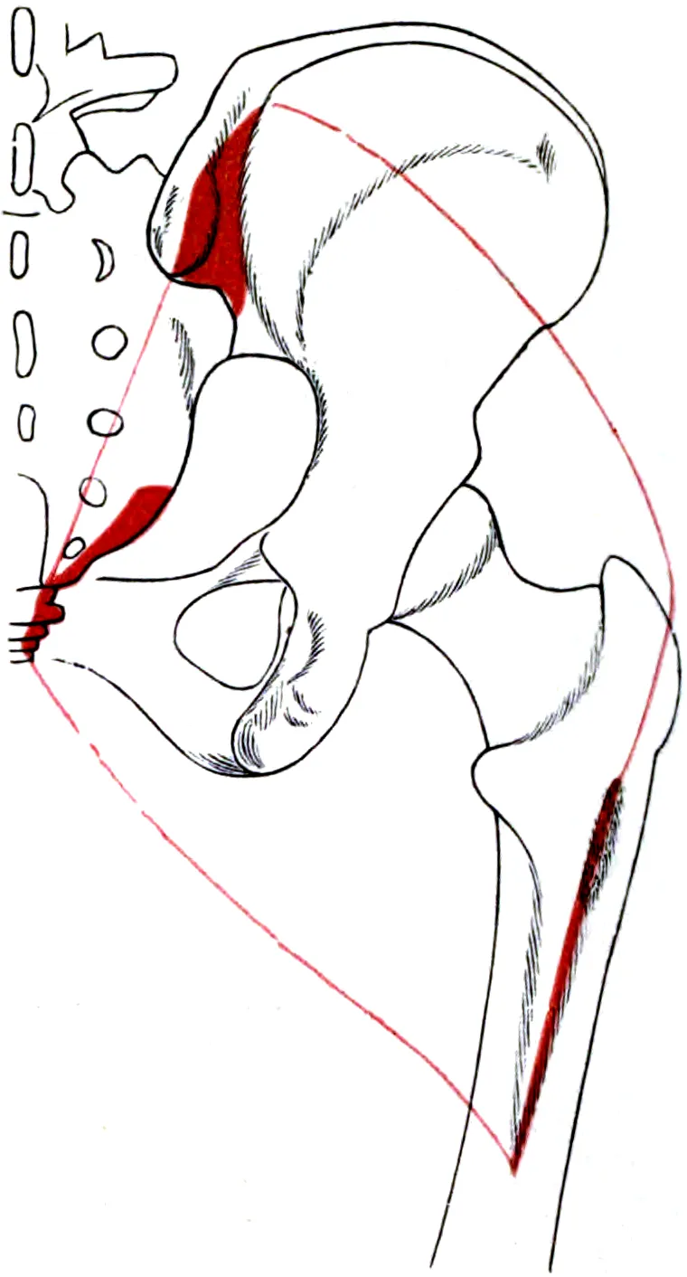
Where it sits and what it attaches to
The gluteal muscles of the buttock rank among the body’s biggest and strongest muscles1. The gluteus maximus is the largest of the three1. The gluteus medius lies beneath it, and the gluteus minimus lies beneath the medius1. Its nerve supply comes from the inferior gluteal nerve5.
In 11 embalmed legs from donors, its fibres started from the same set of places in each one6:
- The ilium, the upper part of the hip bone6.
- The sacrum and coccyx, the bones at the base of the spine6.
- The thoracolumbar fascia, a sheet of tough tissue over the lower back6.
- The flat tendon sheet of the erector spinae, the long back muscles6.
- The fascia covering the gluteus medius6.
- Two ligaments: the dorsal sacroiliac and the sacrotuberous ligaments6.
From there its fibres run down and outward toward the femur (thigh bone)5. The fibres nearest the skin help form part of the iliotibial tract, a band of tissue on the outer thigh5. The deeper fibres attach to the femur at a spot called the gluteal tuberosity5. In six donor bodies, the muscle had a large insertion into the fascia lata, a sheet of tough connective tissue7. It was so large that the authors suggest the iliotibial tract could count as one of its tendons7.
What it moves
The muscle’s line and leverage suit it to extend the hip, which moves the thigh backward2. It also turns the thigh outward and helps with hip abduction, lifting the leg out to the side2. It shares hip extension with the hamstrings, the muscles at the back of the thigh8. In the first half of rising from a full forward bend, most of the movement happens at the hip2. The gluteus maximus and the hamstrings produce it2.
Different parts of the muscle seem to favour different jobs9. A study used imaging in living people to estimate what each region could do9:
| Region of the muscle | What its fibres seemed suited to |
|---|---|
| Lower part | Hip extension and outward rotation9 |
| Upper part | Hip abduction9 |
| Lower, outer fibres | Pulling the thigh inward, the opposite of abduction9 |
A dissection study took apart 27 muscles from 21 human legs10. The gluteus maximus paired long fibres with a very large muscle10. The authors describe it as able to make very large forces, move through long distances and shorten at high speed10.
What it does in walking and running
In recordings of its electrical activity (electromyography, EMG), the gluteus maximus was mostly quiet during level and uphill walking3. Its activity rose sharply during running3. In running, its main jobs were to control forward bending of the trunk over the standing leg and to slow the swinging leg3. On the standing side, it may also help control hip bending and extend the thigh3.
A computer model built from five donor specimens looked at the iliotibial band in running11. It estimated that pull from the gluteus maximus and the tensor fasciae latae, in the upper outer thigh, stretches the band a lot1,11. That stretch stores elastic energy in the band11.
Breakdown
The studies gathered here describe the gluteus maximus shrinking with disuse, and being smaller alongside some conditions2,4.
What disuse does to it
In one study, 24 healthy people were kept in head-down tilt bed rest for 60 days4. Their gluteus maximus volume fell by 9.2% by day 594. Daily sessions of artificial gravity did not lessen that loss4.
How it changes with low back pain
Several studies link low back pain with smaller or weaker hip muscles2,12,13:
| Study | What it found in people with low back pain |
|---|---|
| Systematic review of 16 studies | Findings were disputed, but most high-quality studies showed smaller hip muscles, including the gluteus maximus2 |
| CT study of 68 women | A smaller gluteus maximus, adjusted for height, in the 36 with long-term back pain12 |
| Systematic review of 54 studies | Weaker hip extensors and hip abductors during set tests and daily tasks13 |
How it changes with hip and knee problems
A meta-analysis looked at 13 cross-sectional studies of hip joint problems, including hip osteoarthritis, a long-term disease of a joint14. It pooled their data where possible14. In people with osteoarthritis in one hip, low to very low quality evidence found a smaller gluteus maximus on the side with symptoms14. Compared with matched people without hip osteoarthritis, low to very low quality evidence found no difference in hip muscle size14.
A review of five studies looked at females with patellofemoral pain (pain around or behind the kneecap)15. It found strong evidence of weaker hip extension than in healthy people15. It found no evidence that hip extension was weaker than on their unaffected side15.
Recovery
How it grows with training
In healthy adults, resistance training tends to make the gluteus maximus grow5. A meta-analysis pooled studies of healthy adults aged 18 to 45, each lasting at least five weeks5. It found a moderate effect on the muscle’s hypertrophy, meaning growth in size5.
In one trial, 17 men trained squats twice a week for 10 weeks17. Full squats grew the muscle more than half squats did17:
| Squat training for 10 weeks | Rise in gluteus maximus volume |
|---|---|
| Full squats | About 6.7%17 |
| Half squats | About 2.2%17 |
In the same trial, adductor (inner thigh) volume also rose, while hamstring volume did not change17. In another trial, untrained college-aged adults did 9 weeks of hip thrusts or back squats18. Gluteal muscle size rose by a similar amount with each exercise18. Strength gains favoured the exercise each group trained, while deadlift strength rose similarly in both18.
Growth was not seen in one trial in people with hip osteoarthritis19. In 27 people with hip osteoarthritis, a 12-week gluteal program was compared with a sham program19. Gluteus maximus volume did not change significantly19. Hip strength, including extension, improved by a similar amount in both groups19.
How it comes back after bed rest
Resistance exercise protected the gluteal muscles in one bed rest study20. In its 24 men, kept in bed for 60 days, resistance exercise with or without vibration stopped or reduced gluteal muscle loss20. In the group that did no exercise, the upper gluteus maximus was still 7.4% below its starting volume 14 days later20. By 90 days after bed rest, volume was no longer significantly lower in any group20.
How long changes can last
Some changes last longer21. In 20 people with osteoarthritis in one hip, the muscles were scanned for two years after hip replacement21. At two years, the gluteus maximus on the operated side was less dense on CT scans than on the healthy side21. The authors read this as lasting muscle wasting21.
Connections
The muscles it works with
- Hamstrings. They share hip extension with the gluteus maximus8. Their origin lies underneath it16.
- Gluteus medius and minimus. They lie in layers beneath it1.
- Tensor fasciae latae. This muscle shares an attachment with it, since both insert into the iliotibial band22. It helps steady the outer knee by pulling the band tight1.
In a study of 38 women, early in a forward bend, the low back muscles and one hamstring switched on before the gluteus maximus23. The low back muscles and that hamstring switched on in a similar order in the 19 with long-term back pain and the 19 without23.
How it links to the back and pelvis
In 11 donor legs, about 70% of the muscle’s force-producing size crossed the sacroiliac joint, where the spine’s base meets the pelvis6. The authors conclude it may press the joint together and may help pass load between the legs and trunk6.
The muscle also connects to the thoracolumbar fascia over the lower back24,25:
- In 10 donor bodies, researchers loaded the back layer of this fascia to mimic muscle pull24.
- From this, the authors infer that gluteus maximus contraction tightens its outer layer in life24.
- Below about the fourth lumbar vertebra, a bone of the lower spine, the tension crossed to the other side24.
- One proposal: the latissimus dorsi, the broad muscle of the lower back, and the opposite gluteus maximus both tighten it, linking pelvis and trunk25.
- Low in the back, the erector spinae’s tendon sheet fuses into a thick layer that spreads to join the gluteus maximus25.
A systematic review looked at a chain of the latissimus dorsi, the opposite gluteus maximus and the outer quadriceps26. The quadriceps are the four muscles on the front of the thigh. It found moderate evidence, from three studies, for force passing at one of its two links26. The studies used methods too different to compare easily26.
Its nerve
The inferior gluteal nerve supplies the gluteus maximus5. A meta-analysis of 20 studies found this nerve ran below the piriformis, a small muscle deep in the buttock, in about 91% of cases27.
Exercises that load it
Some of the studies below give activity as a percentage of the strongest contraction a person can make on purpose28,29. These findings come from single studies or reviews of the exercise:
| Exercise | What studies found for the gluteus maximus |
|---|---|
| Back squat | A medium or wide stance has been found to raise its activity30 |
| Back squat | Activity rose from shallow to medium depth; deeper than that is disputed30 |
| Back squat | A low bar position can go with more gluteal activity than a high bar31 |
| Deadlift | Activity was higher when the knees were more bent32 |
| Deadlift | A review found the erector spinae and quadriceps more active than it33 |
| Glute bridge hold | More active than the tensor fasciae latae in a two-leg bridge, but less when one leg was lifted34 |
| Glute bridge hold | Squeezing the knees together raised activity from about 20% to 27% of maximum28 |
| Glute bridge hold | Activity did not differ across four knee angles35 |
| Wall sit | A single-leg wall squat gave the highest activity of five one-leg exercises36 |
| Farmer’s carry | Activity averaged 114% of maximum on the right side in one study29 |
| Side plank | With the top leg lifted, activity was above 70% of maximum37 |
Gluteus maximus works with to extend the hip
Gluteus maximus arises partly from the tendon sheet of
Gluteus maximus proposed to tense the back fascia with
Gluteus maximus shares a proposed back chain with
Gluteus maximus is loaded by
Sources
- Betts JG, Young KA, Wise JA, Johnson E, Poe B, Kruse DH, Korol O, Johnson JE, Womble M, DeSaix P (2022). Anatomy and Physiology 2e. OpenStax. Link
- Pourahmadi M, Asadi M, Dommerholt J, Yeganeh A (2020). Changes in the macroscopic morphology of hip muscles in low back pain.. Journal of Anatomy. doi:10.1111/joa.13086 PMID 31475359
- Lieberman DE, Raichlen DA, Pontzer H, Bramble DM, Cutright-Smith E (2006). The human gluteus maximus and its role in running.. Journal of Experimental Biology. doi:10.1242/jeb.02255 PMID 16709916
- Tran V, De Martino E, Hides J, Cable G, Elliott JM, Hoggarth M, Zange J, Lindsay K, Debuse D, Winnard A, Beard D, Cook JA, Salomoni SE, Weber T, Scott J, Hodges PW, Caplan N (2021). Gluteal Muscle Atrophy and Increased Intramuscular Lipid Concentration Are Not Mitigated by Daily Artificial Gravity Following 60-Day Head-Down Tilt Bed Rest.. Frontiers in Physiology. doi:10.3389/fphys.2021.745811 PMID 34867450
- Krause Neto W, Krause TLV, Gama EF (2025). The impact of resistance training on gluteus maximus hypertrophy: a systematic review and meta-analysis.. Frontiers in Physiology. doi:10.3389/fphys.2025.1542334 PMID 40276368
- Barker PJ, Hapuarachchi KS, Ross JA, Sambaiew E, Ranger TA, Briggs CA (2014). Anatomy and biomechanics of gluteus maximus and the thoracolumbar fascia at the sacroiliac joint.. Clinical Anatomy. doi:10.1002/ca.22233 PMID 23959791
- Stecco A, Gilliar W, Hill R, Fullerton B, Stecco C (2013). The anatomical and functional relation between gluteus maximus and fascia lata.. Journal of Bodywork and Movement Therapies. doi:10.1016/j.jbmt.2013.04.004 PMID 24139012
- Lee DK, Oh JS (2018). Relationship between hamstring length and gluteus maximus strength with and without normalization.. Journal of Physical Therapy Science. doi:10.1589/jpts.30.116 PMID 29410579
- Takahashi K, Kawama R, Wakahara T (2025). End-divergent architecture diversifies within-muscle mechanical action in human gluteus maximus in vivo.. Journal of Biomechanics. doi:10.1016/j.jbiomech.2024.112488 PMID 39740392
- Ward SR, Eng CM, Smallwood LH, Lieber RL (2009). Are current measurements of lower extremity muscle architecture accurate?. Clinical Orthopaedics and Related Research. doi:10.1007/s11999-008-0594-8 PMID 18972175
- Eng CM, Arnold AS, Lieberman DE, Biewener AA (2015). The capacity of the human iliotibial band to store elastic energy during running.. Journal of Biomechanics. doi:10.1016/j.jbiomech.2015.06.017 PMID 26162548
- Amabile AH, Bolte JH, Richter SD (2017). Atrophy of gluteus maximus among women with a history of chronic low back pain.. PLoS One. doi:10.1371/journal.pone.0177008 PMID 28715424
- Pizol GZ, Miyamoto GC, Cabral CMN (2024). Hip biomechanics in patients with low back pain, what do we know? A systematic review.. BMC Musculoskeletal Disorders. doi:10.1186/s12891-024-07463-5 PMID 38807086
- Lawrenson PR, Crossley KM, Vicenzino BT, Hodges PW, James G, Croft KJ, King MG, Semciw AI (2019). Muscle size and composition in people with articular hip pathology: a systematic review with meta-analysis.. Osteoarthritis and Cartilage. doi:10.1016/j.joca.2018.10.008 PMID 30389399
- Prins MR, van der Wurff P (2009). Females with patellofemoral pain syndrome have weak hip muscles: a systematic review.. Australian Journal of Physiotherapy. doi:10.1016/s0004-9514(09)70055-8 PMID 19226237
- Grimaldi A, Mellor R, Nasser A, Vicenzino B, Hunter DJ (2024). Current and future advances in practice: tendinopathies of the hip.. Rheumatology Advances in Practice. doi:10.1093/rap/rkae022 PMID 38601140
- Kubo K, Ikebukuro T, Yata H (2019). Effects of squat training with different depths on lower limb muscle volumes.. European Journal of Applied Physiology. doi:10.1007/s00421-019-04181-y PMID 31230110
- Plotkin DL, Rodas MA, Vigotsky AD, McIntosh MC, Breeze E, Ubrik R, Robitzsch C, Agyin-Birikorang A, Mattingly ML, Michel JM, Kontos NJ, Lennon S, Frugé AD, Wilburn CM, Weimar WH, Bashir A, Beyers RJ, Henselmans M, Contreras BM, Roberts MD (2023). Hip thrust and back squat training elicit similar gluteus muscle hypertrophy and transfer similarly to the deadlift.. Frontiers in Physiology. doi:10.3389/fphys.2023.1279170 PMID 37877099
- Rostron ZPJ, Zacharias A, Semciw AI, Kingsley M, Pizzari T, Woodley SJ, Green R (2022). Effects of a targeted resistance intervention compared to a sham intervention on gluteal muscle hypertrophy, fatty infiltration and strength in people with hip osteoarthritis: analysis of secondary outcomes from a randomised clinical trial.. BMC Musculoskeletal Disorders. doi:10.1186/s12891-022-05907-4 PMID 36309690
- Miokovic T, Armbrecht G, Felsenberg D, Belavy DL (2011). Differential atrophy of the postero-lateral hip musculature during prolonged bedrest and the influence of exercise countermeasures.. Journal of Applied Physiology. doi:10.1152/japplphysiol.01105.2010 PMID 21233337
- Rasch A, Byström AH, Dalén N, Martinez-Carranza N, Berg HE (2009). Persisting muscle atrophy two years after replacement of the hip.. Journal of Bone and Joint Surgery (British Volume). doi:10.1302/0301-620X.91B5.21477 PMID 19407289
- Duke MPR, Jones A, Takla A, Johnson MI (2025). The Relationship Between Tensor Fascia Latae and Gluteus Maximus Has the Potential to Indicate Early Intra-articular and Degenerative Pathologies of the Femoral-Acetabular Joint: A Narrative Review.. International Journal of Sports Physical Therapy. doi:10.26603/001c.129974 PMID 40041534
- Leinonen V, Kankaanpää M, Airaksinen O, Hänninen O (2000). Back and hip extensor activities during trunk flexion/extension: effects of low back pain and rehabilitation.. Archives of Physical Medicine and Rehabilitation. doi:10.1016/s0003-9993(00)90218-1 PMID 10638873
- Vleeming A, Pool-Goudzwaard AL, Stoeckart R, van Wingerden JP, Snijders CJ (1995). The posterior layer of the thoracolumbar fascia. Its function in load transfer from spine to legs.. Spine. PMID 7701385
- Willard FH, Vleeming A, Schuenke MD, Danneels L, Schleip R (2012). The thoracolumbar fascia: anatomy, function and clinical considerations.. Journal of Anatomy. doi:10.1111/j.1469-7580.2012.01511.x PMID 22630613
- Krause F, Wilke J, Vogt L, Banzer W (2016). Intermuscular force transmission along myofascial chains: a systematic review.. Journal of Anatomy. doi:10.1111/joa.12464 PMID 27001027
- Tempski J, Fibiger G, Majka K, Pękala J, Łazarz D, Malinowski K, Walocha JA, Pękala P (2025). Clinical anatomy of the inferior gluteal nerve - systematic review and meta-analysis.. Annals of Anatomy. doi:10.1016/j.aanat.2025.152661 PMID 40220798
- Jang EM, Kim MH, Oh JS (2013). Effects of a Bridging Exercise with Hip Adduction on the EMG Activities of the Abdominal and Hip Extensor Muscles in Females.. Journal of Physical Therapy Science. doi:10.1589/jpts.25.1147 PMID 24259933
- Hindle BR, Lorimer A, Winwood P, Keogh JWL (2019). The Biomechanics and Applications of Strongman Exercises: a Systematic Review.. Sports Medicine - Open. doi:10.1186/s40798-019-0222-z PMID 31820223
- Straub RK, Powers CM (2024). A Biomechanical Review of the Squat Exercise: Implications for Clinical Practice.. International Journal of Sports Physical Therapy. doi:10.26603/001c.94600 PMID 38576836
- Glassbrook DJ, Helms ER, Brown SR, Storey AG (2017). A Review of the Biomechanical Differences Between the High-Bar and Low-Bar Back-Squat.. Journal of Strength and Conditioning Research. doi:10.1519/JSC.0000000000002007 PMID 28570490
- Escamilla RF, Francisco AC, Kayes AV, Speer KP, Moorman CT 3rd (2002). An electromyographic analysis of sumo and conventional style deadlifts.. Medicine and Science in Sports and Exercise. doi:10.1097/00005768-200204000-00019 PMID 11932579
- Martín-Fuentes I, Oliva-Lozano JM, Muyor JM (2020). Electromyographic activity in deadlift exercise and its variants. A systematic review.. PLoS One. doi:10.1371/journal.pone.0229507 PMID 32107499
- Khosrokiani Z, Letafatkar A, Sheikhi B, Thomas AC, Aghaie-Ataabadi P, Hedayati MT (2022). Hip and Core Muscle Activation During High-Load Core Stabilization Exercises.. Sports Health. doi:10.1177/19417381211015225 PMID 34060953
- Hirose N, Tsuruike M (2018). Differences in the Electromyographic Activity of the Hamstring, Gluteus Maximus, and Erector Spinae Muscles in a Variety of Kinetic Changes.. Journal of Strength and Conditioning Research. doi:10.1519/JSC.0000000000002747 PMID 30102684
- Ayotte NW, Stetts DM, Keenan G, Greenway EH (2007). Electromyographical analysis of selected lower extremity muscles during 5 unilateral weight-bearing exercises.. Journal of Orthopaedic and Sports Physical Therapy. doi:10.2519/jospt.2007.2354 PMID 17366959
- Boren K, Conrey C, Le Coguic J, Paprocki L, Voight M, Robinson TK (2011). Electromyographic analysis of gluteus medius and gluteus maximus during rehabilitation exercises.. International Journal of Sports Physical Therapy. PMID 22034614
Changes
- Added a figure showing where it sits, links to the exercise pages that load it, and tighter wording in a few places.
- First published.