Patella
Also called kneecap
The kneecap, a bone that sits inside the tendon at the front of the knee. How it slides on the thigh bone and carries load, how it breaks or slips out of place, and how bone responds to use.
- Region
- Knee
- Muscles
- quadriceps
- Updated
- October 22, 2026
- Reviewed
- October 22, 2026 · how entries are checked
Function
A bone inside a tendon
The patella is the body’s largest sesamoid bone1. It sits within the tendon of the quadriceps, the group of four muscles on the front of the thigh that straighten the knee1. The kneecaps are the only sesamoid bones that every person has1. Other sesamoid bones differ in number and position from one person to the next1.
Sesamoid bones form in tendons where a joint creates a lot of pressure1. They help those tendons cope with squeezing forces1.
Where it meets the thigh bone
The knee is made of three joints inside one joint capsule, the sleeve of tough tissue that encloses a joint1. One is the patellofemoral joint, between the patella and the lower femur1. The other two are the inner and outer tibiofemoral joints, between the femur and the tibia1.
At the front of the lower femur, two smooth surfaces meet in a wide groove called the patellar surface1. The patella fits against this groove1. It slides up and down in the groove as the knee moves1.
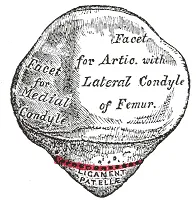
What it protects
The patella acts as a bony shield for the deeper parts of the knee joint2. Because it sits against the femur’s groove, it keeps the quadriceps tendon from rubbing on the end of the femur1. This limits friction on that tendon2.
It also raises the tendon off the knee joint, which gives the quadriceps more leverage to straighten the knee1,2. A study of knees from body donors found it is not a simple pulley passing on equal force6. The ratio of force above and below the kneecap changed with knee angle6.
How much load it carries
A systematic review sums up the research on a question in a checkable way. One such review pooled the peak force across this joint in healthy people3. The figures are averages, as multiples of body weight3.
| Activity, healthy people | Peak force (times body weight) |
|---|---|
| Walking | 0.93 |
| Stair descent | 2.83 |
| Stair ascent | 3.23 |
| Running | 5.23 |
The same review concluded that more knee bend, as in squatting, puts more force on this joint than less bend, as in walking3. The cartilage behind the kneecap, a firm, rubbery body tissue softer than bone, squashes with use7. MRI is a scan that shows soft tissue in detail. In an MRI study of 12 volunteers, its volume fell 2.8% after walking and 5.9% after knee bends7. In that study, more intense loading produced more squashing7.
Breakdown
Fractures
A fracture is a broken bone. A review of 756 patellar fractures over ten years found 13.1 per 100,000 people per year, rising with age8. In males the rate peaked at ages 10 to 19, and in females at ages 60 to 808.
A national register study looked at 3194 patella fractures in adults4. It found:
- a median age of 67, with 64% of patients female4;
- most caused by low-energy injury, 70% from a fall from standing height4;
- 56% of breaks running across the bone and 26% running up and down it4;
- 67% managed without an operation4.
A meta-analysis looked at adults whose patella fractures were fixed with surgery9. Across 24 studies, 33.6% had a further operation, including removal of the fixing hardware9. Infection was reported in 3.2% across 18 studies, and nonunion (a broken bone that fails to join and heal) in 1.3% across 159.
Dislocation
A Cochrane review is a systematic review done to the Cochrane Collaboration’s published standards. One describes a patellar dislocation as the kneecap coming fully out of its groove on the femur10. Most episodes of kneecap instability follow a twisting injury on a bent knee without contact5. The kneecap moves outward across the femur, often also damaging the inner soft tissues that hold it and the joint cartilage5.
One population cohort counted 609 first-time outward dislocations over 21 years5. Adjusted for age and sex, it found 23.2 per 100,000 person-years, a count of people multiplied by the years each was followed5. The rate was similar in males and females and highest at ages 14 to 185. Imaging showed osteochondral injury, damage to the cartilage of a joint and the bone just beneath it, in 16.7%5. Most people in that cohort had a single episode5.
When it happens again
Studies of repeat dislocation report these findings5,11:
- In the cohort of 609, followed for a mean of 12.3 years, 30.0% dislocated again5.
- A meta-analysis of 17 studies found an overall repeat rate of 33.6%11.
- In studies within that review, repeat rates were 7.7 to 13.8% with no risk factors and 70.4 to 78.5% with three11.
The meta-analysis linked younger age and open growth plates to a higher chance of repeat dislocation11. Growth plates are layers of cartilage near the ends of growing bones, where the bones grow longer. It also linked trochlear dysplasia, a shallow or badly shaped groove at the lower end of the thigh bone11. Patella alta, a kneecap that sits higher on the knee than usual, was linked too5,11.
A greater distance from the groove to the tibial tuberosity was a further factor11. The tibial tuberosity is a bump on the front of the shinbone, just below the knee. Sex and earlier dislocation of the other knee were not linked11.
A Cochrane review of 10 trials in 519 people with patellar dislocation was uncertain whether surgery improves outcome over non-surgical care10. It rated the evidence very low certainty10. In 8 of its studies, it took an illustrative 348 per 1000 repeat dislocations over two to nine years without surgery10. Against that, it estimated 157 fewer per 1000 after surgery, on very low-certainty evidence10.
Pain around the kneecap
A 2016 international consensus statement defined patellofemoral pain as pain around or behind the patella12. By that definition, the pain gets worse with at least one activity that loads the joint on a bent, weight-bearing knee12. It gives squatting, stairs, running and jumping as examples12.
A meta-analysis of 23 studies reported that 22.7% of the general population had it over a year, and 28.9% of adolescents13. The consensus statement put its yearly rate of new cases in the military at 3.8% in men and 6.5% in women12. A review of 24 studies found 16 factors reported as linked to a poor outcome14. The most reported was symptoms lasting longer than 4 months14.
A meta-analysis of 18 prospective studies, which follow people forward in time, looked for risk factors15. It found strong to moderate evidence that age, height, weight and Q-angle were not risk factors for it15. The Q-angle is the angle between the line of the thigh and the line of the shin, seen from the front. Moderate evidence showed hip weakness was not a risk factor either, but in adolescents greater hip abduction strength was15. Hip abduction means moving the thigh out to the side.
The same meta-analysis found moderate evidence that weaker quadriceps was a risk factor in the military15. Another meta-analysis found lower hip strength in adults who already had the pain, and suggested the weakness may follow the pain16.
Wear of the joint
Osteoarthritis is a long-term joint disease in which the cartilage lining the joint wears away. A meta-analysis of 32 studies found it in the patellofemoral joint in 25% of people in population-based groups over age 2017. In groups picked because they had symptoms, over age 30, the figure was 39%17.
Recovery
How bone responds to load
A review of bone mechanics reports that bone adapts its structure to the loads it usually carries18. In mechanostat theory, strain below a minimum level leads to bone loss, and strain above it to bone formation18. A meta-analysis pooled 10 randomised trials of supervised exercise lasting 6 months or more19. Leg bones grew stronger where loaded in children, though not in adults19.
One case study treated the patella as a bone loaded almost entirely by the quadriceps20. In that 26-year-old woman, a year of one-legged strength training raised one estimate of strain in the bone by 47%20. Its bone density did not change20.
After injury and disuse
In the same case study, she tore a knee ligament, a tough band of tissue that holds bones together at a joint20. After the tear, her patella lost bone for about 4 months20. Its density then rose over the recovery period, until it came into balance with her muscle strength by the 3-year end point20.
A 2010 systematic review looked at bone after injury to the anterior cruciate ligament, the front one of two ligaments that cross inside the knee21. Bone density fell in the injured leg, at sites including the patella21. It had not returned to pre-injury levels even after ligament surgery21. Loss was greater with limited weight bearing and long periods of disuse or immobilisation, keeping a limb still21.
How a broken bone heals
A review reports that fracture repair usually restores bone’s structure and function, but about 10% of fractures do not heal normally22. A textbook says clotting blood forms a fracture haematoma, the blood clot at the break, in about six to eight hours1. A callus, new tissue that bridges the break, begins to form within about 48 hours and steadies it1. Over the next weeks its cartilage turns into spongy bone, and reshaping often leaves no outer sign of the break1. In one study of 39 patients with surgically fixed patella fractures, the bone had knitted after a mean of 6.9 months23.
Cartilage in trained and untrained people
In the MRI study, cartilage squashing after knee bends did not differ significantly between 14 athletes and 14 untrained volunteers7. The athletes were seven sprinters and seven professional weight lifters7.
Connections
Tendons and muscles
The four quadriceps muscles join into one tendon, the quadriceps tendon, which fixes into the base of the patella24. Below the kneecap it carries on as the patellar ligament, the band that runs down to the shinbone1. That band fixes onto the tibial tuberosity on the tibia1. So the patella anchors both tendons2. Working through them, the quadriceps straightens the knee1.
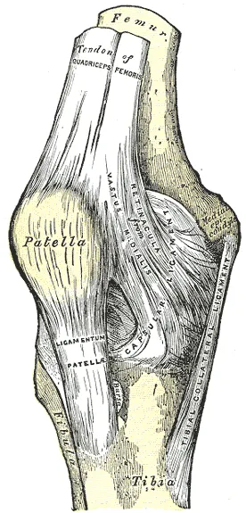
How it tracks in its groove
As the knee straightens, the quadriceps pulls the patella upward and outward1. The outward pull is greater in females, because of their larger Q-angle1. A textbook describes the same angle as how far the femur slants out from vertical, normally 10 to 15 degrees1. A large lip on the outer side of the femur’s groove normally offsets the outward pull and helps the kneecap track1.
Exercises that load it
One study tested 35 exercises in 20 healthy people25. A computer model estimated the highest peak load on this joint in a one-leg decline squat25. That estimate was 8.2 times body weight, against 0.6 for walking25. These figures come from a different study from the table above. Related exercise pages: back squat, decline squat.
Patella is part of
Patella forms a joint with
Patella is attached to by
Patella is pulled by
Patella is loaded by
Sources
- Betts JG, Young KA, Wise JA, Johnson E, Poe B, Kruse DH, Korol O, Johnson JE, Womble M, DeSaix P (2022). Anatomy and Physiology 2e. OpenStax. Link
- Cox CF, Sinkler MA, Black AC, Launico MV, Hubbard JB (2023). Anatomy, Bony Pelvis and Lower Limb, Knee Patella. StatPearls. PMID 30137819
- Hart HF, Patterson BE, Crossley KM, Culvenor AG, Khan MCM, King MG, Sritharan P (2022). May the force be with you: understanding how patellofemoral joint reaction force compares across different activities and physical interventions-a systematic review and meta-analysis.. British Journal of Sports Medicine. doi:10.1136/bjsports-2021-104686 PMID 35115309
- Kruse M, Wolf O, Mukka S, Brüggemann A (2022). Epidemiology, classification and treatment of patella fractures: an observational study of 3194 fractures from the Swedish Fracture Register.. European Journal of Trauma and Emergency Surgery. doi:10.1007/s00068-022-01993-0 PMID 35644894
- Sanders TL, Pareek A, Hewett TE, Stuart MJ, Dahm DL, Krych AJ (2018). Incidence of First-Time Lateral Patellar Dislocation: A 21-Year Population-Based Study.. Sports Health. doi:10.1177/1941738117725055 PMID 28795924
- Huberti HH, Hayes WC, Stone JL, Shybut GT (1984). Force ratios in the quadriceps tendon and ligamentum patellae.. Journal of Orthopaedic Research. doi:10.1002/jor.1100020108 PMID 6491798
- Eckstein F, Lemberger B, Gratzke C, Hudelmaier M, Glaser C, Englmeier KH, Reiser M (2005). In vivo cartilage deformation after different types of activity and its dependence on physical training status.. Annals of the Rheumatic Diseases. doi:10.1136/ard.2004.022400 PMID 15647438
- Larsen P, Court-Brown CM, Vedel JO, Vistrup S, Elsoe R (2016). Incidence and Epidemiology of Patellar Fractures.. Orthopedics. doi:10.3928/01477447-20160811-01 PMID 27536953
- Dy CJ, Little MT, Berkes MB, Ma Y, Roberts TR, Helfet DL, Lorich DG (2012). Meta-analysis of re-operation, nonunion, and infection after open reduction and internal fixation of patella fractures.. Journal of Trauma and Acute Care Surgery. doi:10.1097/TA.0b013e31825168b6 PMID 22902732
- Smith TO, Gaukroger A, Metcalfe A, Hing CB (2023). Surgical versus non-surgical interventions for treating patellar dislocation.. Cochrane Database of Systematic Reviews. doi:10.1002/14651858.CD008106.pub4 PMID 36692346
- Huntington LS, Webster KE, Devitt BM, Scanlon JP, Feller JA (2020). Factors Associated With an Increased Risk of Recurrence After a First-Time Patellar Dislocation: A Systematic Review and Meta-analysis.. American Journal of Sports Medicine. doi:10.1177/0363546519888467 PMID 31825650
- Crossley KM, Stefanik JJ, Selfe J, Collins NJ, Davis IS, Powers CM, McConnell J, Vicenzino B, Bazett-Jones DM, Esculier JF, Morrissey D, Callaghan MJ (2016). 2016 Patellofemoral pain consensus statement from the 4th International Patellofemoral Pain Research Retreat, Manchester. Part 1: Terminology, definitions, clinical examination, natural history, patellofemoral osteoarthritis and patient-reported outcome measures.. British Journal of Sports Medicine. doi:10.1136/bjsports-2016-096384 PMID 27343241
- Smith BE, Selfe J, Thacker D, Hendrick P, Bateman M, Moffatt F, et al (2018). Incidence and prevalence of patellofemoral pain: A systematic review and meta-analysis.. PloS one. doi:10.1371/journal.pone.0190892 PMID 29324820
- Matthews M, Rathleff MS, Claus A, McPoil T, Nee R, Crossley K, Vicenzino B (2017). Can we predict the outcome for people with patellofemoral pain? A systematic review on prognostic factors and treatment effect modifiers.. British Journal of Sports Medicine. doi:10.1136/bjsports-2016-096545 PMID 27965435
- Neal BS, Lack SD, Lankhorst NE, Raye A, Morrissey D, van Middelkoop M (2019). Risk factors for patellofemoral pain: a systematic review and meta-analysis.. British Journal of Sports Medicine. doi:10.1136/bjsports-2017-098890 PMID 30242107
- Rathleff MS, Rathleff CR, Crossley KM, Barton CJ (2014). Is hip strength a risk factor for patellofemoral pain? A systematic review and meta-analysis.. British Journal of Sports Medicine. doi:10.1136/bjsports-2013-093305 PMID 24687010
- Kobayashi S, Pappas E, Fransen M, Refshauge K, Simic M (2016). The prevalence of patellofemoral osteoarthritis: a systematic review and meta-analysis.. Osteoarthritis and Cartilage. doi:10.1016/j.joca.2016.05.011 PMID 27188684
- Hart NH, Nimphius S, Rantalainen T, Ireland A, Siafarikas A, Newton RU (2017). Mechanical basis of bone strength: influence of bone material, bone structure and muscle action.. Journal of musculoskeletal & neuronal interactions. PMID 28860414
- Nikander R, Sievänen H, Heinonen A, Daly RM, Uusi-Rasi K, Kannus P (2010). Targeted exercise against osteoporosis: A systematic review and meta-analysis for optimising bone strength throughout life.. BMC Medicine. doi:10.1186/1741-7015-8-47 PMID 20663158
- Sievänen H, Heinonen A, Kannus P (1996). Adaptation of bone to altered loading environment: a biomechanical approach using X-ray absorptiometric data from the patella of a young woman.. Bone. doi:10.1016/8756-3282(96)00111-1 PMID 8830989
- Nyland J, Fisher B, Brand E, Krupp R, Caborn DN (2010). Osseous deficits after anterior cruciate ligament injury and reconstruction: a systematic literature review with suggestions to improve osseous homeostasis.. Arthroscopy. doi:10.1016/j.arthro.2010.03.017 PMID 20810081
- Einhorn TA, Gerstenfeld LC (2015). Fracture healing: mechanisms and interventions.. Nature reviews. Rheumatology. doi:10.1038/nrrheum.2014.164 PMID 25266456
- Pesch S, Greve F, Zyskowski M, Müller M, Crönlein M, Biberthaler P, Kirchhoff C, Wurm M (2023). High return to sports rates after operative treatment of patella fractures.. European Journal of Medical Research. doi:10.1186/s40001-023-01359-1 PMID 37736742
- Piagkou M, Triantafyllou G, Georgiev GP, Tsakotos G, Olewnik Ł, Landfald IC, Gonera B (2025). Systematic Review and Prevalence Meta-Analysis of Quadriceps Femoris Morphology: Significance of the Quadriceps Tendon in Anterior Cruciate Ligament Reconstruction.. Journal of Functional Morphology and Kinesiology. doi:10.3390/jfmk10030250 PMID 40700186
- Song K, Scattone Silva R, Hullfish TJ, Silbernagel KG, Baxter JR (2023). Patellofemoral Joint Loading Progression Across 35 Weightbearing Rehabilitation Exercises and Activities of Daily Living.. The American journal of sports medicine. doi:10.1177/03635465231175160 PMID 37272685
Changes
- Links to the new patellar dislocation page
- First published.