Anterior cruciate ligament
Also called ACL, anterior cruciate
The ligament inside the knee that stops the shinbone sliding forward. How it is built, how and in whom it tears, why it heals poorly, and which muscles load or unload it.
- Region
- Knee
- Updated
- September 30, 2026
- Reviewed
- September 30, 2026 · how entries are checked
Function
Where it runs
The anterior cruciate ligament (ACL) is a band of collagen fibres inside the knee1. It starts on top of the tibia (shinbone), at the front inner part of the area between its two joint surfaces1. It runs backward and outward to the inner face of the outer condyle of the femur (thigh bone)1. A condyle is a rounded knob at the end of a bone, where it meets another bone at a joint. The ACL measures about 32 mm long and 7 to 12 mm wide1.
It has two bundles, separated where they meet the femur by a bony ridge called the bifurcate ridge1:
| Bundle | Tightest when | Main role |
|---|---|---|
| Anteromedial (front, inner side) | The knee is bent1 | Resisting forward movement of the tibia1 |
| Posterolateral (back, outer side) | The knee is straight1 | Side-to-side and turning stability, as a backup restraint1 |
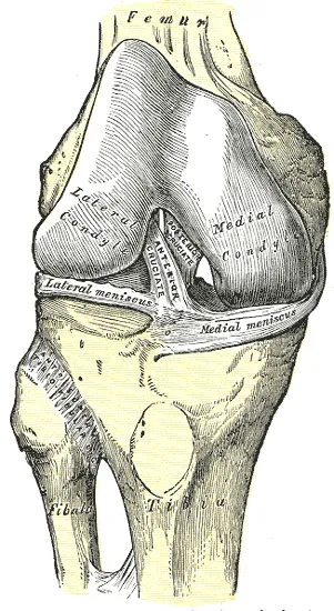
What it holds back
The ACL has long been considered the main passive restraint stopping the tibia sliding forward on the femur3. Together with the posterior cruciate ligament, it forms a cross inside the knee1. The pair stop the tibia moving too far forward or backward as the knee bends and straightens1. The ACL tightens as the knee straightens, and so resists the knee bending backward past straight2. Its direction also helps it steady the knee against turning and side-to-side movement3.
Blood and nerve supply
The ACL gets its blood mainly from the middle genicular artery, one of the arteries of the knee1. In one study of a cadaver (a body donated for study), the ends of the ACL looked better supplied than its middle6. Its attachments to bone added little to that supply6.
The posterior articular nerve, a branch of the tibial nerve (the main nerve at the back of the lower leg), supplies the ACL1. A review with a systematic search reported that mechanoreceptors, nerve endings that sense movement, sit mainly near the ligament’s attachments to bone7. They are believed to add to the sense of where the knee is7.
Breakdown
How it tears
A position statement describes ACL rupture as loading beyond the tissue’s current strength8. It says that strength varies with age, sex, training and injury8. A 2014 review puts more than 70% of ACL injuries as happening without contact, typically when landing from a jump or cutting sideways3. A 2022 review of 45 team-sport studies put the share at 55%: 63% in females and 50% in males4.
The 2014 review says studies have named a mix of three loads as the worst case and main way non-contact tears happen3. One is a shear force (a force that pushes two surfaces to slide across each other) moving the tibia forward3. The others are the knee falling inward and the tibia twisting inward3.
The position statement lists these typical body postures at the moment of injury8:
- a nearly straight knee8
- the knee collapsing inward, with the hip turned in8
- a flat foot8
- the trunk leaning sideways over the standing leg8
The position statement estimates that the ligament ruptures less than 50 milliseconds after the foot first touches the ground8. It says that is too fast for muscles to switch on, whether by choice or by reflex8. In 8 people imaged during a jump landing, ACL strain (stretch as a share of resting length) peaked 55 milliseconds before touchdown, on average9.
A systematic review (a study that searches for all the research on a question in a set, checkable way) studied bone bruises after ACL tears10. It found bruises on the outer top of the tibia in 82.8% of cases and on the outer femoral condyle in 71.4%10. Where the bruises sat, front to back, showed the tibia shifting forward on the femur at the moment of injury10.
Who tears it
A systematic review converted injury studies into yearly rates11:
| Group | ACL injuries per person per year |
|---|---|
| National population studies in Australia | Up to 0.05%11 |
| Amateur sport | Between the two11 |
| Professional basketball, soccer and other football codes | 0.15% to 3.7%11 |
The professional figures exclude small studies, and amateur rates were generally in between11.
One population study counted 1,841 people newly diagnosed with isolated ACL tears (the ACL torn on its own) between 1990 and 20105. Adjusted to the US population, the yearly incidence (new cases in a set time) was 68.6 per 100,000 person-years5. It was higher in males (81.7) than in females (55.3)5. In that study, tears peaked at ages 19 to 25 in males and 14 to 18 in females5.
In athletes, a meta-analysis (a study that pools the results of several earlier studies) found female athletes more likely than males to injure the ACL12. Its authors noted it is unknown whether this holds in every sport or level12. Another meta-analysis put the female rate at about 3 times the male rate in contact sports13. It found similar rates between the sexes in collision, limited-contact and noncontact sports13.
Risk factors in the body
A position statement drew on a systematic review of prospective studies (studies that follow people forward in time)8. It names a previous ACL injury as the most consistent risk factor inside the body8. A meta-analysis of 19 studies found a 15% rate of second ACL injury after reconstruction (surgery to rebuild the ligament)14. That was 7% to the same knee and 8% to the other14. Other factors have been studied:
- Knee shape. The position statement reports strong evidence tying risk to a smaller ACL, a steeper backward tibial slope and a narrower notch between femoral condyles8. It notes these are largely not changeable8.
- Family history. In a nationwide Korean group of 5,184,603 people, risk was 1.79 times higher with an affected parent, sibling or child15. With an affected twin, it was 4.49 times higher15.
A study followed 880 elite female handball and football players16. Trained raters’ single-leg squat scores did not pick out those who later had a non-contact ACL injury16. In 17 healthy adults imaged during a single-leg jump, the knee barely angled inward or outward, under 1 degree on average17. No significant link was found between ACL strain and that angle17.
Damage that comes with it
ACL injuries are associated with torn menisci (pads of cartilage in the knee), damage to joint cartilage and a higher risk of early osteoarthritis3. Osteoarthritis is a long-term joint disease in which the cartilage lining the joint wears away. A meta-analysis of 53 studies found the odds of knee osteoarthritis were about 4 times higher after an ACL injury than in an uninjured knee18.
In 100 patients having surgery for what seemed a lone ACL rupture, MRI (a scan that shows soft tissue in detail) was checked19. In 67%, it also showed injury to the medial collateral ligament area19. The ACL tear page covers how the injury is defined, its course and what trials found; the menisci page covers the torn pads.
Recovery
Why it heals poorly
The ACL has long been thought to heal poorly3. Failure rates of 40% to 100% were reported even after surgeons stitched it back together3. A review contrasts this with the high healing ability of tissues outside the joint, such as the medial collateral ligament3. The reasons it reports from earlier research include the joint fluid, the injury response, differences in the cells, blood supply and loading3.
In torn ligaments outside the joint, a blood clot forms a temporary scaffold for healing3. After an ACL tear, that clot is missing, mainly because joint fluid keeps flowing and spreads the blood away3. Cell and tissue studies show the ACL and the medial collateral ligament grow new vessels and nerves after injury in a comparable way3. Collagen production in the ACL carries on for up to a year after injury3.
What scans and tests show after a tear
A systematic review took in 9 studies of 734 people whose complete ACL rupture was managed without surgery20. All 9 reported some ligaments regaining continuous fibres on MRI20. In the two studies that recorded where the tear was, most of the ligaments that healed had torn near the femur20. The studies were of low quality20.
After a rupture, the number of mechanoreceptors in the ACL falls quickly, according to a review with a systematic search7. The fall is slower where the torn stump sticks to the posterior cruciate ligament7.
How a replacement graft changes
In ACL reconstruction, a tendon (a tough cord of tissue that connects a muscle to a bone) graft takes its place21. A systematic review of human biopsy studies found the graft survives in the joint21. It goes through a series of changes called ligamentization21. Under the microscope, a mature graft may come to resemble a normal ACL, though differences remain in how its finest collagen fibres are laid out21. There is no agreement on how long each stage takes21.
A systematic review of 23 biopsy and imaging studies, with 706 patients, described three phases: early healing, remodelling and ligamentization22. The process was still going on a year after surgery22.
Connections
The joint, bones and ligaments around it
The ACL sits inside the knee joint, joining the tibia to the femur1,2. It crosses the posterior cruciate ligament in an X2. A review of meniscus research describes one study in a knee without a working ACL23. It found the back end of the inner meniscus was the most important structure resisting forward movement of the tibia23.
Muscles that load or unload it
A position statement explains that muscles can raise or lower the load on the ACL, depending on their line of pull and the knee’s position8. It says that in landing and cutting, the quadriceps and gastrocnemius pull the tibia forward8. The hamstrings and soleus pull it back8. A narrative review (a summary of research written without a set, checkable search method) adds detail on each group24:
- The quadriceps and gastrocnemius raise the load, especially when the knee is straight24.
- The hamstrings and soleus appear to unload it, the hamstrings only with the knee bent past about 20 to 30 degrees24.
The position statement names the gluteus medius, gluteus maximus and piriformis as the biggest contributors against the knee-inward turning force8. The piriformis is a small muscle deep in the buttock.
Exercises that load it
A narrative review of exercise studies found the ACL is loaded less with the knee bent 50 to 100 degrees25. It found more forward trunk lean in squats and lunges unloads it25. In 12 recreationally active people, modelled peak ACL force was 24% lower in single-leg squats with moderate forward lean than with little lean26. In 17 healthy adults imaged during a single-leg jump, ACL strain rose as the knee straightened17. Related exercise pages: back squat, single-leg squat and drop landing.
Anterior cruciate ligament holds together
Anterior cruciate ligament attaches to
Anterior cruciate ligament crosses
Anterior cruciate ligament is often injured with
Anterior cruciate ligament has its load raised by
Anterior cruciate ligament is unloaded by
Anterior cruciate ligament is torn in
Anterior cruciate ligament is loaded by
Sources
- Evans J, Mabrouk A, Nielson JL (2023). Anterior Cruciate Ligament Knee Injury.. StatPearls. PMID 29763023
- Betts JG, Young KA, Wise JA, Johnson E, Poe B, Kruse DH, Korol O, Johnson JE, Womble M, DeSaix P (2022). Anatomy and Physiology 2e. OpenStax. Link
- Kiapour AM, Murray MM (2014). Basic science of anterior cruciate ligament injury and repair.. Bone & joint research. doi:10.1302/2046-3758.32.2000241 PMID 24497504
- Chia L, De Oliveira Silva D, Whalan M, McKay MJ, Sullivan J, Fuller CW, et al (2022). Non-contact Anterior Cruciate Ligament Injury Epidemiology in Team-Ball Sports: A Systematic Review with Meta-analysis by Sex, Age, Sport, Participation Level, and Exposure Type.. Sports medicine (Auckland, N.Z.). doi:10.1007/s40279-022-01697-w PMID 35622227
- Sanders TL, Maradit Kremers H, Bryan AJ, Larson DR, Dahm DL, Levy BA, et al (2016). Incidence of Anterior Cruciate Ligament Tears and Reconstruction. The American journal of sports medicine. doi:10.1177/0363546516629944 PMID 26920430
- Toy BJ, Yeasting RA, Morse DE, McCann P (1995). Arterial supply to the human anterior cruciate ligament.. Journal of athletic training. PMID 16558326
- Kosy JD, Mandalia VI (2018). Anterior Cruciate Ligament Mechanoreceptors and their Potential Importance in Remnant-Preserving Reconstruction: A Review of Basic Science and Clinical Findings.. The journal of knee surgery. doi:10.1055/s-0037-1608941 PMID 29228403
- Collings TJ, Diamond LE, Barrett RS, Saxby DJ, Lloyd DG, Whiteley R, et al (2026). An Evidence-Based and Mechanistic Approach to Reducing the Risk of Anterior Cruciate Ligament Injury: An Exercise and Sport Science Australia Position Statement.. Sports medicine (Auckland, N.Z.). doi:10.1007/s40279-026-02450-3 PMID 42133198
- Taylor KA, Terry ME, Utturkar GM, Spritzer CE, Queen RM, Irribarra LA, et al (2011). Measurement of in vivo anterior cruciate ligament strain during dynamic jump landing.. Journal of biomechanics. doi:10.1016/j.jbiomech.2010.10.028 PMID 21092960
- Sohn S, AlShammari SM, Hwang BJ, Kim MS (2024). A Systematic Review of Bone Bruise Patterns following Acute Anterior Cruciate Ligament Tears: Insights into the Mechanism of Injury.. Bioengineering (Basel, Switzerland). doi:10.3390/bioengineering11040396 PMID 38671817
- Moses B, Orchard J, Orchard J (2012). Systematic review: Annual incidence of ACL injury and surgery in various populations.. Research in sports medicine (Print). doi:10.1080/15438627.2012.680633 PMID 22742074
- Norman D, Terrian L, Novosel J, Guzman A, Montalvo AM, Myer GD, et al (2026). Anterior Cruciate Ligament Injury Incidence Across Sex, Sport, and Competition Level: A Systematic Review and Meta-Analysis.. Journal of athletic training. doi:10.4085/1062-6050-0559.24 PMID 41816313
- Montalvo AM, Schneider DK, Webster KE, Yut L, Galloway MT, Heidt RS Jr, et al (2019). Anterior Cruciate Ligament Injury Risk in Sport: A Systematic Review and Meta-Analysis of Injury Incidence by Sex and Sport Classification.. Journal of athletic training. doi:10.4085/1062-6050-407-16 PMID 31009238
- Wiggins AJ, Grandhi RK, Schneider DK, Stanfield D, Webster KE, Myer GD (2016). Risk of Secondary Injury in Younger Athletes After Anterior Cruciate Ligament Reconstruction. The American journal of sports medicine. doi:10.1177/0363546515621554 PMID 26772611
- Ahn HS, Lee DH, Kazmi SZ, Kang T, Lee YS, Sung R, et al (2021). Familial Risk and Its Interaction With Body Mass Index and Physical Activity in Anterior Cruciate Ligament Injury Among First-Degree Relatives: A Population-Based Cohort Study.. The American journal of sports medicine. doi:10.1177/03635465211032643 PMID 34491137
- Petushek E, Nilstad A, Bahr R, Krosshaug T (2021). Drop Jump? Single-Leg Squat? Not if You Aim to Predict Anterior Cruciate Ligament Injury From Real-Time Clinical Assessment: A Prospective Cohort Study Involving 880 Elite Female Athletes.. Journal of Orthopaedic & Sports Physical Therapy. doi:10.2519/jospt.2021.10170 PMID 34192883
- Foody JN, Bradley PX, Spritzer CE, Wittstein JR, DeFrate LE, Englander ZA (2023). Elevated In Vivo ACL Strain Is Associated With a Straight Knee in Both the Sagittal and the Coronal Planes.. The American journal of sports medicine. doi:10.1177/03635465221141876 PMID 36625427
- Poulsen E, Goncalves GH, Bricca A, Roos EM, Thorlund JB, Juhl CB (2019). Knee osteoarthritis risk is increased 4-6 fold after knee injury - a systematic review and meta-analysis.. British journal of sports medicine. doi:10.1136/bjsports-2018-100022 PMID 31072840
- Willinger L, Balendra G, Pai V, Lee J, Mitchell A, Jones M, Williams A (2022). High incidence of superficial and deep medial collateral ligament injuries in 'isolated' anterior cruciate ligament ruptures: a long overlooked injury. Knee Surgery, Sports Traumatology, Arthroscopy. doi:10.1007/s00167-021-06514-x PMID 33661325
- Pitsillides A, Stasinopoulos D, Giannakou K (2021). Healing potential of the anterior cruciate ligament in terms of fiber continuity after a complete rupture: A systematic review.. Journal of bodywork and movement therapies. doi:10.1016/j.jbmt.2021.06.003 PMID 34776148
- Claes S, Verdonk P, Forsyth R, Bellemans J (2011). The “Ligamentization” Process in Anterior Cruciate Ligament Reconstruction. The American journal of sports medicine. doi:10.1177/0363546511402662 PMID 21515806
- van Groningen B, van der Steen MC, Janssen DM, van Rhijn LW, van der Linden AN, Janssen RPA (2020). Assessment of Graft Maturity After Anterior Cruciate Ligament Reconstruction Using Autografts: A Systematic Review of Biopsy and Magnetic Resonance Imaging studies.. Arthroscopy, sports medicine, and rehabilitation. doi:10.1016/j.asmr.2020.02.008 PMID 32875303
- Fox AJ, Bedi A, Rodeo SA (2012). The Basic Science of Human Knee Menisci. Sports health. doi:10.1177/1941738111429419 PMID 23016106
- Maniar N, Cole MH, Bryant AL, Opar DA (2022). Muscle Force Contributions to Anterior Cruciate Ligament Loading.. Sports medicine (Auckland, N.Z.). doi:10.1007/s40279-022-01674-3 PMID 35437711
- Escamilla RF, Macleod TD, Wilk KE, Paulos L, Andrews JR (2012). Cruciate ligament loading during common knee rehabilitation exercises.. Proceedings of the Institution of Mechanical Engineers, Part H. doi:10.1177/0954411912451839 PMID 23025167
- Kulas AS, Hortobágyi T, DeVita P (2012). Trunk position modulates anterior cruciate ligament forces and strains during a single-leg squat.. Clinical Biomechanics. doi:10.1016/j.clinbiomech.2011.07.009 PMID 21839557
Changes
- A supporting claim was narrowed to match its source; the page text is unchanged.