Medial collateral ligament
Also called MCL, tibial collateral ligament, medial collateral ligament of the knee, MCL complex
The broad band on the inner side of the knee that holds it against inward-pushing and twisting forces. How it is built, how and in whom it is injured, and what studies report about how it heals.
- Region
- Knee
- Muscles
- hamstrings, gastrocnemius, semimembranosus
- Updated
- September 30, 2026
- Reviewed
- September 30, 2026 · how entries are checked
Function
Three parts on the inner knee
The MCL runs from the inner epicondyle of the femur to a ridge at the back-inner edge of the tibia1. An epicondyle is a bony bump on the side of a condyle, where muscles or ligaments attach. A condyle is a rounded knob at the end of a bone.
The MCL is 8 to 10 cm long5. It is a complex with three parts4:
- The superficial MCL is the largest ligament on the inner side of the knee4. It is broad, flat and triangular, and widest in its middle6.
- The deep MCL is a thickening of the inner joint capsule, the sleeve of tough tissue that encloses a joint6. It attaches to the inner meniscus (a pad of cartilage in the knee)6.
- The posterior oblique ligament is made of fibrous extensions of the tendon of the semimembranosus, one of the three hamstring muscles4. They strengthen the back-inner part of the capsule4.
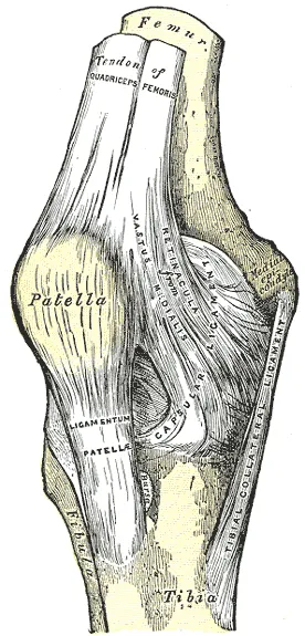
Where it attaches
The superficial MCL joins the femur and tibia through one attachment on the femur and two on the tibia4. Anatomy studies have measured these sites:
| What was measured | What was found |
|---|---|
| Length of the superficial MCL | Averaged 94.8 mm in eight cadaver knees (knees from bodies donated for study)7 |
| Attachment on the femur | Wrapped around the inner epicondyle, centred about 1 mm above it, in 22 knees8 |
| Upper attachment on the tibia | Reaches the front arm of the semimembranosus tendon, 12.2 mm below the joint on average6 |
| Lower attachment on the tibia | 61.2 mm below the knee joint in eight cadaver knees7 |
What it holds steady
The MCL is a broad, strong band whose main job is to steady the knee from side to side1. It resists valgus and helps hold the knee against external rotation (turning a limb outward, away from the middle of the body)1. Its parts share this work by knee angle6,9:
- In a cutting study of 18 cadaver knees, the superficial MCL controlled valgus at each angle tested9. It was the dominant restraint from 30° to 90° of bend9.
- In 28 cadaver knees tested by robot, the superficial MCL was the most important restraint to external rotation and valgus at each angle tested10. The same held for anteromedial rotation (a twist of the shinbone in which its inner side moves forward)10.
- The deep MCL takes about 20% of the load resisting outward turning of the tibia at 0–30° of bend6.
- In the 18-knee cutting study, the deep MCL was a secondary restraint to valgus9.
- The posterior oblique ligament is tight with the knee straight and goes slack as soon as it bends6.
In one cadaver study at 60° of knee bend, the lower superficial MCL took 45% more valgus load than the upper portion6. In 28 cadaver knees, cutting the upper tibial attachment caused no significant rise in laxity while the lower one stayed intact10. Laxity is looseness in a joint or ligament.
How little it stretches in landing
When a robot moved 18 cadaver knees through a recorded drop-jump landing, the ACL’s peak strain far exceeded the MCL’s11. Strain is stretch as a share of resting length. The peaks were 6.1% for the ACL and 0.4% for the MCL11.
Breakdown
How it gets hurt
MCL injury most often comes from a blow to the outside of the knee, pushing it inward2. It can also come from valgus force combined with the tibia turning outward2. The ligament is especially exposed with the knee bent about 20–30°, when the superficial MCL is taut6. Minor trauma can tear the superficial part, while higher-energy injuries can disrupt both layers6.
Studies of professional men’s football show how this plays out2,12:
- In video of 37 MCL injuries, 62% were direct contact, 24% indirect contact and 14% non-contact12.
- Each of the indirect and non-contact injuries showed the knee loaded into valgus12. Most also showed the foot turned outward and hip abduction (the thigh moving out to the side)12.
- In a prospective study (one that follows people forward in time) of 51 teams, 75% of 130 MCL injuries came through contact2. The commonest were being tackled (29%) and tackling (12%)2.
Who injures it, and how often
Most MCL injuries come from valgus loading, outward turning or both, in sports such as football, skiing and ice hockey4. They occur mainly in young athletes4. A 2010 review describes the superficial MCL, deep MCL and posterior oblique ligament as the knee’s most commonly injured ligaments3. One 10-year series recorded 19,530 sports injuries, 7,769 of them to the knee13. MCL lesions made up 7.9% of the injuries documented, against 20.3% for the ACL13.
That series linked MCL injury with judo and skiing13.
| Group studied | What was found |
|---|---|
| 27 European professional football teams over 11 seasons | 346 MCL injuries, 0.33 per 1000 hours, nine times higher in matches than in training14 |
| Cadets at the United States Military Academy, 2005 to 2009 | 128 had isolated MCL injuries (no other structure hurt)15 |
| College athletes at the same academy | Wrestling, judo, hockey and rugby had the highest rates of isolated MCL sprain (a stretched or torn ligament)15 |
| College athletes at the same academy, by sex | Men had about 2.9 times the women’s rate of isolated MCL sprain15 |
| US high school and college athletes | Female athletes had the higher rate in high school, but the lower rate in college16 |
How injuries are graded
MCL injuries are graded I to III by how far the inner side of the joint opens under valgus5. On MRI (a scan that shows soft tissue in detail), each grade has its own look2:
| Grade | Inner joint opening5 | On MRI2 |
|---|---|---|
| I | No laxity; 3–5 mm5 | Fibres intact, with oedema (fluid swelling) around them2 |
| II | Opens at 30° of bend; 6–10 mm5 | Some fibres torn2 |
| III | Opens at 0° and 30°; more than 10 mm5 | Fully torn, or pulled off the bone (avulsion)2 |
In 130 MCL injuries in European professional men’s football, the upper third of the ligament was the most common site (54%)2. The middle third followed (31%), then the lower third (15%)2. Of these, 88% were isolated2. The inner meniscus was the most frequent injury alongside it2.
When the ACL tears too
Studies report different figures for how often the MCL is hurt alongside an ACL tear17,18. A 2019 review puts MCL injuries at about 30% of ACL ruptures17. ACL reconstruction is surgery that rebuilds a torn ACL using a graft. Among 100 people having ACL reconstruction for what seemed an isolated ACL rupture, 67% had MCL complex injury on MRI18. Biomechanical studies show that partial or complete MCL tears raise the load on the ACL6.
Recovery
How well it heals: two views
Sources differ on how completely the MCL heals2,4. One says it has greater potential to heal than the ACL, often to full recovery, because it lies outside the joint with enough blood supply2.
Another source says injured ligaments heal by forming scar rather than regrowing, and the MCL heals fairly well but cannot be fully restored4. One source says that such scar is worse, biologically and mechanically, than the tissue it replaces19.
Tears of the lower superficial MCL have shown poorer healing than upper tears6. This has been linked to a poorer blood supply6. It has also been linked to soft tissue, such as the pes anserinus, getting caught between the torn ends6. The pes anserinus is the spot on the inner upper shinbone where the tendons of three thigh muscles attach together.
What animal studies show
Animal studies have tracked healing over weeks and months4,20:
- In a rabbit MCL model, looseness seemed restored by 14 weeks4. Yet about 65% of the ligament’s strength had returned by 40 weeks, compared with the other side4.
- In dog models, the MCL’s mechanical properties had not fully recovered by 48 weeks after injury20.
A systematic review (a study that searches for all the research on a question) looked at seven studies of surgically injured rat or rabbit MCLs20. It found early evidence that loading after injury raised the ligament’s breaking force and stiffness, compared with unloading it, such as by keeping it still20. Stiffness is how much force it takes to stretch something by a given amount.
Yet three of those studies consistently reported more laxity in the loaded groups20. The review judged these changes slow, with large, precise effects not clear until 14 weeks20. Data from two studies suggest that strength was about 40% of uninjured controls after 12 weeks of loading20.
Time lost from sport
Days lost to MCL injuries, by study and grade2,5,15:
| Group and grade | Days lost |
|---|---|
| Professional men’s football, grade I (average) | 102 |
| Professional men’s football, any grade (average) | 242 |
| Military academy, grade 1 (median) | 13.515 |
| Military academy, higher grades (median) | 2915 |
| Review of 26 reports, grade I (average) | 105 |
| Review of 26 reports, grade II (average) | 355 |
In the study of 130 football injuries, 11 (8.5%) were re-injuries of the same knee within 2 months of return to play2.
Nine years on
One study followed people about nine years after isolated MCL sprains that had no surgery21. The 54 people with grade II sprains generally did well, though some leftover looseness was common21. The 27 with grade III sprains fared much worse21. Their problems included lasting instability, weakness and osteoarthritis (a long-term joint disease in which the cartilage lining the joint wears away) after the injury21.
Connections
The joint and bones
The MCL runs down the inner side of the knee joint, from the femur to the tibia1. Both it and its outer partner, the lateral collateral ligament, sit outside the joint capsule22. In 10 cadaver knees from older donors, the fan-shaped MCL was longer, much wider and thinner than the cord-like lateral collateral ligament23.
The meniscus and other ligaments
The MCL is firmly joined on its deep side to the joint capsule and the inner meniscus22. The deep MCL holds the inner meniscus firmly at its midpoint24. The anchor to the MCL is one reason the inner meniscus moves less than the outer one22. The menisci page covers them. The anterior cruciate ligament takes more load when the MCL is torn6.
Muscles that share the load
The gastrocnemius, the inner hamstrings and the MCL work together to steady the inner knee against valgus5. In a biomechanical study of knees with a complete MCL tear, simulated hamstring pull brought the valgus angle close to that of the intact knee6.
Exercises that load it
Related exercise pages: drop landing, single-leg squat. One study tracked 16 people with ACL deficiency during a single-leg squat, using a two-angle X-ray system25. ACL deficiency means a knee whose ACL is torn or no longer working. At 0–30° of bend on the way down, the MCL was longer before ACL reconstruction than 18 or more months after25.
Medial collateral ligament steadies
Medial collateral ligament joins
Medial collateral ligament is attached to
Medial collateral ligament shares valgus restraint with
Medial collateral ligament is often injured with
Medial collateral ligament is the inner partner of
Medial collateral ligament is loaded by
Sources
- Mabrouk A, Hubbard JB (2025). Anatomy, Bony Pelvis and Lower Limb, Knee Medial Collateral Ligament. StatPearls. PMID 29939557
- Lundblad M, Hägglund M, Thomeé C, Hamrin Senorski E, Ekstrand J, Karlsson J, Waldén M (2019). Medial collateral ligament injuries of the knee in male professional football players: a prospective three-season study of 130 cases from the UEFA Elite Club Injury Study. Knee Surgery, Sports Traumatology, Arthroscopy. doi:10.1007/s00167-019-05491-6 PMID 30949749
- Wijdicks CA, Griffith CJ, Johansen S, Engebretsen L, LaPrade RF (2010). Injuries to the medial collateral ligament and associated medial structures of the knee. The Journal of Bone and Joint Surgery. American Volume. doi:10.2106/JBJS.I.01229 PMID 20439679
- Georgiev GP, Telang M, Landzhov B, Olewnik Ł, Slavchev SA, LaPrade RF, Ruzik K, Tubbs RS (2022). The novel epiligament theory: differences in healing failure between the medial collateral and anterior cruciate ligaments. Journal of Experimental Orthopaedics. doi:10.1186/s40634-021-00440-0 PMID 35028759
- Svantesson J, Piussi R, Weissglas E, Svantesson E, Horvath A, Börjesson E, Williams A, Prill R (2024). Shedding light on the non-operative treatment of the forgotten side of the knee: rehabilitation of medial collateral ligament injuries-a systematic review. BMJ Open Sport & Exercise Medicine. doi:10.1136/bmjsem-2023-001750 PMID 38933372
- Lucidi GA, Solaro L, Grassi A, Alhalalmeh MI, Ratti S, Manzoli L, Zaffagnini S (2024). Current trends in the medial side of the knee: not only medial collateral ligament (MCL). Journal of Orthopaedics and Traumatology. doi:10.1186/s10195-024-00808-9 PMID 39704918
- LaPrade RF, Engebretsen AH, Ly TV, Johansen S, Wentorf FA, Engebretsen L (2007). The anatomy of the medial part of the knee. The Journal of Bone and Joint Surgery. American Volume. doi:10.2106/JBJS.F.01176 PMID 17768198
- Athwal KK, Willinger L, Shinohara S, Ball S, Williams A, Amis AA (2020). The bone attachments of the medial collateral and posterior oblique ligaments are defined anatomically and radiographically. Knee Surgery, Sports Traumatology, Arthroscopy. doi:10.1007/s00167-020-06139-6 PMID 32737529
- Robinson JR, Bull AM, Thomas RR, Amis AA (2006). The role of the medial collateral ligament and posteromedial capsule in controlling knee laxity. The American Journal of Sports Medicine. doi:10.1177/0363546506289433 PMID 16816148
- Wierer G, Milinkovic D, Robinson JR, Raschke MJ, Weiler A, Fink C, Herbort M, Kittl C (2021). The superficial medial collateral ligament is the major restraint to anteromedial instability of the knee. Knee Surgery, Sports Traumatology, Arthroscopy. doi:10.1007/s00167-020-05947-0 PMID 32277264
- Bates NA, Nesbitt RJ, Shearn JT, Myer GD, Hewett TE (2015). Relative strain in the anterior cruciate ligament and medial collateral ligament during simulated jump landing and sidestep cutting tasks: implications for injury risk. The American Journal of Sports Medicine. doi:10.1177/0363546515589165 PMID 26150588
- Buckthorpe M, Pisoni D, Tosarelli F, Danelon F, Grassi A, Della Villa F (2021). Three Main Mechanisms Characterize Medial Collateral Ligament Injuries in Professional Male Soccer-Blow to the Knee, Contact to the Leg or Foot, and Sliding: Video Analysis of 37 Consecutive Injuries. The Journal of Orthopaedic and Sports Physical Therapy. doi:10.2519/jospt.2021.10529 PMID 34784244
- Majewski M, Susanne H, Klaus S (2006). Epidemiology of athletic knee injuries: A 10-year study. The Knee. doi:10.1016/j.knee.2006.01.005 PMID 16603363
- Lundblad M, Waldén M, Magnusson H, Karlsson J, Ekstrand J (2013). The UEFA injury study: 11-year data concerning 346 MCL injuries and time to return to play. British Journal of Sports Medicine. doi:10.1136/bjsports-2013-092305 PMID 23624324
- Roach CJ, Haley CA, Cameron KL, Pallis M, Svoboda SJ, Owens BD (2014). The epidemiology of medial collateral ligament sprains in young athletes. The American Journal of Sports Medicine. doi:10.1177/0363546514524524 PMID 24603529
- Stanley LE, Kerr ZY, Dompier TP, Padua DA (2016). Sex Differences in the Incidence of Anterior Cruciate Ligament, Medial Collateral Ligament, and Meniscal Injuries in Collegiate and High School Sports: 2009-2010 Through 2013-2014.. The American journal of sports medicine. doi:10.1177/0363546516630927 PMID 26940226
- Filbay SR, Grindem H (2019). Evidence-based recommendations for the management of anterior cruciate ligament (ACL) rupture.. Best practice & research. Clinical rheumatology. doi:10.1016/j.berh.2019.01.018 PMID 31431274
- Willinger L, Balendra G, Pai V, Lee J, Mitchell A, Jones M, Williams A (2022). High incidence of superficial and deep medial collateral ligament injuries in 'isolated' anterior cruciate ligament ruptures: a long overlooked injury. Knee Surgery, Sports Traumatology, Arthroscopy. doi:10.1007/s00167-021-06514-x PMID 33661325
- Frank CB (2004). Ligament structure, physiology and function. Journal of musculoskeletal & neuronal interactions. PMID 15615126
- Bleakley C, Netterström-Wedin F (2023). Does mechanical loading restore ligament biomechanics after injury? A systematic review of studies using animal models. BMC Musculoskeletal Disorders. doi:10.1186/s12891-023-06653-x PMID 37349749
- Kannus P (1988). Long-term results of conservatively treated medial collateral ligament injuries of the knee joint. Clinical Orthopaedics and Related Research. PMID 3335084
- Betts JG, Young KA, Wise JA, Johnson E, Poe B, Kruse DH, Korol O, Johnson JE, Womble M, DeSaix P (2022). Anatomy and Physiology 2e. OpenStax. Link
- Wilson WT, Deakin AH, Payne AP, Picard F, Wearing SC (2012). Comparative analysis of the structural properties of the collateral ligaments of the human knee. Journal of Orthopaedic & Sports Physical Therapy. doi:10.2519/jospt.2012.3919 PMID 22030378
- Fox AJ, Bedi A, Rodeo SA (2012). The Basic Science of Human Knee Menisci. Sports health. doi:10.1177/1941738111429419 PMID 23016106
- Agostinone P, Di Paolo S, Lucidi GA, Marchiori G, Bontempi M, Bragonzoni L, Davidoni V, Grassi A, Zaffagnini S (2024). Changes in the Elongation Pattern of the Medial Collateral Ligament During a Single-Leg Squat After Anterior Cruciate Ligament Reconstruction.. Orthopaedic Journal of Sports Medicine. doi:10.1177/23259671241283795 PMID 39507327
- Bilyy A, Win Mar WW, Al Abdeen Al Zuabi Z, Frimpong-Manso HB, Famure S, Solomou G, Brassett C, Pasapula C (2025). The Deltoid-Spring Ligament Complex: A Scoping Review and New Segmental Classification.. Cureus. doi:10.7759/cureus.81715 PMID 40322447
Changes
- A supporting claim was narrowed to match its source; the page text is unchanged.