Lateral collateral ligament
Also called LCL, fibular collateral ligament, lateral collateral ligament of the knee
The cord-like ligament on the outer side of the knee, from the thigh bone to the fibula. What it holds back, how and in whom it is injured, and what studies found about recovery.
- Region
- Knee
- Muscles
- hamstrings, gastrocnemius, popliteus
- Updated
- September 30, 2026
- Reviewed
- September 30, 2026 · how entries are checked
Function
Where it runs
The LCL is a fibrous band1. Its top end is on the lateral epicondyle, a bony bump on the outer femur, and its lower end on the fibular head1. Its accepted anatomical name is the fibular collateral ligament, and lateral collateral ligament is an accepted second name5. It sits outside the joint capsule, the sleeve of tough tissue that encloses the knee6. It is about 70 mm long, 4 to 5 mm wide and 2 to 3 mm thick1.
The medial collateral ligament on the inner side is fan-shaped, but the LCL is cord-like1. It has no attachment to the meniscus (a pad of cartilage in the knee) or to the joint capsule1. In 10 cadaver knees from older donors (bodies donated for study), the medial ligament was significantly longer, much wider and thinner than the LCL7.
Where it attaches
In 10 cadaver knees, the top end attached to the femur on average 1.4 mm above and 3.1 mm behind the lateral epicondyle8. The lower end attached to the head of the fibula, on average 8.2 mm behind its front edge8.
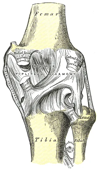
Its blood comes from branches of the popliteal artery, the main artery behind the knee1. Its nerve supply comes from the common fibular (or common peroneal) nerve, a nerve that wraps around the outer side of the knee1. In a study of 36 cadavers, the ligament held three types of nerve ending9.
What it holds back
A review describes the LCL as the knee’s primary restraint against varus load2. The same review says it also resists outward turning of the shin up to 30° of knee bend, and helps less against inward turning2. In 10 cadaver knees, cutting it showed it mattered for resisting varus with the knee straight and at 30° and 60° of bend10. In a cadaver study, its load under varus was fairly steady at 0°, 30° and 60°, and fell significantly at 90°11.
In a cadaver study, it carried load under varus and under turning in either direction, most when the straight knee was turned outward11. In another study of 10 cadaver knees, it mattered for resisting outward rotation with the knee straight, but not at 30° or 60°10. That differs from the review, which gives a role up to 30°2,10.
How it changes as the knee bends
In 10 cadaver knees, the LCL slackened significantly as the knee bent, while the popliteofibular ligament complex did not12. The popliteofibular ligament runs from the tendon of popliteus (a small knee muscle) to the fibular head’s tip, behind the LCL’s attachment5. The authors concluded that the popliteofibular ligament takes over when the knee is bent, because the LCL slackens12. In 10 cadaver knees, the LCL passed through vertical at 70° of bend, leaving it poorly angled to resist outward rotation12.
In a cadaver study, the LCL took more of an outward-rotation load than the popliteus complex at lower knee angles11. The popliteus complex took more at 60° and 90°11.
In 12 cadaver specimens moved passively and through loaded squats, it stayed close to constant length from 0° to 70° of bend13. Its strain (stretch as a share of resting length) stayed under 2%13.
How strong it is
Two cadaver studies measured how much pull the LCL takes before it fails, in newtons (a unit of force)7,12:
| Study | Structure | Force at failure (N) |
|---|---|---|
| 10 knees from older donors | LCL | 3927 |
| 10 cadaver knees | LCL | 30912 |
| 10 cadaver knees | Popliteofibular ligament | 18612 |
In the older donors, the LCL had half the strength of the medial collateral ligament7. The study found no significant difference in how stiff the two were7. Pulled to failure in 10 cadaver knees, it tore at its fibular attachment in 6 and in its middle in 47.
Breakdown
How often, and in which sports
A 10-year series of 19,530 sports injuries recorded these shares of the injuries documented3:
| Ligament injured | Share of injuries documented (%) |
|---|---|
| Anterior cruciate (ACL) | 20.33 |
| Medial collateral | 7.93 |
| Lateral collateral | 1.13 |
In that series, tennis and gymnastics were linked with injury to the LCL3. At the NFL Combine, 23 of 2,285 players checked (1%) showed signs of a past posterolateral corner injury14.
Part of a group: the posterolateral corner
The LCL belongs to the posterolateral corner, with the tendon of biceps femoris (a hamstring muscle) and three other ligaments, including the popliteofibular1.
One prospective study (following people forward in time) used MRI (a scan that shows soft tissue in detail)15. Of 331 acute (recent) knee injuries with bleeding into the joint, posterolateral corner injuries were 16% of the ligament injuries15. In that study, 26 of 30 patients (87%) with a posterolateral corner injury had injured more than one ligament15. A review reports that 28% of these injuries happen alone2. The rest usually come with tears of the cruciate ligaments, which cross inside the knee2.
A review reports isolated corner injuries in under 2% of people with a knee ligament injury2. Among people with significant knee ligament injuries, trauma centres have reported corner injuries of any kind in 16% to 28%2. A 2019 review of ACL rupture calls LCL injuries generally uncommon alongside an ACL tear16.
How it gets hurt
A review names sports injuries, falls and vehicle accidents as the common causes of injury to the outer back of the knee17. Reviews describe these ways the corner is injured2,17:
- a direct varus force to the knee2
- hyperextension (straightening the knee past its normal limit), with or without contact2
- a twist in an injury to several ligaments, such as a knee dislocation (the bones forced fully out of place)2
- a direct hit on the upper shin with the knee straight17
In video of NFL (US professional American football) games, 35 knee injuries involved more than one ligament18. ACL plus LCL was the second most common pattern (15%), after ACL plus medial collateral (65%)18. Combined ACL and PCL, or ACL and LCL, injuries happened most often with varus and inward-turning forces on a straight knee18.
Grades, and when scans and examination disagree
A review describes the usual grading of outer-knee injuries by how much extra the joint opens under varus load2:
| Grade | Extra joint opening |
|---|---|
| I | 0 to 5 mm2 |
| II | 5 to 10 mm2 |
| III | Over 10 mm2 |
In 55 professional athletes with an LCL injury alone on MRI, 76% had grade 1 looseness on examination, 11% grade 2 and one grade 34. Of those 55, 27 had grade 3 injuries on MRI, but just one had grade 3 looseness on examination4.
Injury to a nearby nerve
In 303 patients with knee dislocations, injury to the posterolateral corner went with 42-fold higher odds of injury to the common fibular nerve19. In a review of the records of 61 corner injuries that had surgery, 26% had a peroneal (fibular) nerve injury when first seen20.
Recovery
How ligaments heal
Ligaments are not inert: they respond to many local and whole-body factors that affect how they work21. An injured ligament heals by forming scar22. The scar is weaker and bulkier than normal ligament, and stretches more under load22. Its collagen (the main building material of ligaments) has a different make-up22.
Healing is affected by the size of the gap between the torn ends22. It is also affected by motion in a stable joint, and by whether other ligaments are injured too22. A review describes torn structures at the outer back of the knee turning into fibrous scar after 3 weeks17. From that point, an injury is counted as chronic (long-lasting)17.
Athletes with an LCL injury alone
One study followed 55 elite athletes with an LCL injury alone, managed without surgery4. All returned to play, after a median of 76 days, with a mean of 103 and a range of 0 to 4224. At 2 years, 93% were still playing elite sport, and all had returned to their pre-injury level4. None stopped because of the injury4.
In the same study, 21 of the athletes came back for a second examination at a mean of 44 days4. None had grade 2 or 3 looseness4. None reported the knee feeling unstable4.
Injuries to several ligaments
One study followed 50 NFL athletes with injuries to several knee ligaments23. Of those with ACL plus posterior cruciate (PCL) or LCL tears, 55.6% returned to play23. Of all the athletes with ACL plus PCL or LCL tears, 18.5% returned to their prior level of performance23. Of those with ACL plus medial collateral tears, 43.5% did23.
Connections
The joint and bones it joins
The LCL links the femur to the fibula across the outer side of the knee joint6. The outer meniscus does not attach to it24.
Structures that share its work
Other structures act as secondary restraints to varus load1:
- the popliteus tendon1
- the iliotibial band, a long band of tough tissue down the outer thigh1
- the anterior cruciate and posterior cruciate ligaments1
- the biceps femoris tendon and the tendon of the lateral head of gastrocnemius (the large, two-headed calf muscle)1
The popliteus tendon lies deep to the LCL, and the surface layer of the iliotibial band lies over it1. The outer head of gastrocnemius starts about 14 mm behind the LCL’s attachment on the femur5.
A review describes the biceps femoris tendon, from one of the hamstrings, sending one arm to fix beside the LCL5. A second, sheet-like arm joins its outer back surface5. In cadaver work, the iliotibial band steadies the outer knee together with the LCL, biceps femoris and the popliteus tendon25. The outer knee kept its passive stability as long as at least two of these stayed intact25.
With ligaments cut one by one in cadaver knees, the popliteus complex, LCL and outer back capsule worked as a unit26. No single structure was the primary restraint to the movements studied26. That differs from the review that calls the LCL the primary varus restraint2,26.
Links with the cruciate ligaments
The LCL, popliteus tendon and popliteofibular ligament work together with the posterior cruciate ligament27. In laboratory testing of loaded knees lacking these structures, force in the intact posterior cruciate ligament rose27. In cadaver knees with a rebuilt ACL, cutting the LCL significantly raised force on the new ACL under varus load at 0° and 30°28.
Exercises studied
One study filmed 16 people with ACL deficiency doing a single-leg squat, using a two-plane X-ray system29. No difference was found in LCL length before and after ACL reconstruction29. Related exercise pages: single-leg squat.
Lateral collateral ligament holds together
Lateral collateral ligament attaches to
Lateral collateral ligament pairs with
Lateral collateral ligament works with
Lateral collateral ligament lies next to
Lateral collateral ligament is injured with
Lateral collateral ligament is studied in
Sources
- Yaras RJ, O'Neill N, Mabrouk A, Yaish AM (2024). Lateral Collateral Ligament Knee Injury. StatPearls. PMID 32809682
- Figueroa F, Figueroa D, Putnis S, Guiloff R, Caro P, Espregueira-Mendes J (2021). Posterolateral corner knee injuries: a narrative review. EFORT Open Reviews. doi:10.1302/2058-5241.6.200096 PMID 34532075
- Majewski M, Susanne H, Klaus S (2006). Epidemiology of athletic knee injuries: A 10-year study. The Knee. doi:10.1016/j.knee.2006.01.005 PMID 16603363
- Haslhofer DJ, Jaggard MKJ, Abdul W, Jones M, Mitchell A, Lee J, Ball SV, Williams A (2025). Isolated Clinically Diagnosed Grades 1-2 Lateral Collateral Ligament Injuries in Elite Athletes Do Not Require Surgery. Orthopaedic Journal of Sports Medicine. doi:10.1177/23259671251391357 PMID 41341507
- Angelats X, Carrera A, Tubbs RS, Iwanaga J, Franco M, Kikuchi K, Reina F (2025). Anatomy of the ligaments of the posterolateral corner of the knee: A narrative literature review. Journal of Experimental Orthopaedics. doi:10.1002/jeo2.70562 PMID 41323542
- Betts JG, Young KA, Wise JA, Johnson E, Poe B, Kruse DH, Korol O, Johnson JE, Womble M, DeSaix P (2022). Anatomy and Physiology 2e. OpenStax. Link
- Wilson WT, Deakin AH, Payne AP, Picard F, Wearing SC (2012). Comparative analysis of the structural properties of the collateral ligaments of the human knee. Journal of Orthopaedic & Sports Physical Therapy. doi:10.2519/jospt.2012.3919 PMID 22030378
- LaPrade RF, Ly TV, Wentorf FA, Engebretsen L (2003). The Posterolateral Attachments of the Knee. The American Journal of Sports Medicine. doi:10.1177/03635465030310062101 PMID 14623649
- Yan J, Sasaki W, Hitomi J (2010). Anatomical study of the lateral collateral ligament and its circumference structures in the human knee joint. Surgical and Radiologic Anatomy. doi:10.1007/s00276-009-0547-2 PMID 19693427
- Lasmar RC, Marques de Almeida A, Serbino JW Jr, Mota Albuquerque RF, Hernandez AJ (2010). Importance of the different posterolateral knee static stabilizers: biomechanical study. Clinics (Sao Paulo). doi:10.1590/S1807-59322010000400013 PMID 20454502
- LaPrade RF, Tso A, Wentorf FA (2004). Force measurements on the fibular collateral ligament, popliteofibular ligament, and popliteus tendon to applied loads. The American Journal of Sports Medicine. doi:10.1177/0363546503262694 PMID 15494335
- Sugita T, Amis AA (2001). Anatomic and biomechanical study of the lateral collateral and popliteofibular ligaments. The American Journal of Sports Medicine. doi:10.1177/03635465010290041501 PMID 11476388
- Victor J, Wong P, Witvrouw E, Sloten JV, Bellemans J (2009). How isometric are the medial patellofemoral, superficial medial collateral, and lateral collateral ligaments of the knee?. The American Journal of Sports Medicine. doi:10.1177/0363546509337407 PMID 19589921
- Chahla J, Kennedy NI, Cinque ME, Sanchez G, Logan C, Vopat BG, Beaulieu-Jones B, Price M, Whalen J, LaPrade RF, Provencher MT (2018). Posterolateral Corner Injuries of the Knee at the National Football League Combine: An Imaging and Outcomes Analysis. Arthroscopy. doi:10.1016/j.arthro.2017.08.303 PMID 29146160
- LaPrade RF, Wentorf FA, Fritts H, Gundry C, Hightower CD (2007). A prospective magnetic resonance imaging study of the incidence of posterolateral and multiple ligament injuries in acute knee injuries presenting with a hemarthrosis. Arthroscopy. doi:10.1016/j.arthro.2007.07.024 PMID 18063179
- Filbay SR, Grindem H (2019). Evidence-based recommendations for the management of anterior cruciate ligament (ACL) rupture.. Best practice & research. Clinical rheumatology. doi:10.1016/j.berh.2019.01.018 PMID 31431274
- Shon OJ, Park JW, Kim BJ (2017). Current Concepts of Posterolateral Corner Injuries of the Knee. Knee Surgery & Related Research. doi:10.5792/ksrr.16.029 PMID 29172386
- Ruh E, Kallman T, Lyden E, Greiner J (2026). Knee injury mechanism varies by injury category: Video analysis of multi-ligament knee injuries in the National Football League. Knee Surgery, Sports Traumatology, Arthroscopy. doi:10.1002/ksa.70025 PMID 40905306
- Moatshe G, Dornan GJ, Løken S, Ludvigsen TC, LaPrade RF, Engebretsen L (2017). Demographics and Injuries Associated With Knee Dislocation: A Prospective Review of 303 Patients. Orthopaedic Journal of Sports Medicine. doi:10.1177/2325967117706521 PMID 28589159
- Ridley TJ, McCarthy MA, Bollier MJ, Wolf BR, Amendola A (2018). The incidence and clinical outcomes of peroneal nerve injuries associated with posterolateral corner injuries of the knee. Knee Surgery, Sports Traumatology, Arthroscopy. doi:10.1007/s00167-016-4417-2 PMID 28160014
- Frank CB (2004). Ligament structure, physiology and function. Journal of musculoskeletal & neuronal interactions. PMID 15615126
- Hildebrand KA, Frank CB (1998). Scar formation and ligament healing. Canadian journal of surgery. Journal canadien de chirurgie. PMID 9854530
- Bakshi NK, Khan M, Lee S, Finney FT, Stotts J, Sikka RS, Bedi A (2018). Return to Play After Multiligament Knee Injuries in National Football League Athletes. Sports Health. doi:10.1177/1941738118768812 PMID 29638200
- Fox AJ, Bedi A, Rodeo SA (2012). The Basic Science of Human Knee Menisci. Sports health. doi:10.1177/1941738111429419 PMID 23016106
- Hutchinson LA, Lichtwark GA, Willy RW, Kelly LA (2022). The Iliotibial Band: A Complex Structure with Versatile Functions.. Sports medicine (Auckland, N.Z.). doi:10.1007/s40279-021-01634-3 PMID 35072941
- Pasque C, Noyes FR, Gibbons M, Levy M, Grood E (2003). The role of the popliteofibular ligament and the tendon of popliteus in providing stability in the human knee. The Journal of Bone and Joint Surgery. British volume. doi:10.1302/0301-620x.85b2.12857 PMID 12678372
- Winkler PW, Zsidai B, Wagala NN, Hughes JD, Horvath A, Senorski EH, Samuelsson K, Musahl V (2021). Evolving evidence in the treatment of primary and recurrent posterior cruciate ligament injuries, part 1: anatomy, biomechanics and diagnostics.. Knee Surgery, Sports Traumatology, Arthroscopy. doi:10.1007/s00167-020-06357-y PMID 33201271
- LaPrade RF, Resig S, Wentorf F, Lewis JL (1999). The effects of grade III posterolateral knee complex injuries on anterior cruciate ligament graft force. A biomechanical analysis. The American Journal of Sports Medicine. doi:10.1177/03635465990270041101 PMID 10424217
- Agostinone P, Di Paolo S, Lucidi GA, Marchiori G, Bontempi M, Bragonzoni L, Davidoni V, Grassi A, Zaffagnini S (2024). Changes in the Elongation Pattern of the Medial Collateral Ligament During a Single-Leg Squat After Anterior Cruciate Ligament Reconstruction.. Orthopaedic Journal of Sports Medicine. doi:10.1177/23259671241283795 PMID 39507327
Changes
- A supporting claim was narrowed to match its source; the page text is unchanged.