Iliotibial band
Also called IT band, iliotibial tract, ITB
The long band of tough tissue down the outer thigh, from the hip bone to the shin. How it is built and anchored, what it does at the hip and knee, how it gets injured, and what studies found about stretching it.
- Region
- Thigh
- Muscles
- gluteus maximus, tensor fasciae latae
- Updated
- October 22, 2026
- Reviewed
- October 22, 2026 · how entries are checked
Function
Where it runs and how it is anchored
A review describes the iliotibial band as tough, fibrous fascia running from the iliac crest to the outer upper tibia (shinbone)2. It ends on Gerdy’s tubercle, a small bump on the outer front of the upper tibia5. Near the hip it splits into a surface layer and a deep layer1. These enclose the tensor fasciae latae (a small muscle at the upper outer thigh) and anchor it to the iliac crest1,6. The band receives most of the tendon of the gluteus maximus, the large muscle of the buttock1.
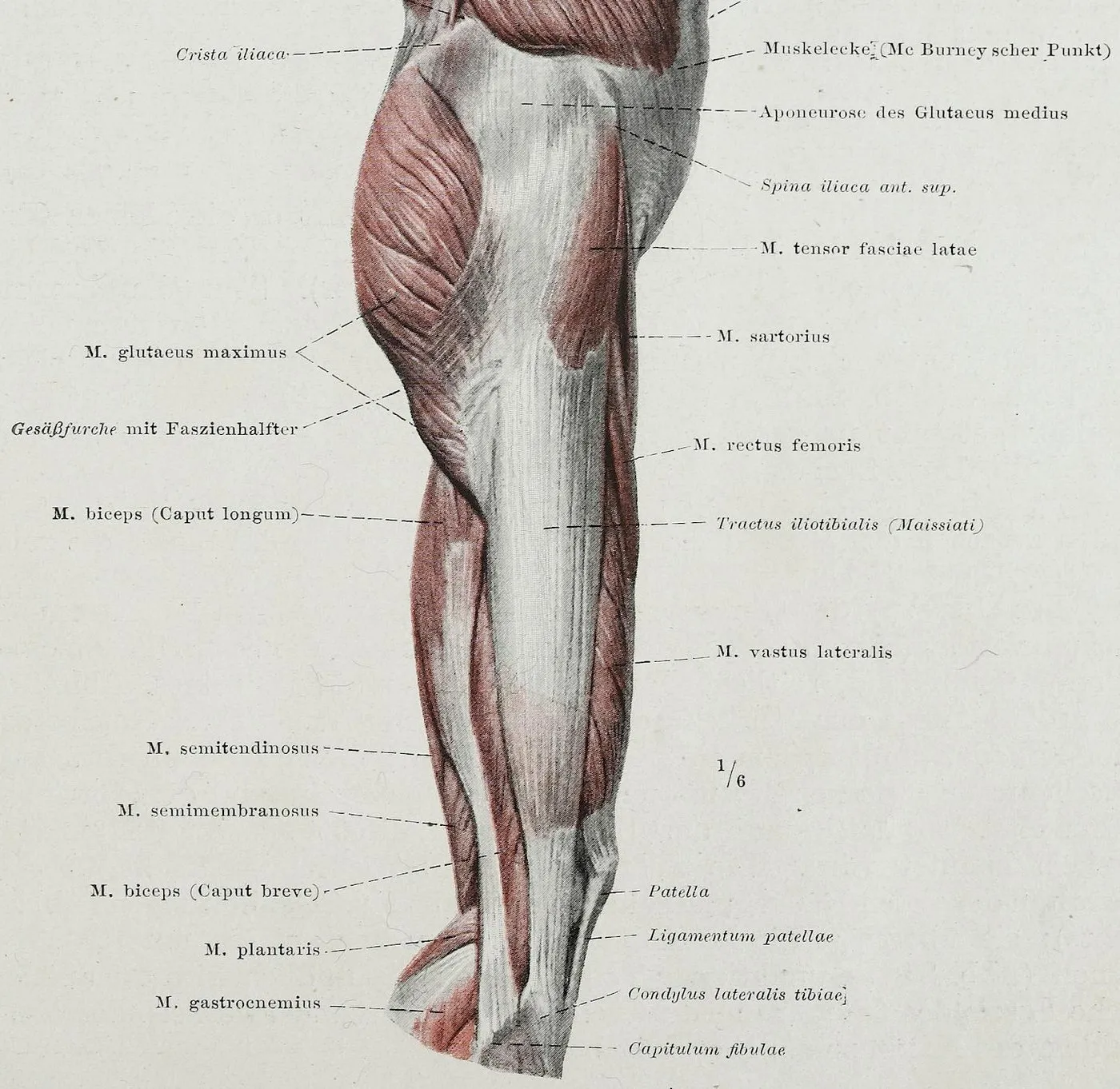
In 20 embalmed cadavers, the band was firmly attached along the linea aspera, a long ridge down the back of the femur (thigh bone)7. That attachment ran from the greater trochanter (the bony bump at the outer hip) down to the outer condyle at the knee7. A condyle is a rounded knob at the end of a bone, where it meets another bone at a joint. Cadaver dissections also show it anchored to the femur shaft by the lateral intermuscular septum, a sheet of tissue between muscle groups1. They show it continuous with the patellar retinacula, bands of tissue at the sides of the kneecap1.
In 15 cadavers, fibrous strands anchored the lower band to the lower femur1. These strands split the band into an upper “tendinous” part and a lower “ligamentous” part1. Only the lower part crosses the knee1.
Where the lower end attaches
A review says all authors agree the band attaches to Gerdy’s tubercle2. It lists four other lower attachments that are often described2:
- a shared attachment with a ligament the review calls the femoral tibial ligament2
- the lower femur above the condyle, through the distal Kaplan fibres (fibrous bands that tie the lower iliotibial band to the thigh bone)2
- along the linea aspera, through the proximal Kaplan fibres2
- the patella (kneecap)2
A review describes the Kaplan fibres as fine fibrous bands running from the band to the septum and lower femur8. In 10 cadaver knees, they formed two separate bundles9. These fixed to the femur about 54 mm and 31 mm above its lateral epicondyle9. That is the outer bony bump at the knee end of the thigh bone.
The band’s size varies a lot between people2. A review reports that, for its thickness and width, the usual spread between people is 19% to 68% of the average2. Some authors argue it should not count as a separate structure at all2. Their reason is that its edges cannot be clearly told apart from the rest of the fascia lata in dissection2.
What it does at the hip and knee
The band is thought to act as a strut in walking, steadying both hip and knee, mainly against side-to-side movement2. Through the band, the tensor fasciae latae generally pulls forward and up to bend the hip2. The gluteus maximus pulls backward to straighten it2.
In a review of cadaver work, all 6 cutting studies reported the band backing up the anterior cruciate ligament against inward rotation3. In a straight knee without an ACL, its attachments do nothing to stop the tibia sliding forward2. Once the knee bends past 30 degrees, they reduce that slide2. One study found the Kaplan fibres provided about 44% of the resistance to inward rotation at 90 degrees of bend, with the ACL aside8. The band’s attachments to the kneecap steady it against slipping out inward2.
How it stretches and stores energy
Tests of dissected bands put its Young’s modulus (how stiff a material is in itself) at about half that of the Achilles tendon2. In 10 unembalmed bands pulled until they failed, the average peak load was about 873 newtons (a unit of force), at 9% stretch10.
In a moderately paced run, a computer model put the band’s stored energy at up to 5% of the run’s positive work2. That is about 14% of what the Achilles tendon adds2. Models built from dissections compared walking on two legs11. They estimated the human band stores 15 to 20 times more elastic energy than the chimpanzee’s, per unit of body mass and stride11.
Shear wave elastography (an ultrasound method that estimates how stiff a tissue is) was done in single-leg standing2. Band stiffness rose with hip adduction (moving the thigh toward the midline) and fell with slight hip abduction2. In 30 adults, 10 degrees of hip adduction raised band stiffness at mid-thigh and near the knee, while tensor fasciae latae stiffness did not rise12.
Breakdown
Iliotibial band syndrome
Runners and cyclists commonly get iliotibial band syndrome, an overuse injury1. A systematic review estimated its incidence (how many new cases appear in a group over a set time) in runners at 5% to 14%4. Another systematic review calls iliotibial band syndrome the second most common running injury, making up about a tenth of running injuries13.
Friction or pressure
A review says the syndrome was long thought of as a friction injury2. Pressure on the nerve-rich fat between the band and bone is now proposed instead2. One anatomy study concluded the band cannot roll over the epicondyle, because it is anchored to the femur1. The authors suggest the movement seen comes from tension shifting between its front and back fibres as the knee bends1.
In the MRI (a scan that shows soft tissue in detail) part of that study, six volunteers were scanned1. At 30 degrees of knee bend, the band pressed against the outer epicondyle of the femur as the tibia turned inward1. With the knee straight, it moved outward1. A review adds that MRI shows the tissues under the lower band squeezed as the band tightens2.
In cadaver dissections, the fat between the band and the femur was rich in blood vessels and nerves1. In some specimens it held Pacinian corpuscles, nerve endings that sense pressure1. In two runners with recent-onset iliotibial band syndrome, MRI changes were in this fat, not in the band1. On anatomical grounds, the authors suggest overuse injury may be linked more to pressure on the fat than to rubbing1.
How running loads it
A model built partly from a study of nine runners with the syndrome puts impingement of the band near footstrike16. Those runners’ knees were bent an average of 21.4 degrees at footstrike16. The same authors propose that downhill running raises the risk because the knee is straighter at footstrike16. They also propose sprinting is less likely to make it worse16.
One prospective study (a study that follows people forward in time) followed female runners17. Those who later had the syndrome showed a higher modelled strain rate, how fast the band stretches, than uninjured runners17. The authors suggest the rate of stretch, rather than its size, may be a cause17.
A second prospective study followed female recreational runners for two years18. The 18 who developed the syndrome had shown more hip adduction and inward knee turning when running18.
A systematic review looked at 13 studies, one of them prospective13. Female runners who went on to get the syndrome showed more peak hip adduction and inward knee turning during stance13. Another systematic review found limited or conflicting evidence on the causes in runners4. It was unclear whether weak hip abductors play a major role4.
Other injuries
On MRI after ACL rupture, reported rates of Kaplan fibre injury range from 6.4% to 61%8.
Higher up, a 2013 imaging review describes injuries of the upper band as an under-recognised source of hip pain19. Tendinopathy (local tendon pain with activities that load it) at the band’s origin is described as a possibly overlooked cause of front outer hip pain20.
Recovery
How the Kaplan fibres heal
A review reports fewer Kaplan fibre injuries in people scanned more than 90 days after ACL rupture8. It reads this as a possible sign of healing over time8. A case series (a report on a group of patients with the same condition, without a comparison group) looked at 89 people with ACL injury21. Kaplan fibre injury was seen in 30.3%21. Nine months after ACL reconstruction (surgery to rebuild the ligament), MRI showed the fibres rejoined in 14 of the 27 injured (51.9%)21.
What stretching does to the band
From pull tests on 10 unembalmed bands, the authors conclude the band can withstand a lot of tension10. They add that stretching forces used in clinics likely stay within its elastic range, without lasting lengthening10. A 2022 review argues that stretching now and then is unlikely to change the band’s length or mechanical properties22. One randomised trial (a study that assigns people to groups by chance) had 30 adults12. One session of stretching or foam rolling did not change band stiffness12.
How the band changes over time
In 14 recreational runners with the syndrome, band stiffness was higher after 6 weeks of care than at the start23. Their pain and function improved over the same time23.
In 33 donors aged 4 months to 93 years, the band’s tensile properties did not depend on age, apart from maximum strain in males24.
Connections
Bones and muscles
The band links the iliac crest of the pelvis with the tibia2. In six dissected cadavers, the gluteus maximus had a large insertion into the fascia lata25. The authors suggest the iliotibial tract could count as one of its tendons25. The tensor fasciae latae sits in the upper outer front of the thigh, between the band’s surface and deep fibres6. It helps steady the outer knee by pulling the band taut26.
The outer knee
In cadaver work, the band steadies the outer knee together with three partners2. These are the fibular (outer) collateral ligament, the biceps femoris (a hamstring) and the tendon of the popliteus (a small muscle behind the knee)2. The knee kept its passive stability as long as at least two of these were intact2. The popliteus tendon lies deep to the lateral collateral ligament, and the band’s surface layer lies over it27.
Related pages
Related exercise pages: side-lying hip abduction, where hip position is the link28. In 18 unfixed cadavers and 12 people having hip replacement, force under the band at the greater trochanter rose with adduction and fell with abduction28. That study did not test the exercise itself28. A separate page covers the standing IT band stretch.
Iliotibial band starts on
Iliotibial band is anchored to
Iliotibial band attaches to
Iliotibial band receives most of the tendon of
Iliotibial band crosses
Iliotibial band backs up
Iliotibial band steadies the outer knee with
Iliotibial band has been studied in people with
Iliotibial band is tightened by the hip position of
Sources
- Fairclough J, Hayashi K, Toumi H, Lyons K, Bydder G, Phillips N, Best TM, Benjamin M (2006). The functional anatomy of the iliotibial band during flexion and extension of the knee: implications for understanding iliotibial band syndrome.. Journal of anatomy. doi:10.1111/j.1469-7580.2006.00531.x PMID 16533314
- Hutchinson LA, Lichtwark GA, Willy RW, Kelly LA (2022). The Iliotibial Band: A Complex Structure with Versatile Functions.. Sports medicine (Auckland, N.Z.). doi:10.1007/s40279-021-01634-3 PMID 35072941
- van der Wal WA, Meijer DT, Hoogeslag RAG, LaPrade RF (2024). The Iliotibial Band is the Main Secondary Stabilizer for Anterolateral Rotatory Instability and both a Lemaire Tenodesis and Anterolateral Ligament Reconstruction Can Restore Native Knee Kinematics in the Anterior Cruciate Ligament Reconstructed Knee: A Systematic Review of Biomechanical Cadaveric Studies.. Arthroscopy. doi:10.1016/j.arthro.2023.05.005 PMID 37207919
- van der Worp MP, van der Horst N, de Wijer A, Backx FJ, Nijhuis-van der Sanden MW (2012). Iliotibial band syndrome in runners. Sports medicine (Auckland, N.Z.). doi:10.2165/11635400-000000000-00000 PMID 22994651
- Hyland S, Graefe SB, Varacallo MA (2023). Anatomy, Bony Pelvis and Lower Limb, Iliotibial Band (Tract). StatPearls. PMID 30725782
- Trammell AP, Nahian A, Pilson H (2023). Anatomy, Bony Pelvis and Lower Limb: Tensor Fasciae Latae Muscle. StatPearls. PMID 29763045
- Falvey EC, Clark RA, Franklyn-Miller A, Bryant AL, Briggs C, McCrory PR (2010). Iliotibial band syndrome: an examination of the evidence behind a number of treatment options.. Scandinavian journal of medicine & science in sports. doi:10.1111/j.1600-0838.2009.00968.x PMID 19706004
- Poursalehian M, Hajiaghajani S, Ayati Firoozabadi M, Dehghani Ashkezari D, Mortazavi SMJ (2024). Kaplan fibers of iliotibial band: a comprehensive review of current literature.. EFORT open reviews. doi:10.1530/eor-24-0017 PMID 39360787
- Godin JA, Chahla J, Moatshe G, Kruckeberg BM, Muckenhirn KJ, Vap AR, Geeslin AG, LaPrade RF (2017). A Comprehensive Reanalysis of the Distal Iliotibial Band: Quantitative Anatomy, Radiographic Markers, and Biomechanical Properties.. The American journal of sports medicine. doi:10.1177/0363546517707961 PMID 28609131
- Seeber GH, Wilhelm MP, Sizer PS, Guthikonda A, Matthijs A, Matthijs OC, Lazovic D, Brismée JM, Gilbert KK (2020). THE TENSILE BEHAVIORS OF THE ILIOTIBIAL BAND - A CADAVERIC INVESTIGATION.. International journal of sports physical therapy. doi:10.26603/ijspt20200451 PMID 32566381
- Eng CM, Arnold AS, Biewener AA, Lieberman DE (2015). The human iliotibial band is specialized for elastic energy storage compared with the chimp fascia lata.. The Journal of experimental biology. doi:10.1242/jeb.117952 PMID 26026035
- Pepper TM, Brismée JM, Sizer PS, Kapila J, Seeber GH, Huggins CA, Hooper TL (2021). The Immediate Effects of Foam Rolling and Stretching on Iliotibial Band Stiffness: A Randomized Controlled Trial.. International journal of sports physical therapy. doi:10.26603/001c.23606 PMID 34123517
- Aderem J, Louw QA (2015). Biomechanical risk factors associated with iliotibial band syndrome in runners: a systematic review.. BMC musculoskeletal disorders. doi:10.1186/s12891-015-0808-7 PMID 26573859
- Bolia IK, Gammons P, Scholten DJ, Weber AE, Waterman BR (2020). Operative Versus Nonoperative Management of Distal Iliotibial Band Syndrome-Where Do We Stand? A Systematic Review.. Arthroscopy, sports medicine, and rehabilitation. doi:10.1016/j.asmr.2020.04.001 PMID 32875305
- Nemeth WC, Sanders BL (1996). The lateral synovial recess of the knee: anatomy and role in chronic Iliotibial band friction syndrome.. Arthroscopy. doi:10.1016/s0749-8063(96)90197-8 PMID 8902132
- Orchard JW, Fricker PA, Abud AT, Mason BR (1996). Biomechanics of iliotibial band friction syndrome in runners.. The American journal of sports medicine. doi:10.1177/036354659602400321 PMID 8734891
- Hamill J, Miller R, Noehren B, Davis I (2008). A prospective study of iliotibial band strain in runners.. Clinical biomechanics (Bristol, Avon). doi:10.1016/j.clinbiomech.2008.04.017 PMID 18583001
- Noehren B, Davis I, Hamill J (2007). ASB Clinical Biomechanics Award Winner 2006. Clinical biomechanics (Bristol, Avon). doi:10.1016/j.clinbiomech.2007.07.001 PMID 17728030
- Huang BK, Campos JC, Michael Peschka PG, Pretterklieber ML, Skaf AY, Chung CB, Pathria MN (2013). Injury of the gluteal aponeurotic fascia and proximal iliotibial band: anatomy, pathologic conditions, and MR imaging.. Radiographics : a review publication of the Radiological Society of North America, Inc. doi:10.1148/rg.335125171 PMID 24025934
- Buttinger JN, Romero JM, Sellon JL, Lachman N, Kruse RC, Boettcher BJ (2024). Iliotibial Band Origin Tendinopathy Is an Underrecognized Cause of Anterolateral Hip Pain: A Narrative Review and Clinical Commentary.. Current sports medicine reports. doi:10.1249/jsr.0000000000001176 PMID 38838686
- Lord BR, Devitt BM, Hookway SR, Klemm HJ, Webster KE, Whitehead TS, Feller JA (2023). The Resolution of Kaplan Fiber Injuries Is Observed in a Majority of Cases at 9 Months After Acute Primary Anterior Cruciate Ligament Reconstruction: A Radiological Study.. The American journal of sports medicine. doi:10.1177/03635465231180859 PMID 37434516
- Friede MC, Innerhofer G, Fink C, Alegre LM, Csapo R (2022). Conservative treatment of iliotibial band syndrome in runners: Are we targeting the right goals?. Physical therapy in sport : official journal of the Association of Chartered Physiotherapists in Sports Medicine. doi:10.1016/j.ptsp.2021.12.006 PMID 35007886
- Friede MC, Klauser A, Fink C, Csapo R (2020). Stiffness of the iliotibial band and associated muscles in runner's knee: Assessing the effects of physiotherapy through ultrasound shear wave elastography.. Physical therapy in sport : official journal of the Association of Chartered Physiotherapists in Sports Medicine. doi:10.1016/j.ptsp.2020.06.015 PMID 32769015
- Zwirner J, Babian C, Ondruschka B, Schleifenbaum S, Scholze M, Waddell NJ, Hammer N (2019). Tensile properties of the human iliotibial tract depend on height and weight.. Medical engineering & physics. doi:10.1016/j.medengphy.2019.05.001 PMID 31133525
- Stecco A, Gilliar W, Hill R, Fullerton B, Stecco C (2013). The anatomical and functional relation between gluteus maximus and fascia lata.. Journal of Bodywork and Movement Therapies. doi:10.1016/j.jbmt.2013.04.004 PMID 24139012
- Betts JG, Young KA, Wise JA, Johnson E, Poe B, Kruse DH, Korol O, Johnson JE, Womble M, DeSaix P (2022). Anatomy and Physiology 2e. OpenStax. Link
- Yaras RJ, O'Neill N, Mabrouk A, Yaish AM (2024). Lateral Collateral Ligament Knee Injury. StatPearls. PMID 32809682
- Birnbaum K, Siebert CH, Pandorf T, Schopphoff E, Prescher A, Niethard FU (2004). Anatomical and biomechanical investigations of the iliotibial tract.. Surgical and Radiologic Anatomy. doi:10.1007/s00276-004-0265-8 PMID 15378277
Changes
- Links to the new standing IT band stretch page
- A supporting claim was narrowed to match its source; the page text is unchanged.