Achilles tendon
Also called calcaneal tendon
The tendon that joins the calf muscles to the heel bone. How it stores and returns energy, how it wears or tears, how slowly it adapts and heals, and what it works with.
- Region
- Foot & ankle
- Muscles
- gastrocnemius, soleus
- Updated
- October 22, 2026
- Reviewed
- October 22, 2026 · how entries are checked
Revised September 28, 2026: Restructured for easier reading (key points, subheadings, tables, a figure and a chart), and several sentences narrowed to match their sources more exactly.
Function
How the tendon joins muscle to heel
The Achilles tendon links the calf muscles to the calcaneus (heel bone)1,2. A tendon is a tough cord of tissue that passes force from a working muscle to bone, and that pull makes the body move3. The Achilles is described as the body’s biggest and strongest tendon9. The muscles at the back of the lower leg mainly produce plantar flexion, which means pointing the foot down1.
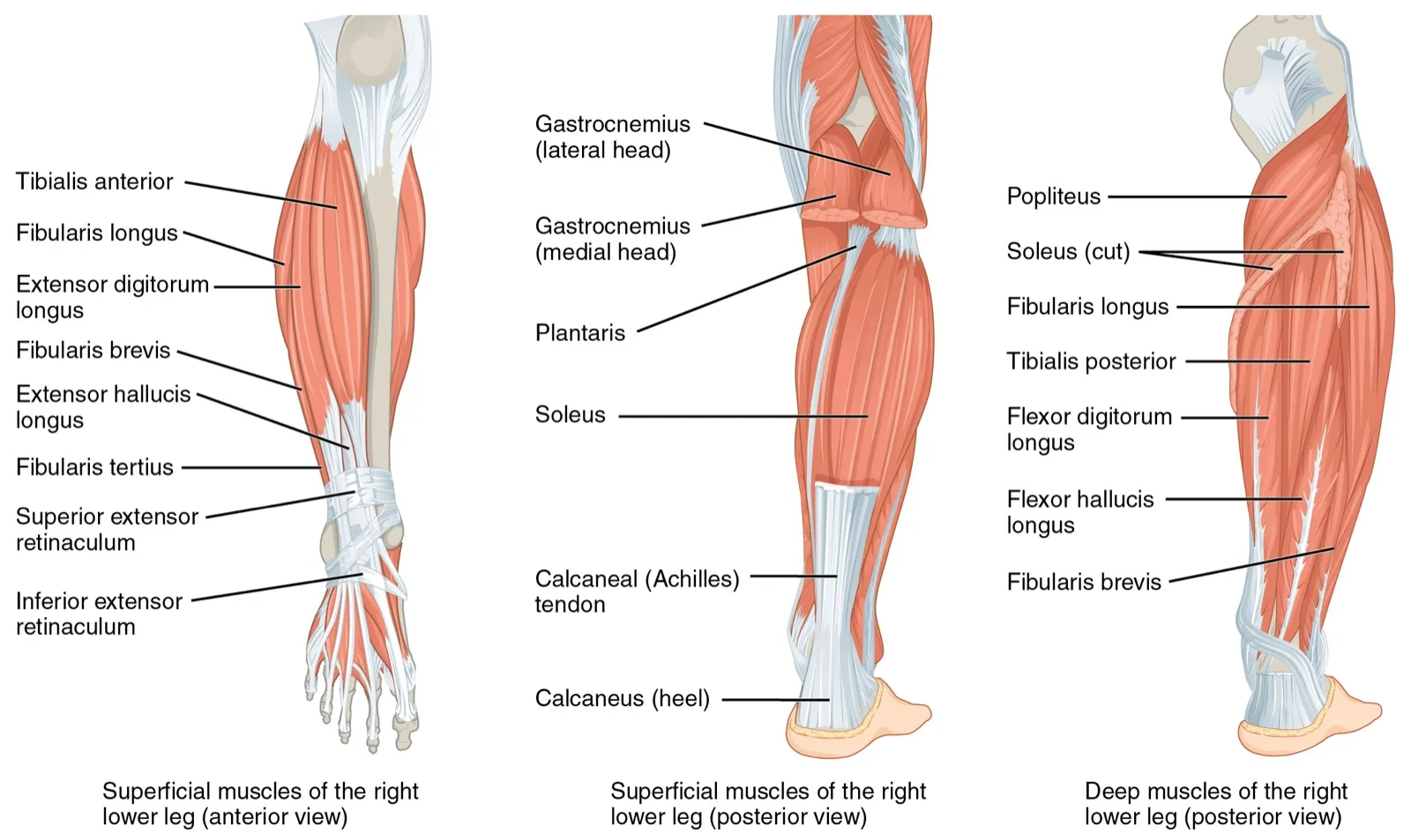
Tendons are made of a kind of connective tissue called dense regular connective tissue1. In it, strands of collagen, a strong rope-like protein, are packed side by side in bundles1. That layout makes the tissue strong under tension and hard to stretch along the fibres1.
How three strands make one tendon
The tendon is built from three strands of fibre, each from a different part of the calf muscles2. The gastrocnemius, the calf muscle nearest the skin, has two parts called heads, inner and outer, that both run from thigh bone to heel bone2,10,11. Fibres come from the soleus, a flat muscle lying deeper, and from both heads of the gastrocnemius1,2. Each strand is called a subtendon2. In 106 legs from 53 male donors, the three subtendons made up these shares of the tendon at the heel2:
| Subtendon from | Share of the tendon at the heel |
|---|---|
| Outer gastrocnemius head | About 44%2 |
| Inner gastrocnemius head | About 28%2 |
| Soleus | About 28%2 |
On the way down to the heel, the subtendons wind around each other2. One meta-analysis put the average twist at about 46.5 degrees12. The amount of twist varied from one person to the next12.
How the tendon stores and returns energy
The tendon also acts like a spring, storing energy and giving it back4,5. In one study, the tendon gave back 7.8 to 11.3 joules of stored elastic energy during push-off in treadmill walking and running4. In ten men hopping on one leg, the tendon’s recoil returned about 38 joules per hop5. That was around 16% of the total work of each hop5. In the same hopping study, Achilles stiffness, meaning the force it takes to stretch the tendon by a set amount, varied between the ten men5.
How load changes with the task
One study of 11 people estimated peak tendon load in kilonewtons4:
| Task | Estimated peak tendon load |
|---|---|
| Walking | About 2.0 kilonewtons4 |
| Running slowly | About 2.3 kilonewtons4 |
| Running fast | About 2.6 kilonewtons4 |
In this study, peak stretch went from about 4.0 to 4.9% of the tendon’s length4. Speed had a significant effect on force4.
A study of eight healthy young adults estimated it in multiples of body weight6:
| Task | Estimated peak tendon load |
|---|---|
| Seated heel raise | About half of body weight6 |
| Forward hop on one leg | 7.3 times body weight6 |
Breakdown
What tendinopathy is and where it sits
The Dutch guideline on Achilles tendinopathy states that, by current consensus, tendinopathy means local pain in a tendon that comes with activities that load it13. The guideline groups Achilles tendinopathy by where it sits13. Insertional tendinopathy lies within 2 cm of the heel attachment13. Midportion tendinopathy lies higher up, more than 2 cm above it13.
Who develops tendinopathy
In one cohort study of 3,379 recreational runners, 4.2% developed Achilles tendinopathy14. Among the new cases, about 64% were midportion and 28% insertional14. The rest involved both regions14.
In 2009, a Dutch study counted how many of 57,725 registered patients saw their family doctor for midportion Achilles problems15. The rate was 1.85 per 1,000 registered patients15. Among adults aged 21 to 60, the figure was 2.35 per 1,00015.
Another study looked at 58 patients with Achilles tendon pain who met criteria for surgery16. Of these, 31% had no direct link to sport or vigorous physical activity16.
What raises the odds is not well settled
One systematic review found limited evidence for nine factors that may raise the odds of tendinopathy9. These included earlier tendinopathy or a leg fracture9. Another was weaker strength in the muscles that point the foot down, tested at a fixed speed9. Twenty-six other suggested factors showed no link, including being overweight, foot posture when standing still, and activity level9.
In the runner study, tendinopathy in the past year was the strongest factor linked to a new case14. Its odds ratio, how many times higher the odds of an outcome are in one group than another, was near 6.514.
How often the tendon ruptures
An acute rupture is a sudden tearing of the tendon17,18. Sudden tears may be grouped as a range: tiny tears, partial tears where the tendon is partly broken, interstitial tears inside the tendon, and complete ruptures17,18. A meta-analysis pooled 28 studies of rupture based on registries or representative samples7. Overall, it found 15.7 ruptures per 100,000 person-years, which could be 100,000 people followed for one year7. Rupture is not spread evenly across people or over time7:
| Group | What the pooled studies found |
|---|---|
| Over time | 6.1 in 1979 and 31.1 by 2021, an average rise of about 2.7% a year7 |
| By sex | Males had about three times the rate of females7 |
| By age | Rates peaked at ages 30 to 49 in males and 40 to 49 in females7 |
| By activity | About 68% of ruptures were linked to sport7 |
Recovery
How healthy tendon adapts to load
Healthy tendon adapts to repeated load19. A meta-analysis of exercise studies in healthy adults aged 18 to 50, each lasting at least eight weeks, found that loading made tendons stiffer19. Their Young’s modulus, the stiffness of the material itself, also rose19. Cross-sectional area, the size of a slice cut straight across the tendon, grew by a smaller amount19. Gains in stiffness depended on how heavy the load was, not on the type of muscle contraction19.
Most of those programmes ran for 8 to 14 weeks19. Programmes of 12 weeks or longer showed larger effects, though the difference was not statistically significant19.
What happens when load is taken away
A review concluded that keeping a limb still lowers the tendon’s mechanical properties in animals, and that human data largely mirror this3. Even one to two weeks of unloading a limb seems to reduce tendon stiffness, without changing its thickness3. In one study of five healthy people, one leg was kept off the ground for four weeks20. Afterwards, that leg’s Achilles tendon had a Young’s modulus about 10% lower20.
Why tendon heals slowly
A review describes tendon as tissue with a poor blood supply that heals poorly compared with bone or skin8. Carbon dating of Achilles samples suggested the tendon core forms while a person is growing taller3,21. After that, the core appeared to be essentially not replaced, unlike muscle, which is renewed continuously3,21.
After a sudden tendon injury, healing is described in three stages in a row22:
A review of tendon healing states that healed tendon does not get back its original mechanical properties22.
How long the effects last
In one study, on average 14 years after surgical repair, the tendon was about 12 mm, or 6%, longer than on the other leg23. In that study, a longer tendon went with smaller calf muscles and lasting loss of strength23. In another study, calf strength and endurance deficits remained seven years after rupture24. Apart from heel-rise height, calf performance did not improve further after the two-year mark24.
The Dutch guideline says recovery from tendinopathy can also be slow, taking months to years13. It states that most people recover, but symptoms may persist at least up to ten years in 23 to 37%13.
Connections
The calf muscles it serves
The Achilles tendon joins the soleus and both heads of the gastrocnemius to the heel1,2. Together these three are called the triceps surae, and they do most of the work of pointing the foot down11. In 48 healthy people pushing the foot down as hard as possible, none of the three knee angles tested singled out one calf muscle alone25.
What studies of the calf raise show
The calf raise is one exercise that loads this tendon6.
- In one study of resistance-trained men, the authors suggested that calf raises can recruit the soleus and both gastrocnemius heads11.
- In its 22 trained men, activation of these muscles did not differ significantly between the calf raise and the leg press11.
- A separate study of 33 people looked at a standing heel raise with the toes turned out10. The inner head of the gastrocnemius was more active than the outer head10.
- A narrative review found support for the calf-raise test as a way to assess the tendon and calf muscles as one unit26.
Other muscles that work in the heel raise
Other lower-leg muscles take part in the heel raise too27. The tibialis posterior and flexor digitorum longus are two of the four deep muscles at the back of the leg1. They lie beneath the calf muscles that end in the Achilles tendon1. The fibularis (peroneus) longus instead is one of two muscles on the outer side of the leg, with the fibularis brevis1. Within the front and back muscle groups of the lower leg, the muscles toward either side turn the foot in or out and rotate it1.
In one study of 14 healthy men, these muscles’ activity in heel raises differed between the foot straight, turned in and turned out27:
| Muscle | Where it lies | Most active in heel raises with the foot |
|---|---|---|
| Tibialis posterior | Deep, back of the leg | Turned in 30 degrees1,27 |
| Flexor digitorum longus | Deep, back of the leg | Turned in 30 degrees1,27 |
| Fibularis longus | Outer side of the leg | Turned out 30 degrees1,27 |
How the tendon links to the arch of the foot
The plantar fascia is a band of tissue in the sole of the foot that runs from the heel bone to the toes1,28. It is a main support of the foot’s long arch1,28. The Achilles tendon and plantar fascia both help spread load through the foot, absorb shock and hold up the arch during push-off28.
One study measured 19 feet from donors28. The Achilles tendon’s cross-sectional area at the heel went strongly with that of the plantar fascia along its heel attachment28. The authors read this as a sign of a working link between the two28. The same study could not confirm full fibre continuity between the two28.
A small muscle beside it
The plantaris is a calf muscle that runs at a slant between the gastrocnemius and the soleus1. Some people have two, and no plantaris was found in about 7% of donor bodies1. In a review of 200 ankle MRI (magnetic resonance imaging) scans, its tendon was directly related to the Achilles attachment in 42.5% of cases29.
Achilles tendon is loaded by
Sources
- Betts JG, Young KA, Wise JA, Johnson E, Poe B, Kruse DH, Korol O, Johnson JE, Womble M, DeSaix P (2022). Anatomy and Physiology 2e. OpenStax. Link
- Pękala PA, Henry BM, Ochała A, Kopacz P, Tatoń G, Młyniec A, Walocha JA, Tomaszewski KA (2017). The twisted structure of the Achilles tendon unraveled: A detailed quantitative and qualitative anatomical investigation.. Scandinavian Journal of Medicine & Science in Sports. doi:10.1111/sms.12835 PMID 28139009
- Magnusson SP, Kjaer M (2019). The impact of loading, unloading, ageing and injury on the human tendon.. The Journal of Physiology. doi:10.1113/JP275450 PMID 29920664
- Kharazi M, Bohm S, Theodorakis C, Mersmann F, Arampatzis A (2021). Quantifying mechanical loading and elastic strain energy of the human Achilles tendon during walking and running.. Scientific Reports. doi:10.1038/s41598-021-84847-w PMID 33712639
- Lichtwark GA, Wilson AM (2005). In vivo mechanical properties of the human Achilles tendon during one-legged hopping.. The Journal of Experimental Biology. doi:10.1242/jeb.01950 PMID 16326953
- Baxter JR, Corrigan P, Hullfish TJ, O'Rourke P, Silbernagel KG (2021). Exercise Progression to Incrementally Load the Achilles Tendon.. Medicine and Science in Sports and Exercise. doi:10.1249/MSS.0000000000002459 PMID 32658037
- Kotsifaki R, Malliaras P, Byron C, Marques J, Korakakis V (2026). Incidence, Temporal Trends, and Surgical Shift of Achilles Tendon Rupture: A Systematic Review and Meta-analysis.. Sports Medicine. doi:10.1007/s40279-026-02397-5 PMID 41933260
- Tempfer H, Traweger A (2015). Tendon Vasculature in Health and Disease.. Frontiers in Physiology. doi:10.3389/fphys.2015.00330 PMID 26635616
- van der Vlist AC, Breda SJ, Oei EHG, Verhaar JAN, de Vos RJ (2019). Clinical risk factors for Achilles tendinopathy: a systematic review.. British Journal of Sports Medicine. doi:10.1136/bjsports-2018-099991 PMID 30718234
- Cibulka M, Wenthe A, Boyle Z, Callier D, Schwerdt A, Jarman D, Strube MJ (2017). Variation in medial and lateral gastrocnemius muscle activity with foot position.. International Journal of Sports Physical Therapy. PMID 28515978
- Gentil P, Souza D, Santana M, Alves RR, Campos MH, Pinto R, Bottaro M (2020). Multi- and Single-Joint Resistance Exercises Promote Similar Plantar Flexor Activation in Resistance Trained Men.. International Journal of Environmental Research and Public Health. doi:10.3390/ijerph17249487 PMID 33352879
- Łazarz DP, Yika ADC, Pękala JR, Walocha JA, Pękala PA (2024). Clinical anatomy of the human Achilles subtendons twist - meta-analysis.. Annals of Anatomy. doi:10.1016/j.aanat.2024.152271 PMID 38677622
- de Vos RJ, van der Vlist AC, Zwerver J, Meuffels DE, Smithuis F, van Ingen R, van der Giesen F, Visser E, Balemans A, Pols M, Veen N, den Ouden M, Weir A (2021). Dutch multidisciplinary guideline on Achilles tendinopathy.. British Journal of Sports Medicine. doi:10.1136/bjsports-2020-103867 PMID 34187784
- Chen W, Cloosterman KLA, Bierma-Zeinstra SMA, van Middelkoop M, de Vos RJ (2024). Epidemiology of insertional and midportion Achilles tendinopathy in runners: A prospective cohort study.. Journal of Sport and Health Science. doi:10.1016/j.jshs.2023.03.007 PMID 36963760
- de Jonge S, van den Berg C, de Vos RJ, van der Heide HJ, Weir A, Verhaar JA, Bierma-Zeinstra SM, Tol JL (2011). Incidence of midportion Achilles tendinopathy in the general population.. British Journal of Sports Medicine. doi:10.1136/bjsports-2011-090342 PMID 21926076
- Rolf C, Movin T (1997). Etiology, histopathology, and outcome of surgery in achillodynia.. Foot & Ankle International. doi:10.1177/107110079701800906 PMID 9310767
- Wang CC, Chen PY, Yang KC, Wang CL, Chen IH (2024). Current treatment concepts for Achilles tendon rupture.. Tzu Chi Medical Journal. doi:10.4103/tcmj.tcmj_113_23 PMID 38406567
- Fenech M, Ajjikuttira A, Edwards H (2024). Ultrasound assessment of acute Achilles tendon rupture and measurement of the tendon gap.. Australasian Journal of Ultrasound in Medicine. doi:10.1002/ajum.12384 PMID 38784700
- Bohm S, Mersmann F, Arampatzis A (2015). Human tendon adaptation in response to mechanical loading: a systematic review and meta-analysis of exercise intervention studies on healthy adults.. Sports Medicine - Open. doi:10.1186/s40798-015-0009-9 PMID 27747846
- Kinugasa R, Hodgson JA, Edgerton VR, Shin DD, Sinha S (2010). Reduction in tendon elasticity from unloading is unrelated to its hypertrophy.. Journal of Applied Physiology. doi:10.1152/japplphysiol.00384.2010 PMID 20616227
- Heinemeier KM, Schjerling P, Heinemeier J, Magnusson SP, Kjaer M (2013). Lack of tissue renewal in human adult Achilles tendon is revealed by nuclear bomb (14)C.. The FASEB Journal. doi:10.1096/fj.12-225599 PMID 23401563
- Darrieutort-Laffite C, Blanchard F, Soslowsky LJ, Le Goff B (2024). Biology and physiology of tendon healing.. Joint Bone Spine. doi:10.1016/j.jbspin.2024.105696 PMID 38307405
- Heikkinen J, Lantto I, Piilonen J, Flinkkilä T, Ohtonen P, Siira P, Laine V, Niinimäki J, Pajala A, Leppilahti J (2017). Tendon Length, Calf Muscle Atrophy, and Strength Deficit After Acute Achilles Tendon Rupture. The Journal of Bone and Joint Surgery. American Volume. doi:10.2106/JBJS.16.01491 PMID 28926379
- Brorsson A, Grävare Silbernagel K, Olsson N, Nilsson Helander K (2018). Calf Muscle Performance Deficits Remain 7 Years After an Achilles Tendon Rupture.. The American Journal of Sports Medicine. doi:10.1177/0363546517737055 PMID 29068725
- Hébert-Losier K, Schneiders AG, García JA, Sullivan SJ, Simoneau GG (2011). Peak triceps surae muscle activity is not specific to knee flexion angles during MVIC.. Journal of Electromyography and Kinesiology. doi:10.1016/j.jelekin.2011.04.009 PMID 21669538
- Hébert-Losier K, Schneiders AG, Newsham-West RJ, Sullivan SJ (2009). Scientific bases and clinical utilisation of the calf-raise test.. Physical Therapy in Sport. doi:10.1016/j.ptsp.2009.07.001 PMID 19897168
- Akuzawa H, Imai A, Iizuka S, Matsunaga N, Kaneoka K (2017). The influence of foot position on lower leg muscle activity during a heel raise exercise measured with fine-wire and surface EMG.. Physical Therapy in Sport. doi:10.1016/j.ptsp.2017.08.077 PMID 28950148
- Singh A, Zwirner J, Templer F, Kieser D, Klima S, Hammer N (2021). On the morphological relations of the Achilles tendon and plantar fascia via the calcaneus: a cadaveric study.. Scientific Reports. doi:10.1038/s41598-021-85251-0 PMID 33727610
- Szaro P, Cifuentes Ramirez W, Borkmann S, Bengtsson A, Polaczek M, Ciszek B (2020). Distribution of the subtendons in the midportion of the Achilles tendon revealed in vivo on MRI.. Scientific Reports. doi:10.1038/s41598-020-73345-0 PMID 33004938
Changes
- Links to the new Achilles tendon rupture page
- Revised: study sizes and populations now stated in the key points and load section; linked to the calf raise page.
- Revised. Restructured for easier reading (key points, subheadings, tables, a figure and a chart), and several sentences narrowed to match their sources more exactly.
- First published.