Plantar fascia
Also called plantar aponeurosis
The band of tough tissue along the sole, from the heel bone to the toes. How it holds up the arch, what walking and running ask of it, how it thickens or tears, and what studies found about how it changes.
- Region
- Foot & ankle
- Updated
- October 8, 2026
- Reviewed
- October 8, 2026 · how entries are checked
Function
Where it runs
Anatomy textbooks use “plantar fascia” and “plantar aponeurosis” for the same tissue6. An aponeurosis is a broad, flat sheet of tendon. One anatomical study argued that “plantar fascia” fits better, because its fibres run in several directions as well as lengthwise6.
The plantar fascia starts on the calcaneal tubercle, a small, rounded bony bump on the heel bone1. It runs forward to the forefoot and has three parts: medial (inner), central and lateral (outer)1. The medial and lateral parts attach to two muscles of the sole1,6. These are the abductor hallucis and the abductor of the little toe1. Those two parts are usually classed as fascia, while the thicker central part is considered an aponeurosis1.
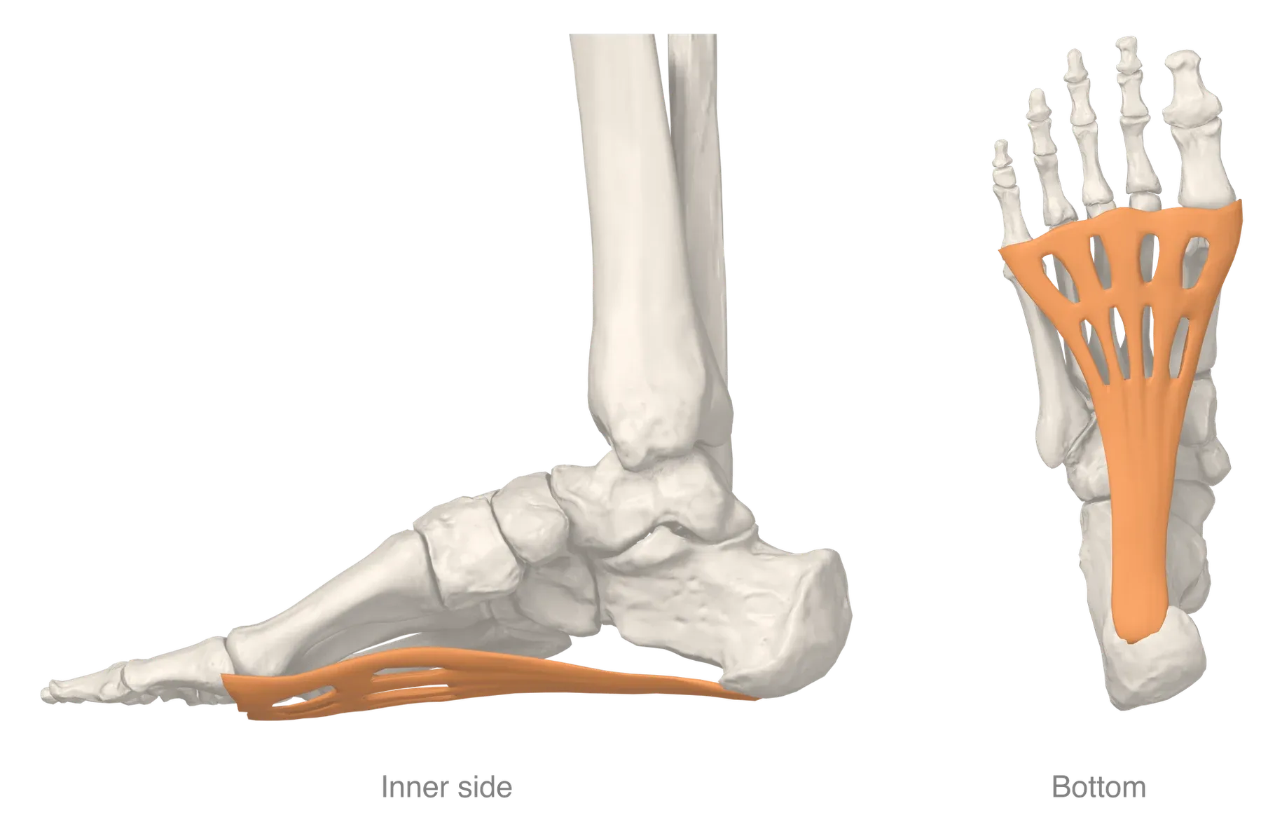
In dissections of 12 feet from unembalmed cadavers, the central part was the thickest6. It arose on the medial tubercle of the heel bone and ran forward over the flexor digitorum brevis muscle6. It then split unevenly into five slips, each ending at a different metatarsophalangeal joint6. Those are the joints at the base of the toes.
On ultrasound (a scan made with sound waves) in 21 healthy volunteers without symptoms, thickness averaged 3.3 mm7.
What it is made of
Under the microscope, cadaver plantar fascia was mostly type I collagen fibres running from heel to toes6. Collagen is a strong, rope-like protein, and type I is a thick, strong form of it. A study of cadaver tissue found few elastic fibres6. Its authors concluded that the plantar fascia is, in general, not an elastic tissue6.
Type II collagen, the collagen of cartilage, was found only in samples taken near the heel6.
How it holds up the arch
Together with the bones of the foot’s long arch, the plantar fascia makes a truss shaped like a triangle1. When the foot bears weight, the fascia keeps the ends of the arch from spreading apart and the arch from collapsing1.
In 12 fresh-frozen cadaver feet loaded along the shin, the fascia was compared with the plantar ligaments and the spring ligament2. The fascia added more to arch stability than either2. Cutting the fascia reduced arch stiffness by 25%2.
The windlass and the spring
The plantar fascia is central to two mechanisms of the arch: the windlass and the arch-spring8. Hicks described the windlass in 19548. Bending the toes up pulls on the stiff fascia, which slides around the heads of the metatarsals, the long bones of the midfoot8. That draws the heel bone toward the toes, shortening and raising the arch8.
In its ideal form, the windlass assumes a fascia of nearly constant length8. In the arch-spring, the fascia stretches and then shortens through the step, storing and releasing elastic energy8.
One study filmed the foot bones with high-speed X-ray during running8. The fascia held a nearly constant length while the toes bent up in push-off, then shortened late in stance8. Stance is the part of each step when the foot is on the ground. The authors thought the shortening likely added to arch recoil8.
What walking and running ask of it
The peak forces below come from cadaver feet or from models3,10,11.
| Activity | Where the figure comes from | Peak force in the fascia |
|---|---|---|
| Walking | Seven cadaver feet in a walking simulation | 96% of body weight on average10 |
| Walking | A foot model of three people walking | 1.5 times body weight on average11 |
| Running | A lower-limb model of five running trials | 1.3 to 2.9 times body weight3 |
In the cadaver simulation, the fascia’s tension correlated with force in the Achilles tendon10. The authors concluded that it passes large forces between the hindfoot and forefoot10.
In running, the arch works as a spring alongside the leg tendons12. One paper reports an estimate by Ker and colleagues, made by loading cadaver feet as in running13. By that estimate, the arch could store and return about 17% of the mechanical work of running13.
Computer models of eight people running at 3.1 metres per second gave a figure for the fascia itself14. They estimated that it stored a mean of 3.1 joules (a unit of energy) in stance and released it late in stance14.
A modelling study of treadmill runners estimated a peak strain of about 1.8% in shod running15. Strain is how far something stretches as a share of its resting length. The authors caution that the model’s figures may not match the real strain in the tissue15.
Breakdown
Change at the heel attachment
A review describes plantar fasciitis as a disorder mainly of the enthesis4. It notes that inflammation is rarely seen in tissue from chronic cases4. A review of tissue from 50 cases of heel spur surgery found degeneration and fragmentation of the fascia16. Its authors argue the condition is a degenerative “fasciosis” without inflammation16. Degenerative means to do with gradual wear and breakdown of a tissue.
A systematic review (a study that searches for all the research on a question in a set, checkable way) looked at imaging studies17. It describes the typical finding in chronic plantar heel pain as a spindle-shaped thickening close to the heel attachment17. A meta-analysis (a study that pools the results of several earlier studies) covered 21 ultrasound studies18. The fascia was 2.00 mm thicker in people with heel pain18.
One study compared 87 patients with plantar fasciitis and 21 healthy volunteers7. Shear wave velocity, a measure of stiffness, was lower in the patients’ fascia, and stiffness did not correlate with thickness7.
A review found the scientific support for a role of arch mechanics equivocal4. It suggests that being pulled apart under tension may not be the main way the tissue fails4.
In runners and other groups
A systematic review of running injuries covered eight studies and about 3500 runners19. It ranked plantar fasciitis among the three main running-related injuries19. Reported incidence (how many new cases appear in a group over a set time) ran from 4.5% to 10.0%19.
A systematic review with meta-analyses of 51 studies looked at clinical factors20. A body mass index (a measure of weight for height) above 27 was the only one significantly associated with plantar fasciopathy20. The effect was strongest in non-athletic people20. The plantar heel pain page covers who the condition affects and what trials found.
Ruptures
A systematic review of rupture found 17 studies covering 124 patients, 12 of them case reports5. In a series of 18 athletes with a rupture, most of them runners, all but 2 ruptures were in the medial portion21.
Two studies report a link with corticosteroid (a drug that copies a type of hormone the body makes) injection22,23:
- Among 765 patients seen for plantar fasciitis over three years, 51 had a rupture, and 44 of those ruptures were associated with an injection22.
- In a retrospective study (a study that looks back at past records) of 286 patients with plantar fasciitis, 35 had a rupture23. Steroid injection was the only assessed factor associated with rupture23.
After ruptures associated with injection, the original heel pain was usually relieved22. Patients went on to new problems, such as arch strain and midfoot strain22.
Recovery
After a single run
After a 10 km run, shear wave velocity at its heel end fell by 4.0% in ten male recreational distance runners24. It fell by 21.9% in 10 untrained men24. The authors describe the decrease as transient and specific to that site24.
In sixteen healthy adults, repeated maximal 400 m sprints reduced the fascia’s thickness and its stiffness at the heel origin25. After 30 minutes of rest, thickness no longer differed from before the run, but stiffness was still lower25.
Whether training changes it
A sports-medicine consensus statement notes that most research on how connective tissue remodels with exercise comes from tendons26. For the plantar fascia, the studies here compare groups at one point in time or look at a single run27,28.
A cross-sectional study (a study that looks at a group once) compared 10 track distance runners, who train counterclockwise, with 10 untrained people27. The runners had a stiffer fascia in the left foot than the right, and the untrained people showed no side difference27. Thickness did not differ between sides or groups27. The authors suggest this may reflect adaptation to uneven loading27.
For the smaller fall in the distance runners after the 10 km run, the authors suggest possible reasons28. These include adaptation that makes the fascia more resilient, or running mechanics28.
After rupture
In the systematic review of rupture, 94.4% of the 124 patients were managed without surgery5. In the series of 18 athletes, all were back in activity within 2 to 26 weeks, 9.1 weeks on average21. None was re-injured or needed surgery over a mean follow-up of 42 months21.
Connections
Heel bone and Achilles tendon
The medial process of the calcaneal tuberosity, part of the heel bone, gives attachment to the plantar fascia29. Studies have shown that most plantar spurs of the calcaneus lie deep to the fascia30.
In dissected cadaver feet, the fascia continued over the heel bone as a thin band about 1 to 2 mm thick6. This band matched the bone’s covering layer, which was continuous with the sheath around the Achilles tendon6. Complete fibre continuity between tendon and fascia was not confirmed under the microscope in a cadaver study31.
In a computer model of the standing foot, both body weight and Achilles tendon load raised tension in the fascia32. The Achilles load had about twice the straining effect32.
Foot muscles and toes
The flexor digitorum brevis, one of the foot muscles, lies immediately beneath the central band of the fascia33. In dissected cadaver feet, sheets of tissue ran from the deep side of the fascia between the three superficial muscles of the sole6. The authors of a walking and running study concluded that the muscles within the sole work in parallel with the fascia34. They add that these muscles may limit strain on it34.
Related pages
Related exercise pages: running, the plantar fascia stretch, the calf stretch and the soleus stretch12,35,36. In 12 cadaver lower-leg specimens, bending the ankle and the toe joints up together stretched the inner band of the fascia more than either alone35. In 20 healthy people, the plantar fascia was stiffer with the knee bent to 90 degrees than with it fully straight36.
Plantar fascia starts on
Plantar fascia winds around the heads of
Plantar fascia is strained by load in
Plantar fascia lies over
Plantar fascia has been studied in people with
Plantar fascia is loaded by
Plantar fascia is stretched by
Sources
- Chen DW, Li B, Aubeeluck A, Yang YF, Huang YG, Zhou JQ, Yu GR (2014). Anatomy and biomechanical properties of the plantar aponeurosis: a cadaveric study.. PloS one. doi:10.1371/journal.pone.0084347 PMID 24392127
- Huang CK, Kitaoka HB, An KN, Chao EY (1993). Biomechanical evaluation of longitudinal arch stability.. Foot & ankle. doi:10.1177/107110079301400609 PMID 8406252
- Scott SH, Winter DA (1990). Internal forces of chronic running injury sites.. Medicine and science in sports and exercise. PMID 2381304
- Wearing SC, Smeathers JE, Urry SR, Hennig EM, Hills AP (2006). The pathomechanics of plantar fasciitis.. Sports medicine (Auckland, N.Z.). doi:10.2165/00007256-200636070-00004 PMID 16796396
- Debus F, Eschbach D, Ruchholtz S, Peterlein CD (2020). Rupture of plantar fascia: Current standard of therapy. Foot and ankle surgery : official journal of the European Society of Foot and Ankle Surgeons. doi:10.1016/j.fas.2019.05.006 PMID 31176530
- Stecco C, Corradin M, Macchi V, Morra A, Porzionato A, Biz C, De Caro R (2013). Plantar fascia anatomy and its relationship with Achilles tendon and paratenon.. Journal of anatomy. doi:10.1111/joa.12111 PMID 24028383
- Baur D, Schwabl C, Kremser C, Taljanovic MS, Widmann G, Sconfienza LM, Sztankay J, Feuchtner G, Klauser AS (2021). Shear Wave Elastography of the Plantar Fascia: Comparison between Patients with Plantar Fasciitis and Healthy Control Subjects.. Journal of clinical medicine. doi:10.3390/jcm10112351 PMID 34072045
- Welte L, Kelly LA, Kessler SE, Lieberman DE, D'Andrea SE, Lichtwark GA, Rainbow MJ (2021). The extensibility of the plantar fascia influences the windlass mechanism during human running.. Proceedings. Biological sciences. doi:10.1098/rspb.2020.2095 PMID 33468002
- Welte L, Kelly LA, Lichtwark GA, Rainbow MJ (2018). Influence of the windlass mechanism on arch-spring mechanics during dynamic foot arch deformation.. Journal of the Royal Society, Interface. doi:10.1098/rsif.2018.0270 PMID 30111662
- Erdemir A, Hamel AJ, Fauth AR, Piazza SJ, Sharkey NA (2004). Dynamic loading of the plantar aponeurosis in walking.. The Journal of bone and joint surgery. American volume. doi:10.2106/00004623-200403000-00013 PMID 14996881
- Caravaggi P, Pataky T, Goulermas JY, Savage R, Crompton R (2009). A dynamic model of the windlass mechanism of the foot: evidence for early stance phase preloading of the plantar aponeurosis.. The Journal of experimental biology. doi:10.1242/jeb.025767 PMID 19617443
- Ker RF, Bennett MB, Bibby SR, Kester RC, Alexander RM (1987). The spring in the arch of the human foot.. Nature. doi:10.1038/325147a0 PMID 3808070
- Stearne SM, McDonald KA, Alderson JA, North I, Oxnard CE, Rubenson J (2016). The Foot's Arch and the Energetics of Human Locomotion.. Scientific reports. doi:10.1038/srep19403 PMID 26783259
- Wager JC, Challis JH (2016). Elastic energy within the human plantar aponeurosis contributes to arch shortening during the push-off phase of running.. Journal of biomechanics. doi:10.1016/j.jbiomech.2016.02.023 PMID 26944691
- McDonald KA, Stearne SM, Alderson JA, North I, Pires NJ, Rubenson J (2016). The Role of Arch Compression and Metatarsophalangeal Joint Dynamics in Modulating Plantar Fascia Strain in Running.. PloS one. doi:10.1371/journal.pone.0152602 PMID 27054319
- Lemont H, Ammirati KM, Usen N (2003). Plantar Fasciitis. Journal of the American Podiatric Medical Association. doi:10.7547/87507315-93-3-234 PMID 12756315
- McMillan AM, Landorf KB, Barrett JT, Menz HB, Bird AR (2009). Diagnostic imaging for chronic plantar heel pain: a systematic review and meta-analysis.. Journal of foot and ankle research. doi:10.1186/1757-1146-2-32 PMID 19912628
- Drake C, Whittaker GA, Kaminski MR, Chen J, Keenan AM, Rathleff MS, Robinson P, Landorf KB (2022). Medical imaging for plantar heel pain: a systematic review and meta-analysis.. Journal of foot and ankle research. doi:10.1186/s13047-021-00507-2 PMID 35065676
- Lopes AD, Hespanhol Júnior LC, Yeung SS, Costa LO (2012). What are the Main Running-Related Musculoskeletal Injuries?. Sports Medicine. doi:10.1007/BF03262301 PMID 22827721
- van Leeuwen KD, Rogers J, Winzenberg T, van Middelkoop M (2016). Higher body mass index is associated with plantar fasciopathy/'plantar fasciitis': systematic review and meta-analysis of various clinical and imaging risk factors.. British journal of sports medicine. doi:10.1136/bjsports-2015-094695 PMID 26644427
- Saxena A, Fullem B (2004). Plantar fascia ruptures in athletes.. The American journal of sports medicine. doi:10.1177/0363546503261727 PMID 15090382
- Acevedo JI, Beskin JL (1998). Complications of plantar fascia rupture associated with corticosteroid injection.. Foot & ankle international. doi:10.1177/107110079801900207 PMID 9498581
- Lee HS, Choi YR, Kim SW, Lee JY, Seo JH, Jeong JJ (2014). Risk factors affecting chronic rupture of the plantar fascia.. Foot & ankle international. doi:10.1177/1071100713514564 PMID 24275488
- Shiotani H, Mizokuchi T, Yamashita R, Naito M, Kawakami Y (2020). Acute effects of long-distance running on mechanical and morphological properties of the human plantar fascia.. Scandinavian journal of medicine & science in sports. doi:10.1111/sms.13690 PMID 32306478
- Krumpl L, Schiele NR, Cannavan D, Larkins LW, Brown AF, Bailey JP (2025). Acute Effects of High-Intensity Interval Running on Plantar Fascia Thickness and Stiffness in Healthy Adults.. Journal of applied biomechanics. doi:10.1123/jab.2025-0043 PMID 40537119
- Zügel M, Maganaris CN, Wilke J, Jurkat-Rott K, Klingler W, Wearing SC, Findley T, Barbe MF, Steinacker JM, Vleeming A, Bloch W, Schleip R, Hodges PW (2018). Fascial tissue research in sports medicine: from molecules to tissue adaptation, injury and diagnostics: consensus statement.. British Journal of Sports Medicine. doi:10.1136/bjsports-2018-099308 PMID 30072398
- Shiotani H, Yamashita R, Mizokuchi T, Sado N, Naito M, Kawakami Y (2021). Track distance runners exhibit bilateral differences in the plantar fascia stiffness.. Scientific reports. doi:10.1038/s41598-021-88883-4 PMID 33927340
- Shiotani H, Mizokuchi T, Yamashita R, Naito M, Kawakami Y (2023). Influence of Body Mass on Running-Induced Changes in Mechanical Properties of Plantar Fascia.. Journal of strength and conditioning research. doi:10.1519/jsc.0000000000004536 PMID 37099441
- Gray H; Lewis WH (editor) (1918). Anatomy of the Human Body. Lea & Febiger, Philadelphia (20th edition). Link
- Menz HB, Zammit GV, Landorf KB, Munteanu SE (2008). Plantar calcaneal spurs in older people: longitudinal traction or vertical compression?. Journal of foot and ankle research. doi:10.1186/1757-1146-1-7 PMID 18822162
- Singh A, Zwirner J, Templer F, Kieser D, Klima S, Hammer N (2021). On the morphological relations of the Achilles tendon and plantar fascia via the calcaneus: a cadaveric study.. Scientific Reports. doi:10.1038/s41598-021-85251-0 PMID 33727610
- Cheung JT, Zhang M, An KN (2006). Effect of Achilles tendon loading on plantar fascia tension in the standing foot.. Clinical biomechanics (Bristol, Avon). doi:10.1016/j.clinbiomech.2005.09.016 PMID 16288943
- Zaottini F, Picasso R, Pistoia F, Perez MM, Möller I, Rossi F, Bruns A, Tagliafico AS, Martinoli C (2023). Ultrasound of the plantar foot: a guide for the assessment of plantar intrinsic muscles.. Journal of Ultrasonography. doi:10.15557/jou.2023.0024 PMID 37732108
- Kelly LA, Lichtwark G, Cresswell AG (2015). Active regulation of longitudinal arch compression and recoil during walking and running.. Journal of the Royal Society Interface. doi:10.1098/rsif.2014.1076 PMID 25551151
- Flanigan RM, Nawoczenski DA, Chen L, Wu H, DiGiovanni BF (2007). The influence of foot position on stretching of the plantar fascia.. Foot & ankle international. doi:10.3113/FAI.2007.0815 PMID 17666175
- Liu CL, Zhou JP, Sun PT, Chen BZ, Zhang J, Tang CZ, Zhang ZJ (2020). Influence of different knee and ankle ranges of motion on the elasticity of triceps surae muscles, Achilles tendon, and plantar fascia.. Scientific reports. doi:10.1038/s41598-020-63730-0 PMID 32313166
Changes
- Figure replaced with a render from the AnatomyTOOL 3D model
- First published.