Gastrocnemius
The two-headed calf muscle nearest the skin, which crosses both the knee and the ankle. How it points the foot, how it strains, how it changes with training, stretching and bed rest, and what it works with.
- Region
- Lower leg
- Starts on
- the inner and outer condyles (rounded knobs) of the thigh bone, by two heads1,2,3
- Ends on
- the heel bone (calcaneus), through the Achilles tendon1,2,3,27
- Tendons
- Achilles tendon
- Updated
- September 30, 2026
- Reviewed
- September 30, 2026 · how entries are checked
Function
Where it sits and what it attaches to
The gastrocnemius lies near the surface, in the back (posterior) compartment of the leg1. The soleus, a wide, flat calf muscle, lies beneath it7. Together the two form the triceps surae, the calf muscle group made of the soleus and both gastrocnemius heads1,3.
The muscle has two heads, the medial (inner) and the lateral (outer)2,3. They start on the inner and outer condyles (rounded knobs) of the femur1. They end on the heel bone through the Achilles tendon, a tough cord of tissue joining muscle to bone1. So both heads cross the knee and the ankle2,3.
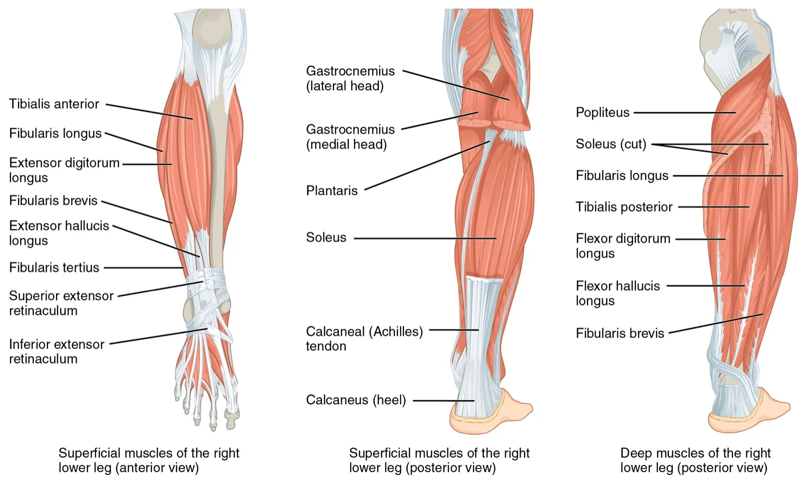
It is supplied by the tibial nerve, the main nerve at the back of the lower leg, which works the calf muscles1. Its blood comes from branches of the popliteal artery, the main artery behind the knee1.
What it moves
The gastrocnemius produces plantar flexion, which means pointing the foot down, away from the shin1. It also helps with flexion, or bending, of the knee1. Through these actions it plays a part in walking, posture and pushing the body forward1. The three triceps surae muscles do most of the work of plantar flexion3.
Because it crosses two joints, both the knee and the ankle positions affect how its force changes with its length8. One study made the muscle contract with electrical pulses in 26 people, at 24 knee and ankle positions8. It made its largest joint moment (turning effect around the ankle) with the knee straight and the foot pulled up toward the shin8. That is also the position where the muscle is longest8. In that study, the full knee and ankle ranges did not appear to stretch it past its best length for pointing the foot8.
How it works in walking and running
One study used ultrasound, a scan made with sound waves, on six men walking on a treadmill at 3 km/h9. While the foot was on the ground, the medial head held almost the same length, with its fascicles (fibre bundles) at about 50 mm9. Its tendon stretched by about 7 mm while the body stood on one leg, and sprang back at push-off9. The authors note that the muscle likely used little energy, while the springy tendon stored and returned energy, and both would help walking economy9.
A second ultrasound study compared treadmill walking at 4.5 km/h with running at 7.5 km/h10. In walking, the medial head’s fascicles stayed fairly steady in length during stance, the part of each step when the foot is on the ground10. In running, they shortened throughout stance while the Achilles tendon and aponeurosis (a flat sheet of tendon) stretched more10. The give in these springy tissues let the fascicles shorten much more slowly10. The fast shortening at take-off came from the tissues springing back10.
Breakdown
Strains of the inner head
A review describes a strain of the medial gastrocnemius as the most common cause of pain in the middle of the calf in athletes11. It says the strain comes from stretching the muscle with the knee straight and the foot pulled up11. The same review describes the main feature as pain at the back and inner side of the calf, often after a feeling of a pop11.
A systematic review of 21 studies looked at 728 muscle injuries that did not come from a blow12. In this review of 728 muscle injuries, calf injuries typically happened as the foot moved up toward the shin with the knee nearly straight12. At the assumed moment of injury, the ankle was more than 10 degrees into dorsiflexion (pulling the foot up)12.
Tennis leg
A review of tennis leg describes injuries where the medial gastrocnemius meets the soleus as common in athletes and active people13. It says they typically come from a sudden overstretch or from many small injuries repeated over time13. One study scanned 141 patients whose doctors had found tennis leg on examination14. Ultrasound showed these findings14:
| Ultrasound finding in 141 patients | Share of patients |
|---|---|
| Tear of the medial gastrocnemius head (94 patients) | 66.7%14 |
| Fluid between the gastrocnemius and soleus, with no muscle tear (30) | 21.3%14 |
| Tear of the plantaris (a small calf muscle) tendon (2) | 1.4%14 |
In that study, the fluid collected between the flat tendon sheets of the medial gastrocnemius and the soleus14. In that study, problems in the medial gastrocnemius appeared more common than problems in the plantaris tendon14.
Strains in football
One 10-year study covered 82 gastrocnemius injuries in elite male Australian football players, seen on MRI (a scan that shows soft tissue in detail)4. Of these, 78% involved the medial head4. Speeding up was the most common running cause (13 cases), and 29 injuries had no specific cause4. In a different study of 184 calf strains in elite Australian Football players, 84.6% were soleus injuries, more than gastrocnemius injuries15.
In a cohort study (one that follows a group over time) of 2,299 male professional footballers, calf muscles made up 13% of muscle injuries16. Calf injuries were the only muscle group whose rate rose with age16.
What is linked with calf strain
A systematic review of ten studies covered 5,397 athletes and 518 calf or lower-leg muscle injuries5. It found older age and a previous calf strain were the strongest risk factors for a future calf muscle injury5. It found no evidence that height, weight, gender or which side was dominant were linked with calf strain5. Evidence linking earlier injuries of the hamstrings, quadriceps (front thigh muscles), adductors (inner thigh muscles) or knee was only limited5. Overall the review found little evidence, and noted a trend for studies at high risk of bias5.
Recovery
How long strains kept players out
In the study of 82 gastrocnemius injuries in elite male Australian football players, the median (middle) time before playing again was 19 days4. In that study, a recent change in training load before the injury, a running cause and older age went with longer recovery4. In the study of 184 calf strains, gastrocnemius injuries took 19.1 days on average before playing again, and soleus injuries 25.4 days15. That difference was not statistically significant15. A review of tennis leg reports that recovery generally comes within 6 to 12 weeks13.
When strains come back
Of the 82 gastrocnemius injuries, 14 were repeat injuries, more than 1 in 64. Of these, 79% came less than 6 months after the first injury4. In 149 calf strains in the Australian Football League, older age and an earlier calf strain raised the risk of another within two seasons17. The MRI findings of the first injury were not linked with a repeat17.
Across 563 calf strains in 359 elite male Australian football players over 10 years, each repeat cost at least 35.6 days on average18. In professional football, time out after a repeat calf muscle injury was significantly longer than after the first19.
How it changes with training and stretching
In 10 healthy men, 18 sessions of eccentric training (muscle work done while the muscle lengthens) over 7 weeks changed the medial head20. The muscle got thicker, and its pennation angle (the slant of its fibres) grew, both at rest and when contracting20. Its fascicles got longer at rest20. The stiffness of its tendon, meaning the force needed to stretch it, also rose20.
In 11 males, six weeks of stretch training made the gastrocnemius 5.6% thicker6. Its fascicles grew 25% longer near the myotendinous junction, where muscle fibres join their tendon, and 5.1% longer mid-muscle6. Its hardest effort, the maximum voluntary contraction, did not change6. The outer head’s fascicles lengthened more than the inner head’s, and the pennation angle fell in the outer head but not the inner head6.
Two pieces of research measured how stretching changed ankle range21,22:
- In 12 people who stretched the calf twice a day for 3 weeks, dorsiflexion range rose 19.9%21. The gain came from more tolerance of stretch and from longer muscle and fascicles, not changes in muscle activity or reflexes21.
- A systematic review pooling five trials found calf stretching gave a small, statistically significant gain of about 2 to 3 degrees22. Its importance in practice was unclear22.
What long bed rest does
The plantar flexors are the muscles that point the foot down, and include both gastrocnemius heads23. Some healthy men spent 90 days in head-down-tilt bed rest, lying with the head tilted slightly below the feet, without exercise24. Their plantar flexor volume fell 29%24. A flywheel is a training device whose resistance comes from a spinning wheel. Men who did flywheel resistance exercise, including a calf press, lost less plantar flexor volume (15%)24.
Connections
The soleus and the Achilles tendon
The soleus lies deep between the two gastrocnemius heads25. Lower down it joins the gastrocnemius to form the Achilles tendon, which attaches to the heel bone25. The soleus crosses only the ankle25. In one study, it worked at similar lengths in standing and seated calf raises, while the two gastrocnemius heads worked at longer lengths when standing26. Minor soleus injuries that are hard to see on ultrasound are often confused with gastrocnemius strains25.
The Achilles tendon is made of fibres from the soleus and from both gastrocnemius heads27. One study looked at 106 limbs from 53 male cadavers (bodies donated for study)27. The outer head’s strand was the largest part of the tendon at the heel, about 44%27. The soleus and inner head strands were each about 28%27.
In publicly available data on NBA basketball players from 1958 to 2024, a previous calf strain went with higher odds of an Achilles tendon rupture28. The odds ratio, how many times higher the odds were, was 2.4928.
Exercises that load it
Studies that record muscle activity with electrical sensors, called electromyography, show how common exercises use the gastrocnemius3,29,30:
| Exercise | What studies found |
|---|---|
| Calf raise | In 22 trained men, soleus and gastrocnemius activity did not differ significantly from the leg press3 |
| Calf raise | In 33 people, the inner head was more active than the outer head with toes turned out, but not with toes turned in2 |
| Eccentric heel drop | In 18 people with Achilles tendinopathy (tendon pain with loading) and 18 without, the gastrocnemius was more active with a straight knee30 |
| Eccentric heel drop | In a modelling study of 19 healthy adults, estimated gastrocnemius activation at peak Achilles tendon force did not differ between straight-knee and bent-knee versions31 |
| Deadlift | In 13 college football players, medial gastrocnemius activity was greater in the conventional (hip-width, hands outside knees) than the sumo (wide, hands inside knees) deadlift29 |
| Back squat | A 2001 review reports moderate to high gastrocnemius activity, generally rising as the knee bends more32 |
| Decline squat | In 10 people doing single-leg squats, gastrocnemius activity rose between 16 and 24 degrees of decline33 |
| Decline squat | In 13 people, calf activity did not differ between a 25-degree decline and a flat surface34 |
| Single-leg Romanian deadlift | In 27 healthy adults, gastrocnemius activity was higher with an elastic barbell than a rigid one35 |
| Glute bridge hold | In 22 futsal (indoor five-a-side football) players in a single-leg bridge, outer head activity fell when the foot was also turned out36 |
The decline squat studies disagree34,37,38. One found more gastrocnemius activity on the board, one less outer head activity at one knee angle, and one no calf difference34,37,38. Related exercise pages: calf raise, eccentric heel drop, deadlift, back squat, decline squat, single-leg Romanian deadlift and glute bridge hold3,29,30,32,35,36,37.
Gastrocnemius pulls through
Gastrocnemius works with
Gastrocnemius attaches to
Gastrocnemius crosses
Gastrocnemius is loaded by
Sources
- Bordoni B, Varacallo MA (2026). Anatomy, Bony Pelvis and Lower Limb: Gastrocnemius Muscle.. StatPearls. PMID 30422541
- Cibulka M, Wenthe A, Boyle Z, Callier D, Schwerdt A, Jarman D, Strube MJ (2017). Variation in medial and lateral gastrocnemius muscle activity with foot position.. International Journal of Sports Physical Therapy. PMID 28515978
- Gentil P, Souza D, Santana M, Alves RR, Campos MH, Pinto R, Bottaro M (2020). Multi- and Single-Joint Resistance Exercises Promote Similar Plantar Flexor Activation in Resistance Trained Men.. International Journal of Environmental Research and Public Health. doi:10.3390/ijerph17249487 PMID 33352879
- Gray C, Pizzari T, Murphy MC, Schache AG, Breidahl WH, Klinken S, Bell T, Green B (2025). Gastrocnemius Muscle Strain Injury Characteristics in Elite Male Australian Football Players: A 10-Year Longitudinal Cohort Study.. The Journal of orthopaedic and sports physical therapy. doi:10.2519/jospt.2025.13526 PMID 40985375
- Green B, Pizzari T (2017). Calf muscle strain injuries in sport: a systematic review of risk factors for injury.. British Journal of Sports Medicine. doi:10.1136/bjsports-2016-097177 PMID 28259848
- Simpson CL, Kim BDH, Bourcet MR, Jones GR, Jakobi JM (2017). Stretch training induces unequal adaptation in muscle fascicles and thickness in medial and lateral gastrocnemii.. Scandinavian journal of medicine & science in sports. doi:10.1111/sms.12822 PMID 28138986
- Betts JG, Young KA, Wise JA, Johnson E, Poe B, Kruse DH, Korol O, Johnson JE, Womble M, DeSaix P (2022). Anatomy and Physiology 2e. OpenStax. Link
- Landin D, Thompson M, Reid M (2015). Knee and Ankle Joint Angles Influence the Plantarflexion Torque of the Gastrocnemius.. Journal of clinical medicine research. doi:10.14740/jocmr2107w PMID 26124905
- Fukunaga T, Kubo K, Kawakami Y, Fukashiro S, Kanehisa H, Maganaris CN (2001). In vivo behaviour of human muscle tendon during walking.. Proceedings. Biological sciences. doi:10.1098/rspb.2000.1361 PMID 11217891
- Lichtwark GA, Bougoulias K, Wilson AM (2007). Muscle fascicle and series elastic element length changes along the length of the human gastrocnemius during walking and running.. Journal of biomechanics. doi:10.1016/j.jbiomech.2005.10.035 PMID 16364330
- Halabchi F, Tavana MM, Seifi V, Mahmoudi Zarandi M (2024). Medial Gastrocnemius Strain: Clinical Aspects and Algorithmic Approach.. Medical journal of the Islamic Republic of Iran. doi:10.47176/mjiri.38.55 PMID 39399607
- Finnern LS, Wilke J, Willwacher S, Pasanen K, Hollander K, Dalos D, Welsch GH, Krosshaug T, Edouard P, Gronwald T, Hoenig T (2026). Qualitative and quantitative situational characteristics of muscle strains in sports: a systematic review and meta-analysis.. British Journal of Sports Medicine. doi:10.1136/bjsports-2025-110327 PMID 41558842
- Schwach M, Gaulin B, Vermorel PH, Horteur C, Tourné Y (2026). Tennis leg: Diagnosis and management - A state-of-the-art review.. Foot (Edinburgh, Scotland). doi:10.1016/j.foot.2026.102267 PMID 42258915
- Delgado GJ, Chung CB, Lektrakul N, Azocar P, Botte MJ, Coria D, Bosch E, Resnick D (2002). Tennis leg: clinical US study of 141 patients and anatomic investigation of four cadavers with MR imaging and US.. Radiology. doi:10.1148/radiol.2241011067 PMID 12091669
- Green B, Lin M, Schache AG, McClelland JA, Semciw AI, Rotstein A, Cook J, Pizzari T (2020). Calf muscle strain injuries in elite Australian Football players: A descriptive epidemiological evaluation.. Scandinavian Journal of Medicine & Science in Sports. doi:10.1111/sms.13552 PMID 31494970
- Ekstrand J, Hägglund M, Waldén M (2011). Epidemiology of muscle injuries in professional football (soccer).. The American Journal of Sports Medicine. doi:10.1177/0363546510395879 PMID 21335353
- Green B, Lin M, McClelland JA, Semciw AI, Schache AG, Rotstein AH, Cook J, Pizzari T (2020). Return to Play and Recurrence After Calf Muscle Strain Injuries in Elite Australian Football Players.. The American Journal of Sports Medicine. doi:10.1177/0363546520959327 PMID 33030961
- Green B, Schache AG, Pizzari T (2025). What is a recurrence? The onset, frequency and time loss impact of recurrent calf muscle strain injuries in elite male Australian football players over a decade.. BMJ Open Sport & Exercise Medicine. doi:10.1136/bmjsem-2025-002865 PMID 40919406
- Ekstrand J, Krutsch W, Spreco A, van Zoest W, Roberts C, Meyer T, Bengtsson H (2020). Time before return to play for the most common injuries in professional football: a 16-year follow-up of the UEFA Elite Club Injury Study.. British Journal of Sports Medicine. doi:10.1136/bjsports-2019-100666 PMID 31182429
- Duclay J, Martin A, Duclay A, Cometti G, Pousson M (2009). Behavior of fascicles and the myotendinous junction of human medial gastrocnemius following eccentric strength training.. Muscle & nerve. doi:10.1002/mus.21297 PMID 19301364
- Blazevich AJ, Cannavan D, Waugh CM, Miller SC, Thorlund JB, Aagaard P, Kay AD (2014). Range of motion, neuromechanical, and architectural adaptations to plantar flexor stretch training in humans.. Journal of applied physiology (Bethesda, Md. : 1985). doi:10.1152/japplphysiol.00204.2014 PMID 24947023
- Radford JA, Burns J, Buchbinder R, Landorf KB, Cook C (2006). Does stretching increase ankle dorsiflexion range of motion? A systematic review.. British journal of sports medicine. doi:10.1136/bjsm.2006.029348 PMID 16926259
- Ward SR, Eng CM, Smallwood LH, Lieber RL (2009). Are current measurements of lower extremity muscle architecture accurate?. Clinical Orthopaedics and Related Research. doi:10.1007/s11999-008-0594-8 PMID 18972175
- Alkner BA, Tesch PA (2004). Knee extensor and plantar flexor muscle size and function following 90 days of bed rest with or without resistance exercise.. European journal of applied physiology. doi:10.1007/s00421-004-1172-8 PMID 15338217
- Trybulski R, Gałęziok K, Matuszczyk F, Halski T, Muracki J (2025). Problems in Diagnosis and Treatment of Soleus Muscle Injuries-Narrative Review and Case Report.. Journal of Clinical Medicine. doi:10.3390/jcm14061955 PMID 40142763
- Kinoshita M, Maeo S, Kobayashi Y, Eihara Y, Ono M, Sato M, Sugiyama T, Kanehisa H, Isaka T (2023). Triceps surae muscle hypertrophy is greater after standing versus seated calf-raise training.. Frontiers in Physiology. doi:10.3389/fphys.2023.1272106 PMID 38156065
- Pękala PA, Henry BM, Ochała A, Kopacz P, Tatoń G, Młyniec A, Walocha JA, Tomaszewski KA (2017). The twisted structure of the Achilles tendon unraveled: A detailed quantitative and qualitative anatomical investigation.. Scandinavian Journal of Medicine & Science in Sports. doi:10.1111/sms.12835 PMID 28139009
- Elsabbagh Z, Le Y, Bcharah G, Ryu D, Badin D, Hsu NN, Thompson JM, Aiyer A (2026). Prior Calf Strain as a Risk Factor for Achilles Tendon Rupture in National Basketball Association Players: A Retrospective Analysis.. Orthopaedic journal of sports medicine. doi:10.1177/23259671261449170 PMID 42293040
- Escamilla RF, Francisco AC, Kayes AV, Speer KP, Moorman CT 3rd (2002). An electromyographic analysis of sumo and conventional style deadlifts.. Medicine and Science in Sports and Exercise. doi:10.1097/00005768-200204000-00019 PMID 11932579
- Reid D, McNair PJ, Johnson S, Potts G, Witvrouw E, Mahieu N (2012). Electromyographic analysis of an eccentric calf muscle exercise in persons with and without Achilles tendinopathy.. Physical therapy in sport : official journal of the Association of Chartered Physiotherapists in Sports Medicine. doi:10.1016/j.ptsp.2011.08.003 PMID 22814448
- Weinert-Aplin RA, Bull AM, McGregor AH (2015). Investigating the Effects of Knee Flexion during the Eccentric Heel-Drop Exercise.. Journal of sports science & medicine. PMID 25983597
- Escamilla RF (2001). Knee biomechanics of the dynamic squat exercise.. Medicine and Science in Sports and Exercise. doi:10.1097/00005768-200101000-00020 PMID 11194098
- Richards J, Thewlis D, Selfe J, Cunningham A, Hayes C (2008). A biomechanical investigation of a single-limb squat: implications for lower extremity rehabilitation exercise.. Journal of athletic training. doi:10.4085/1062-6050-43.5.477 PMID 18833310
- Kongsgaard M, Aagaard P, Roikjaer S, Olsen D, Jensen M, Langberg H, Magnusson SP (2006). Decline eccentric squats increases patellar tendon loading compared to standard eccentric squats.. Clinical Biomechanics. doi:10.1016/j.clinbiomech.2006.03.004 PMID 16675081
- Jeong J, Park I (2025). Comparison of Lower Limb COP and Muscle Activation During Single-Leg Deadlift Using Elastic and Inelastic Barbells.. Sports (Basel). doi:10.3390/sports13080242 PMID 40863751
- Elias G, Jerez-Mayorga D, Intelangelo L (2024). Does the Position of the Ankle Matter During the Single Gluteal Bridge in Futsal Players? An Electromyographic Analysis.. Journal of Sport Rehabilitation. doi:10.1123/jsr.2022-0425 PMID 37917981
- Frohm A, Halvorsen K, Thorstensson A (2007). Patellar tendon load in different types of eccentric squats.. Clinical biomechanics (Bristol, Avon). doi:10.1016/j.clinbiomech.2006.12.006 PMID 17499407
- Lee D, Lee S, Park J (2015). Impact of decline-board squat exercises and knee joint angles on the muscle activity of the lower limbs.. Journal of physical therapy science. doi:10.1589/jpts.27.2617 PMID 26357447
Changes
- First published.