Quadriceps
Also called quadriceps femoris
The group of four muscles at the front of the thigh that straightens the knee. How it pulls through the kneecap, how it strains and weakens, and how quickly it grows and shrinks.
- Region
- Thigh
- Starts on (rectus femoris)
- the anterior inferior iliac spine, a bony point at the front of the pelvis, and the hip socket's rim1,8,36
- Ends on
- the kneecap, through the quadriceps tendon shared by all four, then by the patellar ligament to the shinbone's front bump1
- Tendons
- Quadriceps tendon, Patellar tendon
- Muscles
- rectus femoris, vastus lateralis, vastus medialis, vastus intermedius
- Updated
- October 22, 2026
- Reviewed
- October 22, 2026 · how entries are checked
Function
Four muscles, one tendon
The quadriceps femoris is a group of four muscles in the front compartment of the thigh1. Each one has its own place1:
| Muscle | Where it lies in the thigh |
|---|---|
| Rectus femoris | Down the front1 |
| Vastus lateralis | On the outer side1 |
| Vastus medialis | On the inner side1 |
| Vastus intermedius | Between the other two vasti, beneath the rectus femoris1 |
The four muscles merge into one shared tendon, called the quadriceps tendon1. It attaches to the patella (kneecap) and carries on below it as the patellar ligament1. The patellar ligament fixes to the tibial tuberosity, a bump on the front of the tibia (shinbone)1. The femoral nerve supplies the quadriceps5.
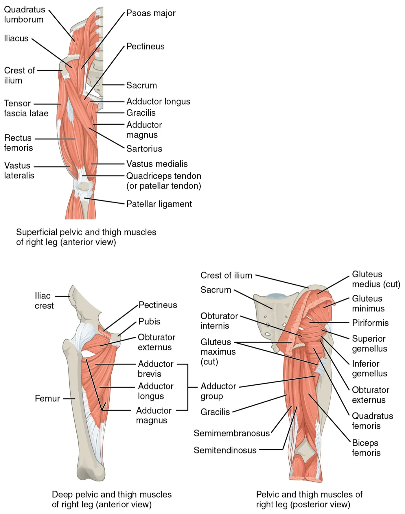
The tendon is usually described as meeting the kneecap in three layers6. Real limbs vary more than that6.
How the kneecap adds leverage
The patella is the largest sesamoid bone in the body, meaning a bone that sits inside a tendon1. It sits in the quadriceps tendon, which crosses the front of the knee on its way to the tibia1. The kneecap anchors the quadriceps tendon above it and the patellar ligament below it7. It also holds the tendon away from the knee joint1. That gives the quadriceps more leverage as it pulls across the knee1.
What the muscles move
Working through the patella and patellar ligament, the quadriceps is a powerful straightener of the knee, a movement called knee extension1. It also acts as a “dynamic ligament”, giving the knee important support and steadiness1. The front thigh muscles also help flex the hip, lifting the thigh forward1.
The rectus femoris is the one quadriceps muscle that crosses two joints8. So it flexes the hip as well as extending the knee8. It starts from two tendon heads on the pelvis8. The direct head, nearer the surface, comes from the anterior inferior iliac spine, a bony point at the front of the pelvis8. The deeper reflected head comes from the ridge around the hip socket8.
Breakdown
How often the quadriceps is injured
A separate page covers quadriceps contusion. A cohort study followed 2,299 male professional footballers from 2001 to 20092. The quadriceps was the third most common site of their muscle injuries2:
| Muscle group | Share of muscle injuries |
|---|---|
| Hamstrings (long muscles at the back of the thigh) | 37%2 |
| Adductors (inner thigh muscles) | 23%2 |
| Quadriceps | 19%2 |
Reviews describe the rectus femoris as the quadriceps muscle injured most often8,9. One systematic review gathered 38 case reports and case series, covering 152 people with rectus femoris injuries9. Of these, 91% were male9. Of the injuries, 80% happened while kicking and 20% while sprinting9. The authors rated the certainty of this evidence as low9.
What raises the odds of a strain
A systematic review pooled 16 studies, with 2,408 quadriceps injuries among 11,719 athletes10. It named four factors as the strongest risks for a future quadriceps injury in sport10:
- An earlier quadriceps injury10.
- A recent hamstring injury10.
- Being the leg used for kicking10.
- Playing in competitive matches10.
The same review found no link between quadriceps injury and age, weight or flexibility10.
How knee injury weakens it
After some knee injuries, people often do not fully switch on the quadriceps by effort11. A systematic review found this common with a torn or rebuilt anterior cruciate ligament (ACL), and with pain at the front of the knee11. The ACL is a ligament inside the knee that joins the thigh bone to the shinbone. It was often seen in both legs, not just the injured one11.
The reviewers describe arthrogenic muscle inhibition as an important underlying factor in lasting quadriceps weakness after knee injury or surgery11. Arthrogenic means arising from a joint.
A meta-analysis of 21 studies compared quadriceps strength after ACL reconstruction with matched people who had no such surgery12. Each gap is given as a standardised mean difference (SMD), the size of a gap between two groups on a common scale. The gap was smaller the longer it had been since surgery12:
| Time since surgery | Strength of the rebuilt leg compared with controls |
|---|---|
| Under 6 months | Clearly lower (SMD −1.42)12 |
| 6 to 18 months | Still lower (SMD −0.92)12 |
| 18 to 48 months | Smaller gap (SMD −0.38), and no difference could not be ruled out12 |
Weak knee extensors are also linked with later knee osteoarthritis, a disease of the knee joint13. A meta-analysis of 11 studies, with 46,819 people followed over time, found this link in both women and men13. The authors rated the evidence as low quality13.
Recovery
How fast the muscle grows with training
One review puts forward the thesis that early size gains with resistance training are mostly swelling, not growth4. It sets out three phases4:
- Up to about 4 sessions, gains in cross-sectional area (the size of a slice cut straight across the muscle) are mostly swelling from muscle damage4.
- After about 10 sessions, modest growth follows4.
- True growth is not seen until after about 18 sessions4.
One study measured the vastus lateralis by ultrasound, a scan made with sound waves, in ten untrained young men doing resistance training14. Its cross-sectional area was about 2.7% larger in week 3 and 10.4% larger after 10 weeks14. The authors propose that the early gain was not purely growth14. It came with swelling, probably from muscle damage, which may explain much of it14. They conclude that early gains in area should not be called growth without some measure of swelling or damage14.
In ten young men who gave vastus lateralis samples, fibre cross-sectional area had risen at 10 weeks of training but not at 315.
In one small study of seven healthy young people, quadriceps cross-sectional area had risen significantly after 20 days of training16. They did hard leg extensions on a flywheel machine three times a week16. At two regions of the muscle, it rose like this16:
| Point in training | Rise in cross-sectional area |
|---|---|
| Day 20 | 3.5% and 5.2%16 |
| Day 35 | 6.5% and 7.4%16 |
By day 35, the most force they could produce was about 39% higher16.
In one trial, squat depth mattered less for this muscle than for others17. Over 10 weeks, 17 men trained either full or half squats17. Knee extensor volume grew by a similar amount in both groups, while rectus femoris volume did not change in either17.
How fast it shrinks without use
Muscle loss while a leg is held still appears fastest in the earliest period3. A review with meta-analysis included 12 studies in healthy volunteers, with 140 people between them3. In healthy quadriceps, the review reports about 6.5% loss by day 14 and 9.1% by day 28, and loss appeared to slow3. The authors note that the evidence overall is limited3.
In thirteen healthy young men with one leg held in a knee brace, loss of thigh muscle showed up within 2 days18. Over the next 5 days it went on at about 0.8% a day18. After the week, MRI scans showed the quadriceps had lost more muscle, 6.7%, against 3.5% for the hamstrings18.
One study followed 9 older and 11 young men through immobilisation (keeping a limb still) and retraining19. Two weeks of immobilisation reduced strength and quadriceps volume in both groups19. After 4 weeks of retraining, both groups had their starting strength back19. The older men regained less muscle volume than the young ones19.
Training during disuse may hold on to some of the muscle20. A meta-analysis of randomised controlled trials compared resistance training with no exercise during a period of disuse20. In the five trials that measured it, training helped keep quadriceps volume20. The authors say more high-quality research is needed to confirm this20.
How long a rectus femoris injury takes
Average time back to sport differed with where the rectus femoris was hurt9. In the review of 152 people, these were the average times after successful care without surgery9:
| Injury site | Average time back to sport |
|---|---|
| Where muscle meets tendon | About 1 month9 |
| Tendon pulled away from the anterior inferior iliac spine | About 2.9 months9 |
| In the free tendon | About 4 months9 |
The authors rated this evidence as low certainty9.
Connections
The hamstrings, its opposite number
The quadriceps extends the knee and the hamstrings bend it, so each opposes the other there1. The rectus femoris opposes the hamstrings at the hip as well as at the knee8. A recent hamstring strain is linked with a later quadriceps injury10. In one group of amateur Australian Rules footballers, more flexible quadriceps went with fewer hamstring strains21. Evidence on hamstring-to-quadriceps strength ratios before hamstring strain is mixed21.
The patellar tendon below it
Below the kneecap, the quadriceps pulls on the tibia through the patellar ligament1. The most common patellar tendon tear follows a lengthening contraction of the quadriceps22. In 15 young men after 9 weeks of knee-extension training, larger quadriceps growth went with larger gains in tendon stiffness23. The tendon changes were not related to strength gains23.
Exercises that load it
Related exercise pages include the hollow hold and those in the table below. In the squat, the hip and knee extensors work against the bend that body weight and load create at both joints24. Quadriceps activity in the squat is moderate to high, and generally rises as the knees bend more25. Other studies compared the quadriceps across exercises and set-ups:
| Exercise | What studies found |
|---|---|
| Back squat | A high bar position gave more leg muscle activity than a low bar26,27 |
| Back squat | Stance width does not appear to change quadriceps activity24 |
| Back squat | Findings on squat depth and quadriceps activity conflict24 |
| Deadlift | The quadriceps and back extensors were more active than the gluteus maximus (the large muscle of the buttock) and biceps femoris, one of the hamstrings1,28 |
| Deadlift | The sumo style gave more vastus medialis and lateralis activity than the conventional style29 |
| Wall squat | Across two wall squat forms and a one-leg squat in 18 people, modelled quadriceps force was about 30 to 720 newtons, hamstrings 15 to 19030 |
| Wall sit | One study tested five static exercises in 24 healthy 18- to 21-year-olds. The authors suggest a 90-second wall sit and a single-leg knee raise fatigued the rectus femoris and two buttock muscles most reliably31 |
| Farmer’s walk | Rectus femoris activity averaged 77% of a maximal effort in one study32 |
| Plank and side plank | In 18 adults, face-down and side exercises done on the toes gave more rectus femoris activity than the same exercises done on the knees33 |
In the deadlift, quadriceps activity was greater when the knees were more bent29. In 23 healthy adults, a one-leg wall squat gave the most vastus medialis oblique activity of five one-leg exercises34. These are the lowest fibres of the vastus medialis. A wall squat with a ball behind the back gave less rectus femoris and vastus lateralis activity than a Spanish squat35.
Quadriceps opposes at the knee
Quadriceps pulls on the tibia through
Quadriceps is loaded by
Sources
- Betts JG, Young KA, Wise JA, Johnson E, Poe B, Kruse DH, Korol O, Johnson JE, Womble M, DeSaix P (2022). Anatomy and Physiology 2e. OpenStax. Link
- Ekstrand J, Hägglund M, Waldén M (2011). Epidemiology of muscle injuries in professional football (soccer).. The American Journal of Sports Medicine. doi:10.1177/0363546510395879 PMID 21335353
- Hardy EJO, Inns TB, Hatt J, Doleman B, Bass JJ, Atherton PJ, Lund JN, Phillips BE (2022). The time course of disuse muscle atrophy of the lower limb in health and disease.. Journal of Cachexia, Sarcopenia and Muscle. doi:10.1002/jcsm.13067 PMID 36104842
- Damas F, Libardi CA, Ugrinowitsch C (2018). The development of skeletal muscle hypertrophy through resistance training: the role of muscle damage and muscle protein synthesis.. European Journal of Applied Physiology. doi:10.1007/s00421-017-3792-9 PMID 29282529
- Kowalska B, Sudoł-Szopińska I (2012). Ultrasound assessment of selected peripheral nerves pathologies. Part II: Entrapment neuropathies of the lower limb.. Journal of Ultrasonography. doi:10.15557/jou.2012.0033 PMID 26673938
- Waligora AC, Johanson NA, Hirsch BE (2009). Clinical anatomy of the quadriceps femoris and extensor apparatus of the knee.. Clinical Orthopaedics and Related Research. doi:10.1007/s11999-009-1052-y PMID 19690926
- Cox CF, Sinkler MA, Black AC, Launico MV, Hubbard JB (2023). Anatomy, Bony Pelvis and Lower Limb, Knee Patella. StatPearls. PMID 30137819
- Begum FA, Kayani B, Chang JS, Tansey RJ, Haddad FS (2020). The management of proximal rectus femoris avulsion injuries.. EFORT Open Reviews. doi:10.1302/2058-5241.5.200055 PMID 33312709
- Bogwasi L, Holtzhausen L, Janse van Rensburg DC, Jansen van Rensburg A, Botha T (2023). Management of proximal rectus femoris injuries - do we know what we're doing?: A systematic review.. Biology of Sport. doi:10.5114/biolsport.2023.116454 PMID 37077795
- Pietsch S, Pizzari T (2022). Risk Factors for Quadriceps Muscle Strain Injuries in Sport: A Systematic Review.. Journal of Orthopaedic & Sports Physical Therapy. doi:10.2519/jospt.2022.10870 PMID 35647880
- Hart JM, Pietrosimone B, Hertel J, Ingersoll CD (2010). Quadriceps activation following knee injuries: a systematic review.. Journal of Athletic Training. doi:10.4085/1062-6050-45.1.87 PMID 20064053
- Brown C, Marinko L, LaValley MP, Kumar D (2021). Quadriceps Strength After Anterior Cruciate Ligament Reconstruction Compared With Uninjured Matched Controls: A Systematic Review and Meta-analysis.. Orthopaedic Journal of Sports Medicine. doi:10.1177/2325967121991534 PMID 33889639
- Øiestad BE, Juhl CB, Culvenor AG, Berg B, Thorlund JB (2022). Knee extensor muscle weakness is a risk factor for the development of knee osteoarthritis: an updated systematic review and meta-analysis including 46 819 men and women.. British Journal of Sports Medicine. doi:10.1136/bjsports-2021-104861 PMID 34916210
- Damas F, Phillips SM, Lixandrão ME, Vechin FC, Libardi CA, Roschel H, Tricoli V, Ugrinowitsch C (2016). Early resistance training-induced increases in muscle cross-sectional area are concomitant with edema-induced muscle swelling.. European Journal of Applied Physiology. doi:10.1007/s00421-015-3243-4 PMID 26280652
- Damas F, Phillips SM, Libardi CA, Vechin FC, Lixandrão ME, Jannig PR, Costa LA, Bacurau AV, Snijders T, Parise G, Tricoli V, Roschel H, Ugrinowitsch C (2016). Resistance training-induced changes in integrated myofibrillar protein synthesis are related to hypertrophy only after attenuation of muscle damage.. The Journal of Physiology. doi:10.1113/JP272472 PMID 27219125
- Seynnes OR, de Boer M, Narici MV (2007). Early skeletal muscle hypertrophy and architectural changes in response to high-intensity resistance training.. Journal of Applied Physiology. doi:10.1152/japplphysiol.00789.2006 PMID 17053104
- Kubo K, Ikebukuro T, Yata H (2019). Effects of squat training with different depths on lower limb muscle volumes.. European Journal of Applied Physiology. doi:10.1007/s00421-019-04181-y PMID 31230110
- Kilroe SP, Fulford J, Jackman SR, van Loon LJC, Wall BT (2020). Temporal Muscle-specific Disuse Atrophy during One Week of Leg Immobilization.. Medicine & Science in Sports & Exercise. doi:10.1249/MSS.0000000000002200 PMID 31688656
- Suetta C, Hvid LG, Justesen L, Christensen U, Neergaard K, Simonsen L, Ortenblad N, Magnusson SP, Kjaer M, Aagaard P (2009). Effects of aging on human skeletal muscle after immobilization and retraining.. Journal of Applied Physiology. doi:10.1152/japplphysiol.00290.2009 PMID 19661454
- Guo X, Zhou Y, Li X, Mu J (2025). Resistance exercise training improves disuse-induced skeletal muscle atrophy in humans: a meta-analysis of randomized controlled trials.. BMC Musculoskeletal Disorders. doi:10.1186/s12891-025-08384-7 PMID 39920735
- Prior M, Guerin M, Grimmer K (2009). An evidence-based approach to hamstring strain injury: a systematic review of the literature.. Sports Health. doi:10.1177/1941738108324962 PMID 23015867
- Tandogan RN, Terzi E, Gomez-Barrena E, Violante B, Kayaalp A (2022). Extensor mechanism ruptures.. EFORT Open Reviews. doi:10.1530/EOR-22-0021 PMID 35638613
- Seynnes OR, Erskine RM, Maganaris CN, Longo S, Simoneau EM, Grosset JF, Narici MV (2009). Training-induced changes in structural and mechanical properties of the patellar tendon are related to muscle hypertrophy but not to strength gains.. Journal of Applied Physiology. doi:10.1152/japplphysiol.00213.2009 PMID 19478195
- Straub RK, Powers CM (2024). A Biomechanical Review of the Squat Exercise: Implications for Clinical Practice.. International Journal of Sports Physical Therapy. doi:10.26603/001c.94600 PMID 38576836
- Escamilla RF (2001). Knee biomechanics of the dynamic squat exercise.. Medicine and Science in Sports and Exercise. doi:10.1097/00005768-200101000-00020 PMID 11194098
- Park JH, Lee SJ, Shin HJ, Cho HY (2022). Influence of Loads and Loading Position on the Muscle Activity of the Trunk and Lower Extremity during Squat Exercise.. International Journal of Environmental Research and Public Health. doi:10.3390/ijerph192013480 PMID 36294056
- Glassbrook DJ, Helms ER, Brown SR, Storey AG (2017). A Review of the Biomechanical Differences Between the High-Bar and Low-Bar Back-Squat.. Journal of Strength and Conditioning Research. doi:10.1519/JSC.0000000000002007 PMID 28570490
- Martín-Fuentes I, Oliva-Lozano JM, Muyor JM (2020). Electromyographic activity in deadlift exercise and its variants. A systematic review.. PLoS One. doi:10.1371/journal.pone.0229507 PMID 32107499
- Escamilla RF, Francisco AC, Kayes AV, Speer KP, Moorman CT 3rd (2002). An electromyographic analysis of sumo and conventional style deadlifts.. Medicine and Science in Sports and Exercise. doi:10.1097/00005768-200204000-00019 PMID 11932579
- Escamilla RF, Zheng N, Imamura R, Macleod TD, Edwards WB, Hreljac A, Fleisig GS, Wilk KE, Moorman CT 3rd, Andrews JR (2009). Cruciate ligament force during the wall squat and the one-leg squat.. Medicine and Science in Sports and Exercise. doi:10.1249/MSS.0b013e3181882c6d PMID 19127183
- Dyer OL, Seeley MA, Wheatley BB (2024). Effects of static exercises on hip muscle fatigue and knee wobble assessed by surface electromyography and inertial measurement unit data.. Scientific Reports. doi:10.1038/s41598-024-61325-7 PMID 38714802
- Hindle BR, Lorimer A, Winwood P, Keogh JWL (2019). The Biomechanics and Applications of Strongman Exercises: a Systematic Review.. Sports Medicine - Open. doi:10.1186/s40798-019-0222-z PMID 31820223
- Escamilla RF, Lewis C, Pecson A, Imamura R, Andrews JR (2016). Muscle Activation Among Supine, Prone, and Side Position Exercises With and Without a Swiss Ball.. Sports Health. doi:10.1177/1941738116653931 PMID 27302152
- Ayotte NW, Stetts DM, Keenan G, Greenway EH (2007). Electromyographical analysis of selected lower extremity muscles during 5 unilateral weight-bearing exercises.. Journal of Orthopaedic and Sports Physical Therapy. doi:10.2519/jospt.2007.2354 PMID 17366959
- Lee JH, Kim S, Heo J, Park DH, Chang E (2022). Differences in the muscle activities of the quadriceps femoris and hamstrings while performing various squat exercises.. BMC Sports Science, Medicine and Rehabilitation. doi:10.1186/s13102-022-00404-6 PMID 35063016
- Eberbach H, Hohloch L, Feucht MJ, Konstantinidis L, Südkamp NP, Zwingmann J (2017). Operative versus conservative treatment of apophyseal avulsion fractures of the pelvis in the adolescents: a systematical review with meta-analysis of clinical outcome and return to sports.. BMC musculoskeletal disorders. doi:10.1186/s12891-017-1527-z PMID 28420360
Changes
- Links to the new quadriceps contusion page
- Added a figure showing where it sits, links to the exercise pages that load it, and tighter wording in a few places.
- First published.