Patellar tendon
Also called patellar ligament
The band below the kneecap that joins it to the shin bone and carries the thigh muscles' pull. How it bears load, how it gets painful or tears, how it adapts, and what it works with.
- Region
- Knee
- Muscles
- quadriceps
- Updated
- September 29, 2026
- Reviewed
- September 29, 2026 · how entries are checked
Function
Where the tendon runs
The four quadriceps muscles on the front of the thigh share one tendon, the quadriceps tendon1. It fixes into the patella and carries on below it as the patellar tendon1. The patella is the body’s largest sesamoid bone, a bone that sits inside a tendon1. So the patella anchors both tendons, one above it and one below9.
From the patella, the tendon runs down to the front of the tibia, just below the knee1. Some anatomy sources call this band the patellar ligament1. It ends on the tibial tuberosity, a raised area on the front of the tibia near its top1.
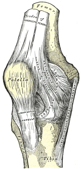
Like any tendon, it passes force from a working muscle to bone, and that pull makes the body move10. Tendons are made of collagen, a strong rope-like protein, packed side by side in bundles1. That layout makes them strong and hard to stretch along the line of the fibres1.
How the kneecap adds leverage
The patella lifts the quadriceps tendon away from the knee joint, giving the quadriceps more leverage for straightening the knee1,9. It increases the patellar tendon’s moment arm (its leverage about the knee)1,9. The quadriceps also acts like a “dynamic ligament”, giving the knee joint support and stability1.
How big the tendon is
The tendon is not the same size all the way along11. In 12 healthy young men, MRI scans (scans that show soft tissue in detail) showed its cross-sectional area grew from top to bottom11:
| Part of the tendon | Cross-sectional area |
|---|---|
| Near the kneecap | About 104 mm²11 |
| Middle | About 118 mm²11 |
| Near the tibia | About 127 mm²11 |
It also differs from side to side12. In 35 healthy tendons from young women and men, the outer side was the thinnest12. It was about 0.34 cm thick, against about 0.41 cm for the inner side and centre12. In the men, the outer side was about 4.8 cm long and the inner side about 4.4 cm12. In the women, the two sides did not differ significantly in length12.
Size differs between people too13. In 37 adults, men’s tendons had 28% more cross-sectional area than women’s, were 11% longer and 35% stiffer13. In the same study, the middle of the tendon was 12% smaller in older adults than in young adults13.
How load changes with the task
One study scored how hard 35 exercises loaded the tendon in 20 healthy adults2. Its loading index combined peak load, how fast the load rose, and the total load over the movement2:
| Exercise | Loading index |
|---|---|
| One-leg squat on a slope | 0.7472 |
| One-leg jump up from a dip | 0.7112 |
| One-leg hop forward | 0.6662 |
| Two-leg jump up from a dip | 0.6102 |
| One-leg drop and jump | 0.5992 |
| Two-leg drop and jump | 0.5632 |
| Two-leg full squat | 0.4282 |
| Two-leg squat to 60 degrees | 0.2242 |
The study sorted exercises into three tiers, and the Spanish squat, the lunge and the Bulgarian squat fell in the middle one2.
Other studies looked at how the way a movement is done changes tendon load14,15:
- Weight on the bar. In 10 trained athletes, back squats went to about 60 degrees of knee bend14. Strain rose from 4.7% to 8.2% as the weight rose from 40% to 80% of maximum14. That maximum is the one-repetition maximum, the heaviest weight a person can lift once14.
- The person. At 80% of the one-repetition maximum, strain varied widely between people, from 5.1% to 10.2%14.
- Speed. In 10 trained men, a back squat done as a fast jump gave a higher estimated peak force than a slow squat16.
- A sloped board. Heavy one-leg squats on a sloped board gave higher modelled peak force than on flat ground15.
- Heavier weight on one leg. In that study, going from 70% to 90% of the one-repetition maximum did not raise peak force significantly15.
- Holding still on a slope. Holding a one-leg squat at 90 degrees gave more strain on a 25-degree slope than on flat ground17.
- Landing direction. In 16 male athletes, a two-foot landing after moving forward gave higher estimated tendon force than a landing after a jump straight up18.
In 50 volleyball players, a statistical model linked higher tendon load on landing to stronger quadriceps in male players19. In the male players, faster bending at the ankle and trunk during the landing also went with higher load19.
Breakdown
What patellar tendinopathy feels like
One review describes patellar tendinopathy as an overuse condition that commonly affects athletes4. A review written for clinicians describes two kinds of pain as hallmark features of patellar tendinopathy20. The first is pain at the lower tip of the kneecap20. The second is pain that grows as the demand on the thigh muscles grows20.
How common it is
A systematic review, a study that searches for all the research on a question in a set, checkable way, pooled 28 studies5. Across 28,171 people, the reported rate was 18.3% in athletes and 0.1% in the general population5. It was 24.8% in volleyball players, 20.8% in basketball players and 6.1% in soccer players5. The authors concluded that volleyball and basketball players are most affected5.
The studies decided who had the condition in different ways5. Rates were higher when they rested on people’s own reports than on a clinical assessment5.
Two single surveys give examples, counting athletes with jumper’s knee at the time they were asked21,22. Jumper’s knee is another name for patellar tendinopathy3,4. Among 613 Norwegian elite athletes from nine sports, 14.2% had it21. The share ranged from 0% to 45% between sports21. Among 891 Dutch non-elite athletes from seven sports, 8.5% had it22.
| Group of athletes | Share with jumper’s knee |
|---|---|
| Elite volleyball | 44.6%21 |
| Elite basketball | 31.9%21 |
| Non-elite volleyball | 14.4%22 |
| Non-elite soccer | 2.5%22 |
| Elite cycling and orienteering | 0%21 |
Which changeable factors matter is not well settled
Which changeable factors raise the odds of patellar tendinopathy is not well settled23. A systematic review of 31 studies found no strong evidence for any risk factor that could be changed23. Pooling the data, it linked three factors to the condition: more activity, higher body weight and higher jumps23. But evidence that activity volume and jump height raise the odds was limited or conflicting23.
A second review looked at how jumping athletes land24. It could not find any landing pattern that predicted who would develop the condition24. No studies had followed athletes forward in time, and all had a high risk of bias24.
How the tendon tears
The most common kind of tear comes from a low-energy injury, not a direct blow25. It happens after an eccentric contraction of the quadriceps, when the muscle works while being lengthened25. One review says these low-energy tears peak in the twenties and thirties, in people without a knee replacement25.
Reported rupture rates, per 100,000 person-years (people times the years each was followed), differ by group25,26:
| Group | Ruptures per 100,000 person-years |
|---|---|
| General population | 0.6825 |
| US active-duty military, 2010 to 2015 | 6, counting surgically repaired tears26 |
In the military study, the people who had surgery for this tear were 33.6 years old on average, and 98% were men26. A hospital study over five years instead grouped this rupture with Achilles and quadriceps tendon rupture as injuries of middle age27. Reported risk factors for rupture include25:
- being over 4025
- steroid injections25
- earlier patellar tendinopathy25
- diabetes, obesity and inflammatory arthritis25
Recovery
How the tendon adapts to training
A meta-analysis pooled training studies in healthy people aged 18 to 50 into one combined answer6. Loading programmes of eight weeks or more made tendons stiffer6. The gains depended on how heavy the load was, not the type of muscle contraction6.
In another study, 8 young men did 12 weeks of held knee-extension contractions28. It measured the vastus lateralis, one of the four quadriceps muscles, on the outer side of the thigh1,28. Its tendon and aponeurosis (a flat sheet of tendon) got stiffer with 20-second holds, not 1-second holds28. This was a thigh-muscle tendon, not the patellar tendon itself28.
Three studies measured patellar tendon stiffness7,11,29:
- Nine weeks. In 15 young men, knee-extension training raised tendon stiffness by 24%7. Its Young’s modulus, how stiff the material is in itself, rose 20%7.
- Heavy against light. In 12 men training one leg heavy and one light for 12 weeks, only the heavy leg’s tendon got stiffer11.
- Habit. In 7 fencers and badminton players, the lead leg’s tendon was stiffer than the other leg’s29.
In the study of 12 men, only the heavy-trained leg’s tendon got stiffer11. Its Young’s modulus did not change in either leg11. In the nine-week study, tendon gains did not follow strength gains7. Instead, men whose quadriceps grew more had larger gains in tendon stiffness7.
How the tendon’s size changes
In the study of 12 men, changes in cross-sectional area were regional, not uniform11. After 12 weeks of heavy training, the tendon grew 6% near the kneecap and 4% near the tibia, but not in the middle11. The light-trained leg grew about 7% near the kneecap only11. In the fencers and badminton players, the lead leg’s tendon was larger near the tibia, but not in the middle29.
In 17 male students, 12 weeks of deep squats grew the front thigh muscles more than shallow squats30. Tendon cross-sectional area did not differ between the two groups30.
A change seen after years of training
One study compared young men with four years, 12 weeks, or no heavy strength training8. Both trained groups had stiffer tendons at high force than untrained men8. Only the four-year group had tendons that stretched less across the whole range of force8. The authors took this to mean that this change takes longer8. Tendon size did not differ between groups, even though the four-year group had 56% more muscle volume than untrained men8.
What happens without load, and after injury
A review found that even one to two weeks of unloading a limb seems to lower tendon stiffness, without changing its thickness10. The same review said human data largely match animal studies of this10. Tendon has a poor blood supply and heals poorly compared with bone or skin31. After a sudden tendon injury, healing is described in three stages: inflammation, new tissue growth, and remodelling32. Healed tendon does not get back its original mechanical properties32.
One small study looked at collagen fibrils (very thin strands of collagen that bundle together to make a tendon) in 8 men with patellar tendinopathy33. After 12 weeks of slow, heavy strength training, the fibrils were packed about 70% more densely33. On average, the fibrils were about 26% smaller in area33. In 9 healthy controls, fibril structure did not change33.
Connections
The thigh muscles it serves
The tendon carries the pull of all four quadriceps muscles1:
| Muscle | Where it lies |
|---|---|
| Rectus femoris | At the front1 |
| Vastus lateralis | On the outer side1 |
| Vastus medialis | On the inner side1 |
| Vastus intermedius | Deep, beneath the rectus femoris1 |
The rectus femoris is the only one of the four that crosses both the hip and the knee34. Where the quadriceps tendon meets the patella, it is usually described as three layers35. Yet in 20 donor limbs, only 3 showed that pattern35.
As the knee straightens, the quadriceps pulls the patella up and outward1.
Exercises that load it
Exercises that load this tendon include the back squat, the lunge and the Spanish squat2,14.
Patellar tendon carries force from
Patellar tendon is loaded by
Sources
- Betts JG, Young KA, Wise JA, Johnson E, Poe B, Kruse DH, Korol O, Johnson JE, Womble M, DeSaix P (2022). Anatomy and Physiology 2e. OpenStax. Link
- Scattone Silva R, Song KE, Hullfish TJ, Sprague A, Silbernagel KG, Baxter JR (2024). Patellar Tendon Load Progression during Rehabilitation Exercises: Implications for the Treatment of Patellar Tendon Injuries.. Medicine & Science in Sports & Exercise. doi:10.1249/MSS.0000000000003323 PMID 37847102
- Mabrouk A, Lee CYM, Sherman AL (2026). Patellar Tendinopathy (Jumper's Knee).. StatPearls. PMID 30422564
- Dan M, Phillips A, Johnston RV, Harris IA (2019). Surgery for patellar tendinopathy (jumper's knee).. Cochrane Database of Systematic Reviews. doi:10.1002/14651858.CD013034.pub2 PMID 31546279
- Nutarelli S, da Lodi CMT, Cook JL, Deabate L, Filardo G (2023). Epidemiology of Patellar Tendinopathy in Athletes and the General Population: A Systematic Review and Meta-analysis.. Orthopaedic Journal of Sports Medicine. doi:10.1177/23259671231173659 PMID 37347023
- Bohm S, Mersmann F, Arampatzis A (2015). Human tendon adaptation in response to mechanical loading: a systematic review and meta-analysis of exercise intervention studies on healthy adults.. Sports Medicine - Open. doi:10.1186/s40798-015-0009-9 PMID 27747846
- Seynnes OR, Erskine RM, Maganaris CN, Longo S, Simoneau EM, Grosset JF, Narici MV (2009). Training-induced changes in structural and mechanical properties of the patellar tendon are related to muscle hypertrophy but not to strength gains.. Journal of Applied Physiology. doi:10.1152/japplphysiol.00213.2009 PMID 19478195
- Massey GJ, Balshaw TG, Maden-Wilkinson TM, Folland JP (2018). Tendinous tissue properties after short- and long-term functional overload: Differences between controls, 12 weeks and 4 years of resistance training.. Acta Physiologica. doi:10.1111/apha.13019 PMID 29253326
- Cox CF, Sinkler MA, Black AC, Launico MV, Hubbard JB (2023). Anatomy, Bony Pelvis and Lower Limb, Knee Patella. StatPearls. PMID 30137819
- Magnusson SP, Kjaer M (2019). The impact of loading, unloading, ageing and injury on the human tendon.. The Journal of Physiology. doi:10.1113/JP275450 PMID 29920664
- Kongsgaard M, Reitelseder S, Pedersen TG, Holm L, Aagaard P, Kjaer M, Magnusson SP (2007). Region specific patellar tendon hypertrophy in humans following resistance training.. Acta Physiologica. doi:10.1111/j.1748-1716.2007.01714.x PMID 17524067
- Ito N, Scattone Silva R, Sigurðsson HB, Cortes DH, Silbernagel KG (2023). Challenging the assumption of uniformity in patellar tendon structure: Regional patellar tendon morphology and mechanical properties in vivo.. Journal of Orthopaedic Research. doi:10.1002/jor.25563 PMID 36970753
- Carroll CC, Dickinson JM, Haus JM, Lee GA, Hollon CJ, Aagaard P, Magnusson SP, Trappe TA (2008). Influence of aging on the in vivo properties of human patellar tendon.. Journal of Applied Physiology. doi:10.1152/japplphysiol.00059.2008 PMID 18927271
- Weidlich K, Mersmann F, Domroes T, Schroll A, Bohm S, Arampatzis A (2023). Quantification of patellar tendon strain and opportunities for personalized tendon loading during back squats.. Scientific Reports. doi:10.1038/s41598-023-35441-9 PMID 37248376
- Hagen M, Chebly J, Dhaen B, Fassian N, Salvalaggio M, Catelli DS, Verschueren S, Vanrenterghem J (2024). Peak patellar tendon force progressions during heavy load single-leg squats on level ground and decline board.. Clinical Biomechanics. doi:10.1016/j.clinbiomech.2024.106179 PMID 38219457
- Earp JE, Newton RU, Cormie P, Blazevich AJ (2016). Faster Movement Speed Results in Greater Tendon Strain during the Loaded Squat Exercise.. Frontiers in Physiology. doi:10.3389/fphys.2016.00366 PMID 27630574
- Kongsgaard M, Aagaard P, Roikjaer S, Olsen D, Jensen M, Langberg H, Magnusson SP (2006). Decline eccentric squats increases patellar tendon loading compared to standard eccentric squats.. Clinical Biomechanics. doi:10.1016/j.clinbiomech.2006.03.004 PMID 16675081
- Edwards S, Steele JR, Cook JL, Purdam CR, McGhee DE, Munro BJ (2012). Characterizing patellar tendon loading during the landing phases of a stop-jump task.. Scandinavian Journal of Medicine & Science in Sports. doi:10.1111/j.1600-0838.2010.01119.x PMID 20500557
- Janssen I, Steele JR, Munro BJ, Brown NA (2013). Predicting the patellar tendon force generated when landing from a jump.. Medicine & Science in Sports & Exercise. doi:10.1249/MSS.0b013e31827f0314 PMID 23247704
- Malliaras P, Cook J, Purdam C, Rio E (2015). Patellar Tendinopathy: Clinical Diagnosis, Load Management, and Advice for Challenging Case Presentations.. Journal of Orthopaedic & Sports Physical Therapy. doi:10.2519/jospt.2015.5987 PMID 26390269
- Lian OB, Engebretsen L, Bahr R (2005). Prevalence of jumper's knee among elite athletes from different sports: a cross-sectional study.. The American Journal of Sports Medicine. doi:10.1177/0363546504270454 PMID 15722279
- Zwerver J, Bredeweg SW, van den Akker-Scheek I (2011). Prevalence of Jumper's knee among nonelite athletes from different sports. The American Journal of Sports Medicine. doi:10.1177/0363546511413370 PMID 21737835
- Sprague AL, Smith AH, Knox P, Pohlig RT, Grävare Silbernagel K (2018). Modifiable risk factors for patellar tendinopathy in athletes: a systematic review and meta-analysis.. British Journal of Sports Medicine. doi:10.1136/bjsports-2017-099000 PMID 30054341
- Tayfur A, Haque A, Salles JI, Malliaras P, Screen H, Morrissey D (2022). Are Landing Patterns in Jumping Athletes Associated with Patellar Tendinopathy? A Systematic Review with Evidence Gap Map and Meta-analysis.. Sports Medicine. doi:10.1007/s40279-021-01550-6 PMID 34554424
- Tandogan RN, Terzi E, Gomez-Barrena E, Violante B, Kayaalp A (2022). Extensor mechanism ruptures.. EFORT Open Reviews. doi:10.1530/EOR-22-0021 PMID 35638613
- Fredericks DR, Slaven SE, McCarthy CF, Dingle ME, Brooks DI, Steelman TJ, Donohue MA, Griffin DW, Giuliani JR, Dickens JF (2021). Incidence and Risk Factors of Acute Patellar Tendon Rupture, Repair Failure, and Return to Activity in the Active-Duty Military Population.. The American Journal of Sports Medicine. doi:10.1177/03635465211026963 PMID 34313493
- Clayton RA, Court-Brown CM (2008). The epidemiology of musculoskeletal tendinous and ligamentous injuries.. Injury. doi:10.1016/j.injury.2008.06.021 PMID 19036362
- Kubo K, Kanehisa H, Fukunaga T (2001). Effects of different duration isometric contractions on tendon elasticity in human quadriceps muscles.. The Journal of Physiology. doi:10.1111/j.1469-7793.2001.0649c.xd PMID 11600697
- Couppé C, Kongsgaard M, Aagaard P, Hansen P, Bojsen-Moller J, Kjaer M, Magnusson SP (2008). Habitual loading results in tendon hypertrophy and increased stiffness of the human patellar tendon.. Journal of Applied Physiology. doi:10.1152/japplphysiol.90361.2008 PMID 18556433
- Bloomquist K, Langberg H, Karlsen S, Madsgaard S, Boesen M, Raastad T (2013). Effect of range of motion in heavy load squatting on muscle and tendon adaptations.. European Journal of Applied Physiology. doi:10.1007/s00421-013-2642-7 PMID 23604798
- Tempfer H, Traweger A (2015). Tendon Vasculature in Health and Disease.. Frontiers in Physiology. doi:10.3389/fphys.2015.00330 PMID 26635616
- Darrieutort-Laffite C, Blanchard F, Soslowsky LJ, Le Goff B (2024). Biology and physiology of tendon healing.. Joint Bone Spine. doi:10.1016/j.jbspin.2024.105696 PMID 38307405
- Kongsgaard M, Qvortrup K, Larsen J, Aagaard P, Doessing S, Hansen P, Kjaer M, Magnusson SP (2010). Fibril morphology and tendon mechanical properties in patellar tendinopathy. The American Journal of Sports Medicine. doi:10.1177/0363546509350915 PMID 20154324
- Begum FA, Kayani B, Chang JS, Tansey RJ, Haddad FS (2020). The management of proximal rectus femoris avulsion injuries.. EFORT Open Reviews. doi:10.1302/2058-5241.5.200055 PMID 33312709
- Waligora AC, Johanson NA, Hirsch BE (2009). Clinical anatomy of the quadriceps femoris and extensor apparatus of the knee.. Clinical Orthopaedics and Related Research. doi:10.1007/s11999-009-1052-y PMID 19690926
Changes
- Added a figure showing where it sits, links to the exercise pages that load it, and tighter wording in a few places.
- First published.