Soleus
The wide, flat calf muscle under the gastrocnemius that points the foot down. How it holds the body upright, how it strains, how it grows and wastes, and what it works with.
- Region
- Lower leg
- Starts on
- the back of the fibula's head and its outer border, and the soleal line and inner border of the shinbone2
- Ends on
- the heel bone, through the Achilles tendon it forms with the gastrocnemius2,22
- Tendons
- Achilles tendon
- Updated
- September 30, 2026
- Reviewed
- September 30, 2026 · how entries are checked
Function
Where it sits
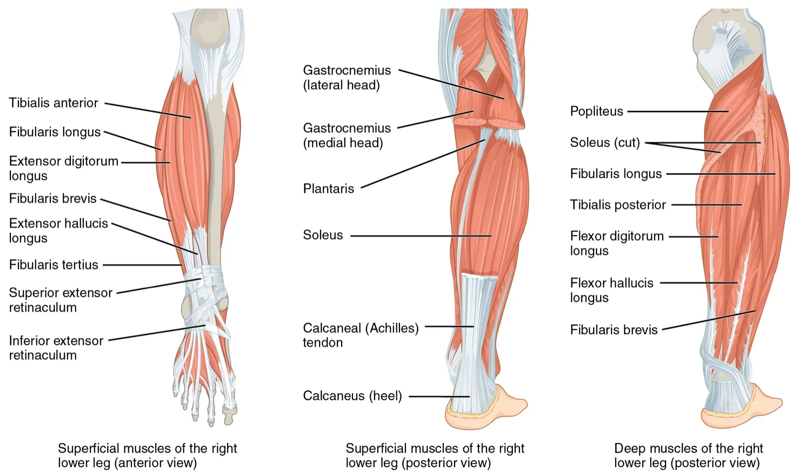
The soleus is a wide, flat muscle that lies under the gastrocnemius, the most superficial calf muscle1. It sits in the outer layer of the back, or calf, compartment of the leg, with the gastrocnemius and the plantaris6. The plantaris is a small calf muscle that runs at a slant between the other two1.
The soleus starts on both bones of the lower leg2. On the fibula (the thin outer bone of the lower leg), it comes from the back of the bone’s head and from its outer edge2. On the tibia (shinbone), it comes from a line on the back of the bone, called the soleal line, and from its inner edge2.
From there, it runs down deep between the two heads of the gastrocnemius2. Lower down it joins the gastrocnemius to form the Achilles tendon, which attaches to the calcaneus (heel bone)2. Its nerve supply comes from branches of the tibial nerve, and its blood from two arteries2.
How it is built inside
Unlike most muscles, the soleus has three tendons running inside it7. It is also split into parts: a back part wraps around a front part2. The way its fibres are laid out changes from the top of the muscle to the bottom2. In living people, five patterns of fibre layout have been seen2.
Most of its fibres are type I fibres, which contract slowly2. In a dissection of 21 legs, the soleus weighed about 276 g and had very short fibres, about 4.4 cm long3. Together, these gave it an exceptionally large physiological cross-sectional area3. That was the largest of the 27 muscles measured, making it the strongest by that measure3.
What it moves
The soleus points the foot down, a movement called plantar flexion2. It crosses only one joint, the ankle2. Plantar flexion is the main job of the calf compartment as a whole6. From its strength, the authors of the dissection study consider the soleus likely the most important muscle at the ankle3.
How it keeps the body upright
In nine people standing quietly, the soleus behaved as an active muscle, shortening against the body’s sway, for about 81% of the time4. One short review calls the soleus central to standing upright8. It describes the muscle pushing the foot down without a break, steadying the body’s centre of mass (balance point)8.
A narrative review, a summary written without a set search method, calls the soleus vital for posture2. It calls it vital for low-effort activity too, such as walking and endurance sports2.
What it does in running
Computer simulations modelled ten people running at 2 to 5 m/s9. At each speed, the soleus did the most to push the body upward9. At 5 m/s, its share peaked at about two bodyweights of ground reaction force, the push of the ground back up on the body9. It also did the most to drive the body forward, and this share rose with speed9.
Breakdown
How often it is strained
A review of soleus injuries on scans says they often go unrecognised7. It calls them a common cause of time out of sport7. In a registry of calf strains in elite Australian Football players from 2014 to 2017, 84.6% of injuries were in the soleus5. Among calf re-injuries in the same players, 91.4% involved the soleus5.
In one Italian Serie A football squad over seven seasons, 17 players had 29 soleus injuries10. Most (83%) happened in training10. The rates per 1,000 hours were10:
| Setting | Soleus injuries per 1,000 hours |
|---|---|
| Training | 0.7610 |
| Matches | 1.0610 |
Where and how it tears
Soleus strains often involve the places where muscle meets tendon or fascia (a sheet of tough connective tissue that wraps muscles)2. One narrative review reports that 56.4% of soleus injuries are at the upper inner tendon2.
The same review says soleus injuries can build up slowly or happen at once2. Repeated sporting movement can tire the muscle and lead to slow-building injury, especially with the knee bent2. A sudden, fast burst of force can cause an injury at once2.
A systematic review of video studies of how muscle strains happened in sport covered calf injuries11. They typically happened with the knee near straight and the foot pulled up toward the shin beyond 10°11. This review did not single out the soleus11. The narrative review, by contrast, links slow-building soleus injury to a bent knee2.
Minor soleus injuries that are hard to see on ultrasound are often confused with a gastrocnemius strain2. They are also confused with Achilles tendinopathy that causes no symptoms2. One review says soleus injuries are often seen as minor discomfort7. It adds that this leads to a fairly quick return to sport with a high risk of injuring it again7.
Who is more likely to strain it
A systematic review of 10 studies covered 5,397 athletes with 518 calf or lower-leg muscle injuries12. Older age and an earlier calf strain were the strongest risk factors for a new calf injury12. It found no evidence linking calf strains to height, weight, sex or which side was dominant12. Evidence for earlier thigh, groin or knee injuries was limited12.
In the Italian football squad, players with soleus injuries were older than those without, at an average of 29.2 against 25.0 years10. Their injuries did not follow sudden jumps in workload measured by GPS tracking10.
Links with shin pain
A systematic review of 17 studies looked at leg muscles and medial tibial stress syndrome (shin splints)13. Higher peak soleus activity during push-off was among the factors most strongly linked to developing it13. The review says studies that follow people forward in time are still needed to confirm this link13. They are also needed to show whether the higher activity is a cause or an effect13. In fifty legs from body donors, the soleus was one of the tissues most often attached to the tibia where shin splint pain occurs14.
Wasting with disuse, spaceflight and age
In ten men during 56 days of bed rest, the soleus had one of the fastest rates of atrophy (muscle wasting)15. The inner gastrocnemius and the vasti (thigh muscles) were also among the fastest to waste15. The triceps surae, the soleus and both gastrocnemius heads together, is described as prone to wasting when inactive16.
Soleus samples came from four male astronauts before and after 17 days in space17. After the flight, their type I fibres made less peak force and shortened faster17. These changes were similar to those seen after 17 days of bed rest17.
One short review reports that with age, tendons get less stiff, force builds more slowly and reflexes weaken8. It says these changes reduce the soleus’s ability to hold balance and help recover from a fall8.
Recovery
How long calf strains keep players out
One study covered 149 calf strains in Australian Football League players18. In soleus injuries, damage to the aponeurosis (a broad, flat sheet of tendon) on MRI (a detailed body scan) was linked to longer time out18.
| Group of elite Australian Football players | Average days before playing again |
|---|---|
| Soleus injuries | 25.45 |
| Gastrocnemius injuries | 19.15 |
| Calf strains (any location) with severe aponeurosis damage on MRI | 31.318 |
| Calf strains (any location) with no aponeurosis damage on MRI | 19.418 |
| Calf strains that recurred before full recovery | 46.719 |
The gap between soleus and gastrocnemius injuries was not statistically significant5. In elite Australian Football players, calf strains that happened while running took about 12 days longer to recover than those from other activities5.
How often calf strains come back
Over ten years, elite male Australian football players had 563 calf strains19. Between 13% and 21.3% came back within two years, depending on how a recurrence was defined19. Each recurrence cost at least 35.6 days on average19. Of the later calf strains, 20% happened before the player had fully recovered19.
In 149 calf strains from the Australian Football League injury registry, older age and an earlier calf strain raised the risk of a repeat18. This covered repeats within two seasons18. MRI findings of the first injury were not linked to a repeat18.
How it grows with training
The triceps surae is described as fairly slow to respond to strength training, compared with other muscles16. In one study, 14 untrained adults trained one leg with standing calf raises and the other with seated calf raises for 12 weeks16. Soleus volume grew 2.1% in the standing leg and 2.9% in the seated leg, amounts the authors report as similar16. The soleus crosses only the ankle2. In this study, it worked at similar lengths in both positions, while the two gastrocnemius heads did not16.
Connections
The gastrocnemius and the Achilles tendon
The soleus and the inner and outer heads of the gastrocnemius make up the triceps surae16. These three muscles do most of the work of pointing the foot down20. The gastrocnemius lies nearer the skin and crosses two joints21. The Achilles tendon is made of fibres from the soleus, which lies deep, and from both gastrocnemius heads, which lie nearer the skin22. In 106 legs from 53 male donors, the soleus strand made up about 28% of the tendon where it meets the heel22.
A review of tennis leg, a calf injury, describes injuries where the inner gastrocnemius meets the soleus as common in athletes and active people23.
Muscles it works with at the ankle and hip
In the nine people standing quietly, the tibialis anterior (the muscle at the front of the shin that pulls the foot up) was also measured4. Its length followed body sway more closely than the soleus or gastrocnemius did4.
Computer simulations modelled eight people walking at four speeds24. At each speed, the soleus was one of the main muscles holding the body up and moving it forward24. The others were the gastrocnemius, gluteus medius, gluteus maximus (the large muscle of the buttock), vasti and hamstrings (back-of-thigh muscles)24.
Exercises that load it
Related exercise pages: calf raise, eccentric heel drop20,25.
| Exercise | What studies found |
|---|---|
| Calf raise | In resistance-trained men, the authors suggested calf raises can recruit the soleus and both gastrocnemius heads20 |
| Calf raise | In 22 trained men, soleus activity did not differ significantly from activity in the leg press20 |
| Pushing the foot down | In 48 healthy people pushing as hard as possible, none of three knee angles singled out the soleus or one gastrocnemius head26 |
| Eccentric heel drop | The bent-knee version is meant to shift load between the gastrocnemius and soleus25 |
| Eccentric heel drop | In 18 people with Achilles tendinopathy and 18 without, soleus activity did not change with knee position27 |
| Eccentric heel drop | In a computer-modelling study of 19 healthy adults, estimated soleus activity did not differ between knee positions25 |
The two heel drop studies disagree on the gastrocnemius25,27. The skin-sensor study found it more active with the knee straight, while the modelling study found no difference25,27. In the modelling study, the soleus’s shortest length was longer in the bent-knee version, while the gastrocnemius heads were shorter25.
Soleus joins to form
Soleus works with
Soleus attaches to
Soleus works with in standing
Soleus works with in walking
Soleus is loaded by
Sources
- Betts JG, Young KA, Wise JA, Johnson E, Poe B, Kruse DH, Korol O, Johnson JE, Womble M, DeSaix P (2022). Anatomy and Physiology 2e. OpenStax. Link
- Trybulski R, Gałęziok K, Matuszczyk F, Halski T, Muracki J (2025). Problems in Diagnosis and Treatment of Soleus Muscle Injuries-Narrative Review and Case Report.. Journal of Clinical Medicine. doi:10.3390/jcm14061955 PMID 40142763
- Ward SR, Eng CM, Smallwood LH, Lieber RL (2009). Are current measurements of lower extremity muscle architecture accurate?. Clinical Orthopaedics and Related Research. doi:10.1007/s11999-008-0594-8 PMID 18972175
- Di Giulio I, Maganaris CN, Baltzopoulos V, Loram ID (2009). The proprioceptive and agonist roles of gastrocnemius, soleus and tibialis anterior muscles in maintaining human upright posture.. The Journal of Physiology. doi:10.1113/jphysiol.2009.168690 PMID 19289550
- Green B, Lin M, Schache AG, McClelland JA, Semciw AI, Rotstein A, Cook J, Pizzari T (2020). Calf muscle strain injuries in elite Australian Football players: A descriptive epidemiological evaluation.. Scandinavian Journal of Medicine & Science in Sports. doi:10.1111/sms.13552 PMID 31494970
- Abuqubo R, Graefe SB, La Rose J (2026). Anatomy, Bony Pelvis and Lower Limb: Leg Posterior Compartment.. StatPearls. PMID 30726025
- Siedi AF, Rolon AU, Bernard N, Bernasconi J, Palmas M, Couto DA, Pascual TA (2022). Posterior Leg Pain: Understanding Soleus Muscle Injuries.. Radiographics. doi:10.1148/rg.210133 PMID 35427175
- Fletcher JR, Strzalkowski NDJ (2026). The neuromechanics of the soleus for fall prevention in aging.. Frontiers in Physiology. doi:10.3389/fphys.2025.1743559 PMID 41574195
- Hamner SR, Delp SL (2013). Muscle contributions to fore-aft and vertical body mass center accelerations over a range of running speeds.. Journal of Biomechanics. doi:10.1016/j.jbiomech.2012.11.024 PMID 23246045
- Benedetti L, Buckthorpe M, Di Paolo S, Della Villa F, Nanni G (2026). The association between GPS-derived external workload metrics and soleus muscle injuries in professional football (soccer) players.. Journal of Experimental Orthopaedics. doi:10.1002/jeo2.70755 PMID 42564880
- Finnern LS, Wilke J, Willwacher S, Pasanen K, Hollander K, Dalos D, Welsch GH, Krosshaug T, Edouard P, Gronwald T, Hoenig T (2026). Qualitative and quantitative situational characteristics of muscle strains in sports: a systematic review and meta-analysis.. British Journal of Sports Medicine. doi:10.1136/bjsports-2025-110327 PMID 41558842
- Green B, Pizzari T (2017). Calf muscle strain injuries in sport: a systematic review of risk factors for injury.. British Journal of Sports Medicine. doi:10.1136/bjsports-2016-097177 PMID 28259848
- Mattock JPM, Steele JR, Mickle KJ (2021). Are Leg Muscle, Tendon and Functional Characteristics Associated with Medial Tibial Stress Syndrome? A Systematic Review.. Sports Medicine - Open. doi:10.1186/s40798-021-00362-2 PMID 34626247
- Beck BR, Osternig LR (1994). Medial tibial stress syndrome. The location of muscles in the leg in relation to symptoms.. The Journal of bone and joint surgery. American volume. doi:10.2106/00004623-199407000-00015 PMID 8027114
- Belavý DL, Miokovic T, Armbrecht G, Richardson CA, Rittweger J, Felsenberg D (2009). Differential atrophy of the lower-limb musculature during prolonged bed-rest.. European Journal of Applied Physiology. doi:10.1007/s00421-009-1136-0 PMID 19680682
- Kinoshita M, Maeo S, Kobayashi Y, Eihara Y, Ono M, Sato M, Sugiyama T, Kanehisa H, Isaka T (2023). Triceps surae muscle hypertrophy is greater after standing versus seated calf-raise training.. Frontiers in Physiology. doi:10.3389/fphys.2023.1272106 PMID 38156065
- Widrick JJ, Knuth ST, Norenberg KM, Romatowski JG, Bain JL, Riley DA, Karhanek M, Trappe SW, Trappe TA, Costill DL, Fitts RH (1999). Effect of a 17 day spaceflight on contractile properties of human soleus muscle fibres.. The Journal of Physiology. doi:10.1111/j.1469-7793.1999.0915u.x PMID 10200437
- Green B, Lin M, McClelland JA, Semciw AI, Schache AG, Rotstein AH, Cook J, Pizzari T (2020). Return to Play and Recurrence After Calf Muscle Strain Injuries in Elite Australian Football Players.. The American Journal of Sports Medicine. doi:10.1177/0363546520959327 PMID 33030961
- Green B, Schache AG, Pizzari T (2025). What is a recurrence? The onset, frequency and time loss impact of recurrent calf muscle strain injuries in elite male Australian football players over a decade.. BMJ Open Sport & Exercise Medicine. doi:10.1136/bmjsem-2025-002865 PMID 40919406
- Gentil P, Souza D, Santana M, Alves RR, Campos MH, Pinto R, Bottaro M (2020). Multi- and Single-Joint Resistance Exercises Promote Similar Plantar Flexor Activation in Resistance Trained Men.. International Journal of Environmental Research and Public Health. doi:10.3390/ijerph17249487 PMID 33352879
- Bordoni B, Varacallo MA (2026). Anatomy, Bony Pelvis and Lower Limb: Gastrocnemius Muscle.. StatPearls. PMID 30422541
- Pękala PA, Henry BM, Ochała A, Kopacz P, Tatoń G, Młyniec A, Walocha JA, Tomaszewski KA (2017). The twisted structure of the Achilles tendon unraveled: A detailed quantitative and qualitative anatomical investigation.. Scandinavian Journal of Medicine & Science in Sports. doi:10.1111/sms.12835 PMID 28139009
- Schwach M, Gaulin B, Vermorel PH, Horteur C, Tourné Y (2026). Tennis leg: Diagnosis and management - A state-of-the-art review.. Foot (Edinburgh, Scotland). doi:10.1016/j.foot.2026.102267 PMID 42258915
- Liu MQ, Anderson FC, Schwartz MH, Delp SL (2008). Muscle contributions to support and progression over a range of walking speeds.. Journal of Biomechanics. doi:10.1016/j.jbiomech.2008.07.031 PMID 18822415
- Weinert-Aplin RA, Bull AM, McGregor AH (2015). Investigating the Effects of Knee Flexion during the Eccentric Heel-Drop Exercise.. Journal of sports science & medicine. PMID 25983597
- Hébert-Losier K, Schneiders AG, García JA, Sullivan SJ, Simoneau GG (2011). Peak triceps surae muscle activity is not specific to knee flexion angles during MVIC.. Journal of Electromyography and Kinesiology. doi:10.1016/j.jelekin.2011.04.009 PMID 21669538
- Reid D, McNair PJ, Johnson S, Potts G, Witvrouw E, Mahieu N (2012). Electromyographic analysis of an eccentric calf muscle exercise in persons with and without Achilles tendinopathy.. Physical therapy in sport : official journal of the Association of Chartered Physiotherapists in Sports Medicine. doi:10.1016/j.ptsp.2011.08.003 PMID 22814448
Changes
- First published.