Posterior cruciate ligament
Also called PCL
The largest ligament in the knee, which stops the shinbone sliding backwards. How it is built, how it gets torn, how often it heals on its own, and what it works with.
- Region
- Knee
- Muscles
- hamstrings, quadriceps
- Updated
- September 30, 2026
- Reviewed
- September 30, 2026 · how entries are checked
Function
Where it runs
The ligament is named for its attachment to the back of the tibia1. The name also refers to the cross it forms with the anterior cruciate ligament inside the joint capsule1. The capsule is the sleeve of tough tissue that encloses the knee1.
It starts on the inner condyle (rounded knob) of the femur, in the intercondylar notch5. The notch is the groove between the two condyles at the lower end of the thigh bone5. It runs down to the back of the tibial plateau (the flat top of the shinbone, where the thigh bone rests)5.
It averages 36 to 38 mm long3. Its cross-sectional area (the size of a slice cut straight across it) is 40 to 60 mm² at its middle3. It sits inside the joint but outside the synovial lining (the thin inner lining of a joint capsule)3. It is about 1.3 to 2 times as thick as the anterior cruciate ligament and about twice as strong5.
Its two footprints (the patches of bone where it attaches) differ in shape and size3:
| Attachment | Shape and place | Area |
|---|---|---|
| Femur | Half-moon shaped, on the side of the inner condyle facing the notch, reaching the notch’s roof3 | About 190 to 230 mm²3 |
| Tibia | Trapezoid shaped, in a groove below the joint surface, midway across, near the back roots of both menisci (knee cartilage pads)3 | About 160 to 220 mm²3 |
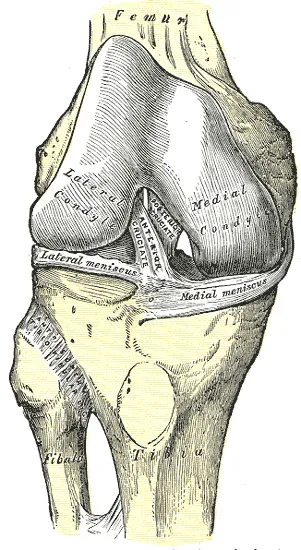
Two bundles and two helpers
The ligament is made of two bundles, the anterolateral (front-outer) and the posteromedial (back-inner)1. The anterolateral bundle is larger and stronger in tension3. The posteromedial bundle is weaker, and its anatomy varies more from person to person3.
The bundles tighten in different patterns as the knee moves, so they resist backward sliding and rotation at different knee angles2. Recent studies call them codominant, meaning neither is the main restraint at a given angle3. Both bundles do more work with the knee bent than straight, and both lengthen as it bends3.
Two meniscofemoral ligaments run beside it3. They link the back of the outer meniscus to the inner femoral condyle, starting near this ligament6. At least one is present in more than 90% of knees3. With the two bundles, they make four fibre bundles that work together against backward and twisting loads on the tibia3.
What it holds back
The ligament stops the tibia sliding backwards under the femur1. One review reports that it provides 95% of the force holding the tibia against backward movement7. Recent studies suggest it also helps resist twisting loads, mainly with the knee bent beyond 90 degrees3. In the bent, weight-bearing knee, as in walking downhill, it keeps the femur from sliding forward off the top of the tibia8.
How its load changes as the knee bends
Laboratory studies found that the force in the ligament changed with knee angle and load9,10:
- A robot pushed the tibia backwards with 22 to 110 N (newtons, a unit of force)9. Force in the ligament ranged from about 6 N (knee straight) to about 112 N (90 degrees, largest load)9.
- In nine donated knees, with a simulated hamstrings (back-of-thigh muscles) pull, the force rose as the knee bent, from about 13 N straight to about 86 N at 90 degrees10.
In the same nine knees, adding a quadriceps (front-of-thigh muscles) pull to the hamstrings pull significantly lowered the force in the ligament10.
What it is made of
Most of both cruciate ligaments is made of bundles of collagen (a strong, rope-like protein)11. Long cells lie between its parallel fibrils (very thin strands of collagen)11.
An anatomical study that injected the blood vessels found the ligament’s blood comes mainly from the middle genicular artery12. That is an artery at the back of the knee that supplies the cruciate ligaments. The injection study found three areas without blood vessels: both bone attachments and the centre of the middle third12.
A review reports that the ligament holds several kinds of nerve ending that can signal movement and position to the brain and spinal cord7.
Breakdown
How it gets torn
Injury can come from a force that pushes the tibia backwards, most commonly with the knee bent2. Because the ligament is larger and stronger than the anterior cruciate, its injuries are associated with high-energy trauma such as vehicle and sport injuries3. Over-bending or over-straightening the knee can also tear it3. Over-straightening injuries are associated with tears near its upper end and crush fractures at the front of the tibial plateau3.
In 494 people with a loose, torn ligament, dashboard injuries (35%) and falls onto the bent knee with the foot pointed (24%) led the causes13.
Who tears it
Males are injured more often than females, which a review links to their greater involvement in trauma3. The same review puts the average age at injury at 28 to 343. The share of injuries caused by sport differs between studies13,14:
| Group studied | Share caused by sport, % |
|---|---|
| 494 people with a loose, torn ligament | 4013 |
| 1,000 reconstructions (surgery to rebuild the ligament) at one centre | 38.815 |
| 1,287 reconstructions in Scandinavia | 35.416 |
| Swedish registry reconstructions of this ligament without the ACL | 6414 |
A population study of 48 people put the yearly incidence (new cases) of isolated, complete tears at 1.8 per 100,00017. Of 2,285 players at the NFL (National Football League) Combine from 2009 to 2015, 69 (3%) showed signs of an injury to it18. The NFL Combine is a yearly event where college American football players are examined and tested before the professional draft.
A case-control study (comparing people with a condition to similar people without it) matched 104 people with tears to 104 controls19. The injured group had a flatter backward slope of the tibial plateau, 5.7 against 8.6 degrees19. In that group, 87.5% of tears came from contact19. Another study linked a shallower inner tibial plateau and a narrower notch with isolated tears and with avulsion fractures at the tibia20. An avulsion is a tear in which a ligament pulls away from the bone it attaches to.
How injuries are graded
Injuries are graded by how far the tibia moves backwards3:
- Grade I: a partial tear, under 5 mm3.
- Grade II: a partial tear, 5 to 10 mm3.
- Grade III: a complete rupture, over 10 mm3.
Often torn with other injuries
A review reports that over 60% of injuries come with other capsule or ligament damage3. The most common pairing, in 15% to 42% of patients, is with the posterolateral corner (the ligaments and tendons at the outer back of the knee)3. The same review reports that over 80% of patients with acute (recent) injuries, alone or with the posterolateral corner, have had a bone bruise3. A bone bruise is an injury inside a bone, without a break, that shows up on an MRI scan.
Other groups also show frequent combined injuries15,18:
- In 1,000 reconstructions at one centre, combined injuries outnumbered isolated ones in sport (58.5%) and traffic (71.7%)15. Handball was the one activity where isolated tears were more common15.
- In that group, a posterolateral corner tear was the most common extra injury, except in skiing and cycling, where a medial collateral ligament injury led15. This is the ligament on the inner side of the knee.
- In 69 NFL Combine players, the medial collateral ligament was also injured in 42%, the anterior cruciate in 11.6% and cartilage in 31.8%18.
A 2019 review reports that this ligament is generally uncommon among the injuries that come with an ACL tear21.
Recovery
It often heals back into one piece
A systematic review of MRI follow-up studies found that 77% to 93% of partial or complete tears regained continuity4. One study followed 48 acutely injured knees22. Its authors concluded that many of these ligaments grow slack but continuous tissue, which might partly hold the tibia22. MRI studies have followed it22,23:
- In 40 knees followed for 3.2 years on average, each low- or mid-grade injury healed with continuity, as did 19 of 22 high-grade ones23.
- In 48 knees after acute injury, 75% showed a continuous ligament22. Those knees had less front-to-back tibial movement (mean 7.6 mm) than knees with a disrupted one (11.4 mm)22.
- In 46 complete tears scanned more than two months after injury, 72% showed continuity, more often when scanned after six months24.
In the 40-knee study, most injuries managed without surgery could heal with continuity even when other knee ligaments were also damaged23. In the 46-tear study, other ligament injuries made regaining continuity less likely24.
What the scans can and cannot show
The same systematic review found a possible link between a ligament regaining continuity on MRI and a stable knee on examination4. But in long-standing injuries, scar or healing in a stretched shape can make the ligament look intact on MRI3.
After surgery
In 22 isolated injuries, surgeons rebuilt the ligament with hamstring tendon grafts (pieces of tissue moved to rebuild a damaged part)25. Repeated MRI scans followed the grafts25. Their look on MRI matured gradually from 6 months, and the part inside the joint matured more slowly25. The grafts needed more than a year to reach a low signal on the scan25.
Connections
Its partner and the bones it joins
With the anterior cruciate ligament, it forms a cross inside the knee that stops the tibia moving too far forward or backward on the femur26. It joins the femur to the tibia5.
Structures that share its work
The posterolateral corner includes the lateral collateral ligament (the ligament on the outer side of the knee), the popliteus tendon and the popliteofibular ligament3. The popliteus is a small muscle at the back of the knee. The popliteofibular ligament joins the popliteus tendon to the top of the fibula. These structures resist backward movement, inward bending and outward twisting of the tibia, and work with this ligament3. In loaded knees tested in the lab, force in an intact posterior cruciate rose when these structures were deficient3.
On the inner side, the medial collateral ligament complex and nearby structures add to backward and twisting looseness when this ligament is deficient3.
In donor knees, force in it rose as the knee bent under a simulated hamstrings pull, and adding a quadriceps pull lowered it10.
Exercises that load it
A 2001 review of the squat (see back squat) reports low to moderate backward shear forces at the knee throughout the lift27. These forces, which push surfaces to slide across each other, are held mainly by this ligament27.
A modelling study (one that estimates forces with a model) looked at 18 people doing the wall sit28. Peak force in the ligament came at 80 to 90 degrees of knee bend28. It averaged about 723 N with the feet farther from the wall, 786 N with them closer, and 414 N in the one-leg squat28. Across 0 to 90 degrees, though, the feet-farther version generally produced greater force28.
A narrative review (a summary without a set search method) ordered exercises from least to most load on this ligament29. The order was level walking, the one-leg squat (see single-leg squat), lunges, the wall squat, the leg press and the two-leg squat29.
Posterior cruciate ligament holds together
Posterior cruciate ligament starts on
Posterior cruciate ligament attaches to
Posterior cruciate ligament crosses
Posterior cruciate ligament works with
Posterior cruciate ligament lies next to
Posterior cruciate ligament is loaded more by
Posterior cruciate ligament is loaded less by
Posterior cruciate ligament is loaded by
Sources
- Cox CF, Graefe SB, Black AC, Bordoni B (2023). Anatomy, Bony Pelvis and Lower Limb: Knee Posterior Cruciate Ligament. StatPearls. PMID 30571037
- Logterman SL, Wydra FB, Frank RM (2018). Posterior Cruciate Ligament: Anatomy and Biomechanics.. Current Reviews in Musculoskeletal Medicine. doi:10.1007/s12178-018-9492-1 PMID 29855794
- Winkler PW, Zsidai B, Wagala NN, Hughes JD, Horvath A, Senorski EH, Samuelsson K, Musahl V (2021). Evolving evidence in the treatment of primary and recurrent posterior cruciate ligament injuries, part 1: anatomy, biomechanics and diagnostics.. Knee Surgery, Sports Traumatology, Arthroscopy. doi:10.1007/s00167-020-06357-y PMID 33201271
- Boks SS, Vroegindeweij D, Koes BW, Hunink MG, Bierma-Zeinstra SM (2006). Follow-up of posttraumatic ligamentous and meniscal knee lesions detected at MR imaging: systematic review.. Radiology. doi:10.1148/radiol.2382050063 PMID 16452395
- Raj MA, Mabrouk A, Varacallo MA (2023). Posterior Cruciate Ligament Knee Injuries. StatPearls. PMID 28613477
- Fox AJ, Bedi A, Rodeo SA (2012). The Basic Science of Human Knee Menisci. Sports health. doi:10.1177/1941738111429419 PMID 23016106
- Katonis P, Papoutsidakis A, Aligizakis A, Tzanakakis G, Kontakis GM, Papagelopoulos PJ (2008). Mechanoreceptors of the posterior cruciate ligament.. Journal of International Medical Research. doi:10.1177/147323000803600302 PMID 18534119
- Betts JG, Young KA, Wise JA, Johnson E, Poe B, Kruse DH, Korol O, Johnson JE, Womble M, DeSaix P (2022). Anatomy and Physiology 2e. OpenStax. Link
- Fox RJ, Harner CD, Sakane M, Carlin GJ, Woo SL (1998). Determination of the in situ forces in the human posterior cruciate ligament using robotic technology. A cadaveric study.. American Journal of Sports Medicine. doi:10.1177/03635465980260030901 PMID 9617402
- Höher J, Vogrin TM, Woo SL, Carlin GJ, Arøen A, Harner CD (1999). In situ forces in the human posterior cruciate ligament in response to muscle loads: a cadaveric study.. Journal of Orthopaedic Research. doi:10.1002/jor.1100170522 PMID 10569489
- Petersen W, Tillmann B (1999). Structure and vascularization of the cruciate ligaments of the human knee joint.. Anatomy and Embryology. doi:10.1007/s004290050283 PMID 10463347
- Petersen W, Tillmann B (1999). Blood and lymph supply of the posterior cruciate ligament: a cadaver study.. Knee Surgery, Sports Traumatology, Arthroscopy. doi:10.1007/s001670050119 PMID 10024962
- Schulz MS, Russe K, Weiler A, Eichhorn HJ, Strobel MJ (2003). Epidemiology of posterior cruciate ligament injuries.. Archives of Orthopaedic and Trauma Surgery. doi:10.1007/s00402-002-0471-y PMID 12734718
- Zsidai B, Horvath A, Winkler PW, Narup E, Kaarre J, Svantesson E, Musahl V, Hamrin Senorski E, Samuelsson K (2022). Different injury patterns exist among patients undergoing operative treatment of isolated PCL, combined PCL/ACL, and isolated ACL injuries: a study from the Swedish National Knee Ligament Registry.. Knee Surgery, Sports Traumatology, Arthroscopy. doi:10.1007/s00167-022-06948-x PMID 35357530
- Schlumberger M, Schuster P, Eichinger M, Mayer P, Mayr R, Immendörfer M, Richter J (2020). Posterior cruciate ligament lesions are mainly present as combined lesions even in sports injuries.. Knee Surgery, Sports Traumatology, Arthroscopy. doi:10.1007/s00167-020-05919-4 PMID 32157362
- Owesen C, Sandven-Thrane S, Lind M, Forssblad M, Granan LP, Årøen A (2015). Epidemiology of surgically treated posterior cruciate ligament injuries in Scandinavia.. Knee Surgery, Sports Traumatology, Arthroscopy. doi:10.1007/s00167-015-3786-2 PMID 26387121
- Sanders TL, Pareek A, Barrett IJ, Kremers HM, Bryan AJ, Stuart MJ, Levy BA, Krych AJ (2017). Incidence and long-term follow-up of isolated posterior cruciate ligament tears.. Knee Surgery, Sports Traumatology, Arthroscopy. doi:10.1007/s00167-016-4052-y PMID 26922055
- Logan CA, Beaulieu-Jones BR, Sanchez G, Chahla J, Kennedy NI, Cinque ME, LaPrade RF, Whalen JM, Vopat BG, Price MD, Provencher MT (2018). Posterior Cruciate Ligament Injuries of the Knee at the National Football League Combine: An Imaging and Epidemiology Study.. Arthroscopy. doi:10.1016/j.arthro.2017.08.304 PMID 29225016
- Bernhardson AS, DePhillipo NN, Daney BT, Kennedy MI, Aman ZS, LaPrade RF (2019). Posterior Tibial Slope and Risk of Posterior Cruciate Ligament Injury.. American Journal of Sports Medicine. doi:10.1177/0363546518819176 PMID 30640507
- Huang WT, Kang K, Wang J, Li T, Dong JT, Gao SJ (2023). Morphological Risk Factors for Posterior Cruciate Ligament Tear and Tibial Avulsion Injuries of the Tibial Plateau and Femoral Condyle.. American Journal of Sports Medicine. doi:10.1177/03635465221131295 PMID 36476119
- Filbay SR, Grindem H (2019). Evidence-based recommendations for the management of anterior cruciate ligament (ACL) rupture.. Best practice & research. Clinical rheumatology. doi:10.1016/j.berh.2019.01.018 PMID 31431274
- Akisue T, Kurosaka M, Yoshiya S, Kuroda R, Mizuno K (2001). Evaluation of healing of the injured posterior cruciate ligament. Arthroscopy. doi:10.1053/jars.2001.21540 PMID 11239346
- Shelbourne KD, Jennings RW, Vahey TN (1999). Magnetic resonance imaging of posterior cruciate ligament injuries: assessment of healing.. American Journal of Knee Surgery. PMID 10626911
- Jung YB, Jung HJ, Yang JJ, Yang DL, Lee YS, Song IS, Lee HJ (2008). Characterization of spontaneous healing of chronic posterior cruciate ligament injury: Analysis of instability and magnetic resonance imaging.. Journal of Magnetic Resonance Imaging. doi:10.1002/jmri.21333 PMID 18504752
- Fujimoto E, Sasashige Y, Masuda Y, Tsuchida Y, Hisatome T, Kashiwagi K, Sasaki H, Touten Y, Kazusa H, Eguchi A, Nagata Y (2014). Serial magnetic resonance imaging study of posterior cruciate ligament reconstruction or augmentation using hamstring tendons.. Orthopaedics & Traumatology: Surgery & Research. doi:10.1016/j.otsr.2014.06.018 PMID 25281553
- Evans J, Mabrouk A, Nielson JL (2023). Anterior Cruciate Ligament Knee Injury.. StatPearls. PMID 29763023
- Escamilla RF (2001). Knee biomechanics of the dynamic squat exercise.. Medicine and Science in Sports and Exercise. doi:10.1097/00005768-200101000-00020 PMID 11194098
- Escamilla RF, Zheng N, Imamura R, Macleod TD, Edwards WB, Hreljac A, Fleisig GS, Wilk KE, Moorman CT 3rd, Andrews JR (2009). Cruciate ligament force during the wall squat and the one-leg squat.. Medicine and Science in Sports and Exercise. doi:10.1249/MSS.0b013e3181882c6d PMID 19127183
- Escamilla RF, Macleod TD, Wilk KE, Paulos L, Andrews JR (2012). Cruciate ligament loading during common knee rehabilitation exercises.. Proceedings of the Institution of Mechanical Engineers, Part H. doi:10.1177/0954411912451839 PMID 23025167
Changes
- A supporting claim was narrowed to match its source; the page text is unchanged.