Menisci of the knee
Also called meniscus, medial meniscus, lateral meniscus
The two C-shaped pads of fibrocartilage between the thigh bone and shinbone. What they are made of, how they spread load, who tears them, and what studies found about how they heal.
- Region
- Knee
- Updated
- September 30, 2026
- Reviewed
- September 30, 2026 · how entries are checked
Function
Two pads between the thigh bone and shinbone
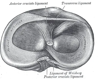
The knee has two menisci, an inner (medial) one and an outer (lateral) one1. Each is thin along its inner edge and thick along its outer edge1. The upper surface is cupped to fit the rounded femoral condyle (the knob at the end of the thigh bone) above it2. The lower surface is flat, to sit on the top of the tibia2.
The two menisci differ in shape, cover and movement2:
| Feature | Medial meniscus | Lateral meniscus |
|---|---|---|
| Shape | Semicircular, about 35 mm front to back, much broader at the back2 | Almost circular, about the same width all round2 |
| Share of its side of the tibia covered | About 60%2 | About 80%2 |
| Movement across the tibia, one study of 5 cadaver (donated body) knees | 5.1 mm on average2 | 11.2 mm on average2 |
How they are anchored
The menisci attach to the tibia, and both can move as the knee moves1. The medial one moves less, because its outer edge is anchored to the joint capsule (the sleeve of tough tissue that encloses a joint) and the inner collateral ligament1. Each has a front and a back end, called horns, and both horns of each meniscus attach to the tibia2. The medial front horn sits in front of the anterior cruciate ligament (ACL, the front one of two ligaments that cross inside the knee)2. Its back horn attaches behind, near the posterior cruciate ligament (the back one of the two)2.
A band of fibrous tissue, the transverse ligament, joins the two front horns2. The medial meniscus is tied to the tibia by part of the joint capsule2. At its midpoint, a thickening of the capsule called the deep medial collateral ligament ties it more firmly to the femur2. The lateral meniscus is only loosely attached to the capsule2. It has no attachment to the lateral collateral ligament, the ligament on the outer side of the knee2.
The back horn of the lateral meniscus is linked to the femur by the meniscofemoral ligaments2. Sources differ on them2,5. One review reports that a knee may have none, one, two or four, and that their function is unknown2. Another source reports at least one in more than 90% of knees, working with the posterior cruciate ligament against backward and twisting loads5.
What they are made of
Meniscal tissue is about 72% water and 22% collagen, with cells set in it2. Most of that collagen is type I, which sets the menisci apart from the hyaline cartilage covering the bone ends2. Collagen is mainly what gives the menisci their strength under pulling loads2. Proteoglycans, molecules that hold water in the tissue, give much of its resistance to squeezing2.
In the deep layers, collagen fibres run around the meniscus, parallel to its outer edge2. Near the surface they run more across the meniscus, and “tie” fibres woven between the deep fibres hold the structure together2. The cells are called fibrochondrocytes, because they look like a mix of fibre-making cells and cartilage cells2.
What they do
A review of meniscus research reports that the menisci steady the knee, spread its load, absorb shock, and help oil and nourish it2. When a person stands, the menisci are squeezed, and the ring-shaped fibres turn that squeeze into tension, called hoop stress2. The horn attachments stop the menisci being pushed out sideways2. A review reports that about 70% of the load on the knee’s outer side passes through the menisci, and 50% on the inner side2. In a straight knee, half the load through the menisci passes through the back horns, rising to 85% at a right-angle bend2.
Because the front and back horns move by different amounts, the menisci curve more tightly as the knee bends2. This matches the tighter curve at the back of the femur and keeps contact with both bones2.
A review cites a study finding the normal knee absorbs about 20% more shock than knees that have had a meniscus removed2. The review reports that removing the whole lateral meniscus cuts contact area by 40% to 50%2. Contact stress on that side rises to 200% to 300% of normal2.
A review cites a study finding that, without a working anterior cruciate ligament, the medial back horn is the main brake on the tibia sliding forward2. The review reports that removing the medial meniscus in such a knee raised front-to-back movement of the tibia by up to 58% at a right-angle bend2. With the ligament intact, the removal had little effect2.
Breakdown
How tears happen
European consensus reports describe traumatic tears, which follow a knee injury with sudden pain, and degenerative lesions (areas of damaged tissue), which develop slowly, typically as a horizontal split in the meniscus of a middle-aged or older person3,6. The meniscus tear page covers these definitions and what trials found.
A review describes how the meniscus becomes stiffer and less elastic with age2. At areas of cystic degenerative change, a twisting force from the femur can shear the surface layers off the deep layer, splitting it horizontally2. The same review notes that the back inner corner moves least2. It proposes that trapping the medial back horn between femur and tibia in full bending may be one way tears happen2.
Who tears a meniscus
Studies in different groups report these rates3,7,8:
- In the general population, 0.5 to 0.7 acute injuries per 1,000 people a year, more in men than women3.
- In US military personnel from 1998 to 2006, 8.27 acute injuries per 1,000 person-years (people counted times years followed)7. Those over 40 were more than four times as likely to be injured as those under 207.
- In US high school athletes in 22 sports, 5.1 injuries per 100,000 athlete exposures, over four times higher in competition than practice8.
In 392 adults aged 18 to 60 with tears seen at arthroscopy (keyhole surgery inside a joint), about a third came from sport9. Most tears from injuries outside sport happened in daily life, and half of those happened while squatting or rising from a squat9.
Risk factors studies have linked
A meta-analysis of 11 studies of people over 16 found strong evidence for these risk factors10:
- for sudden tears, playing soccer or rugby10
- for degenerative tears, age over 60, male sex, kneeling and squatting at work, and climbing more than 30 flights of stairs10
A second meta-analysis linked work-related kneeling, squatting, stair climbing and lifting 10 kg or more with meniscal lesions11. The overall quality of that evidence was moderate to low11. It linked professional football with about five times the risk11. A review of 16 studies linked a steeper backward slope of the top of the tibia with more lateral tears alongside ACL tears12.
Tears alongside ligament injuries
With a fresh anterior cruciate ligament (ACL) tear, lateral tears are more common than medial ones, apart from one type called ramp tears, the 2019 consensus reports3. With a long-standing ACL tear, medial tears become more common with time and age3.
Tears, pain and osteoarthritis
The 2019 consensus states that traumatic tears can cause pain by acting on nerve endings and raising inflammatory signals in the joint3. A study of a population sample used MRI (a scan that shows soft tissue in detail) in 991 adults aged 50 to 9013. Of those with a tear, 61% had had no knee pain, aching or stiffness in the previous month13.
A meta-analysis of 53 studies found the odds of knee osteoarthritis about six times higher after meniscal injury4. Results varied widely between those studies4. The 2016 consensus states it is still unclear whether degenerative lesions cause osteoarthritis or result from it6.
Recovery
Blood supply shapes healing
The menisci get their blood mainly from the geniculate arteries, branches of the popliteal artery (the main artery behind the knee)2. Only the outer rim is well supplied: about the outer 10% to 30% of the medial meniscus and 10% to 25% of the lateral2.
The rest, 65% to 75% of each meniscus, is fed from the synovial fluid (the slippery fluid inside the joint)2. A review states that this feeding needs the joint to be loaded and unloaded again and again, as joint cartilage does2. In a study of menisci from 20 cadavers, nearly the whole meniscus had a blood supply at birth2. After age 50, only the outer quarter of the base did2.
A review names the outer rim with blood the red zone and the inner part the white zone2. The 2019 consensus advises against sorting tears this way, because blood supply changes through life3. It uses measured zones, 0 to 3, instead3.
What studies found about healing
The 2019 consensus found no results on whether isolated tears heal on their own3. It reports that repaired tears in the outer zone 1 healed at a significantly better rate than those in zone 23:
| Zone of the repaired tear | Healing rate |
|---|---|
| Zone 1 (outer) | 87% to 91%3 |
| Zone 2 (next zone in) | 59% to 79%3 |
A meta-analysis of repairs of traumatic tears, in patients whose bones had stopped growing, found failure rose with time14. Failure here meant symptoms, no healing on MRI, or another operation14:
| Time after repair | Share of repairs failed |
|---|---|
| Up to 1 year | 12%14 |
| 2 to 3 years | 15%14 |
| 4 to 6 years | 19%14 |
How the tissue responds to running
One study used ultrasound (a scan made with sound waves) in 100 recreational runners15. Straight after a marathon, the medial meniscus bulged out of the joint 38% further15. It stayed raised through day 3 and was back to baseline from day 415. A larger rise in the bulge straight after the race was linked with a higher knee adduction (the lower leg tilting toward the body’s midline) load, higher body mass index and more monthly running15.
A systematic review of 14 MRI studies of healthy marathon runners found changes in knee cartilage and meniscus after long runs16. Five studies found volume fell at first, and ten found T2 (an MRI measure that changes when tissue is damaged or swollen) returned to baseline in the short term16. Another MRI measure may stay raised for months16.
Connections
The menisci sit in the knee joint, attached to the tibia1. The medial one is also tied to the femur by the deep medial collateral ligament2. The medial front horn attaches in front of the anterior cruciate ligament2. The back roots of both menisci sit close to the tibial attachment of the posterior cruciate ligament5.
The deep part of the medial collateral ligament attaches to the medial meniscus17. The lateral collateral ligament has no attachment to the meniscus18. Like the articular cartilage on the bone ends, the parts of the menisci fed from the joint fluid depend on repeated loading and unloading for that feeding2.
Related exercise pages: deadlift. A narrative review found few studies linking the deadlift to specific injuries, and knee meniscus injuries were among those reported19.
Menisci of the knee forms part of
Menisci of the knee attaches to
Menisci of the knee cushions
Menisci of the knee works with
Menisci of the knee lies next to
Menisci of the knee is bound to
Menisci of the knee is torn in
Menisci of the knee is loaded by
Sources
- Betts JG, Young KA, Wise JA, Johnson E, Poe B, Kruse DH, Korol O, Johnson JE, Womble M, DeSaix P (2022). Anatomy and Physiology 2e. OpenStax. Link
- Fox AJ, Bedi A, Rodeo SA (2012). The Basic Science of Human Knee Menisci. Sports health. doi:10.1177/1941738111429419 PMID 23016106
- Kopf S, Beaufils P, Hirschmann MT, Rotigliano N, Ollivier M, Pereira H, Verdonk R, Darabos N, Ntagiopoulos P, Dejour D, Seil R, Becker R (2020). Management of traumatic meniscus tears: the 2019 ESSKA meniscus consensus.. Knee surgery, sports traumatology, arthroscopy. doi:10.1007/s00167-020-05847-3 PMID 32052121
- Poulsen E, Goncalves GH, Bricca A, Roos EM, Thorlund JB, Juhl CB (2019). Knee osteoarthritis risk is increased 4-6 fold after knee injury - a systematic review and meta-analysis.. British journal of sports medicine. doi:10.1136/bjsports-2018-100022 PMID 31072840
- Winkler PW, Zsidai B, Wagala NN, Hughes JD, Horvath A, Senorski EH, Samuelsson K, Musahl V (2021). Evolving evidence in the treatment of primary and recurrent posterior cruciate ligament injuries, part 1: anatomy, biomechanics and diagnostics.. Knee Surgery, Sports Traumatology, Arthroscopy. doi:10.1007/s00167-020-06357-y PMID 33201271
- Beaufils P, Becker R, Kopf S, Englund M, Verdonk R, Ollivier M, Seil R (2017). Surgical management of degenerative meniscus lesions: the 2016 ESSKA meniscus consensus.. Knee surgery, sports traumatology, arthroscopy. doi:10.1007/s00167-016-4407-4 PMID 28210788
- Jones JC, Burks R, Owens BD, Sturdivant RX, Svoboda SJ, Cameron KL (2012). Incidence and risk factors associated with meniscal injuries among active-duty US military service members.. Journal of athletic training. doi:10.4085/1062-6050-47.1.67 PMID 22488232
- Mitchell J, Graham W, Best TM, Collins C, Currie DW, Comstock RD, Flanigan DC (2016). Epidemiology of meniscal injuries in US high school athletes between 2007 and 2013.. Knee surgery, sports traumatology, arthroscopy. doi:10.1007/s00167-015-3814-2 PMID 26506845
- Drosos GI, Pozo JL (2004). The causes and mechanisms of meniscal injuries in the sporting and non-sporting environment in an unselected population.. The Knee. doi:10.1016/S0968-0160(03)00105-4 PMID 15066629
- Snoeker BA, Bakker EW, Kegel CA, Lucas C (2013). Risk factors for meniscal tears: a systematic review including meta-analysis.. The Journal of orthopaedic and sports physical therapy. doi:10.2519/jospt.2013.4295 PMID 23628788
- Bahns C, Bolm-Audorff U, Seidler A, Romero Starke K, Ochsmann E (2021). Occupational risk factors for meniscal lesions: a systematic review and meta-analysis.. BMC musculoskeletal disorders. doi:10.1186/s12891-021-04900-7 PMID 34911509
- Jiang J, Liu Z, Wang X, Xia Y, Wu M (2022). Increased Posterior Tibial Slope and Meniscal Slope Could Be Risk Factors for Meniscal Injuries: A Systematic Review.. Arthroscopy. doi:10.1016/j.arthro.2022.01.013 PMID 35066109
- Englund M, Guermazi A, Gale D, Hunter DJ, Aliabadi P, Clancy M, et al (2008). Incidental meniscal findings on knee MRI in middle-aged and elderly persons.. The New England journal of medicine. doi:10.1056/NEJMoa0800777 PMID 18784100
- Ow ZGW, Law MSN, Ng CH, Krych AJ, Saris DBF, Debieux P, Wong KL, Lin HA (2021). All-Cause Failure Rates Increase With Time Following Meniscal Repair Despite Favorable Outcomes: A Systematic Review and Meta-analysis.. Arthroscopy. doi:10.1016/j.arthro.2021.05.033 PMID 34058318
- Dobashi S, Iwasaki K, Tanaka S, Hishimura R, Matsuoka M, Ebata T, Sato D, Onodera T, Kondo E, Iwasaki N (2026). Marathon Running Transiently Causes Medial Meniscus Extrusion: Recovery Time Course and Factors Associated With Extrusion.. Scandinavian journal of medicine & science in sports. doi:10.1111/sms.70366 PMID 42683665
- Shu D, Chen F, Guo W, Ding J, Dai S (2022). Acute changes in knee cartilage and meniscus following long-distance running in habituate runners: a systematic review on studies using quantitative magnetic resonance imaging.. Skeletal radiology. doi:10.1007/s00256-021-03943-0 PMID 34854970
- Lucidi GA, Solaro L, Grassi A, Alhalalmeh MI, Ratti S, Manzoli L, Zaffagnini S (2024). Current trends in the medial side of the knee: not only medial collateral ligament (MCL). Journal of Orthopaedics and Traumatology. doi:10.1186/s10195-024-00808-9 PMID 39704918
- Yaras RJ, O'Neill N, Mabrouk A, Yaish AM (2024). Lateral Collateral Ligament Knee Injury. StatPearls. PMID 32809682
- Bengtsson V, Berglund L, Aasa U (2018). Narrative review of injuries in powerlifting with special reference to their association to the squat, bench press and deadlift.. BMJ Open Sport & Exercise Medicine. doi:10.1136/bmjsem-2018-000382 PMID 30057777
Changes
- A supporting claim was narrowed to match its source; the page text is unchanged.