Sacroiliac joint
Also called SI joint
The joint on each side of the pelvis where the sacrum meets the ilium. How it carries the trunk's load, how far it moves, what scans show in people without pain, and how it changes with load and childbirth.
- Region
- Spine
- Muscles
- gluteus maximus, erector spinae, multifidus, transversus abdominis, hamstrings
- Updated
- October 9, 2026
- Reviewed
- October 9, 2026 · how entries are checked
Function
Where the spine meets the pelvis
The sacroiliac joint is where the sacrum meets the ilium, so it links the spine to the pelvis1. The two sacroiliac joints are the largest joints of the axial skeleton (the bones of the head and trunk)2. The first three segments of the sacrum typically form the joint, and its bony shape varies widely between people5.
In adults the joint surface is shaped like an ear, with a short upper limb and a longer lower limb5. Cartilage (a firm, rubbery tissue, softer than bone) covers both surfaces6. The lower portion of the joint is synovial, with a fluid-filled space between the bones that lets them move freely5. The top of its upper limb is more fibrous5.
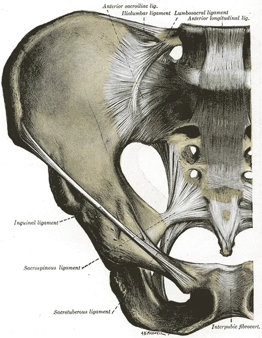
How it carries the trunk
A 1918 anatomy text gives the pelvic girdle’s most important mechanical job as passing the weight of the trunk and arms to the legs6. Body weight falls on the sacrum at the lumbosacral joint, the joint of the lower back and the sacrum6. The back arch of the pelvis then carries it6.
The sacrum is wider at the top than the bottom and wider in front than behind5. It wedges between the two ilia like a keystone5. That shape is adapted to resist shearing, the sliding of one surface across another, under vertical compression5. Body weight drives this wedge down and tends to push the hip bones apart6. The sacroiliac and iliolumbar ligaments, and the ligaments of the pubic symphysis at the front of the pelvis, resist that spreading6.
Rough surfaces and squeezing forces
An examination of 200 preparations found the bony joint surfaces smooth until puberty, with ridges and grooves appearing later5. Sectioned human joints show cartilage-covered ridges and depressions that match across the two surfaces, more pronounced in men than in women7. In a laboratory study, surfaces with both a coarse texture and such ridges had high friction8. A review of the joint’s anatomy states that these interlocking ridges contribute to the joint having the highest friction of any freely movable joint5.
The same review describes two ways a joint can hold steady5:
- Form closure is a theoretical joint whose surfaces fit so closely that it is stable with no extra force5. A sacrum that fitted so perfectly would make movement practically impossible5.
- Force closure needs a sideways squeezing force plus friction to withstand vertical load5. That force comes from tension in ligaments, fasciae and muscles, and from the push of the ground5.
The ligaments that bind it
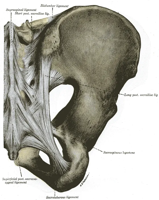
The joint’s core ligaments (tough bands that hold bones together) lie at the front, at the back and between the bones5. They are numerous and strong5. The sacrotuberous, sacrospinous and iliolumbar ligaments are strong accessory ligaments5.
| Ligament | What the sources report |
|---|---|
| Anterior sacroiliac | Numerous thin bands that join the front of the sacrum’s side to the edge of the iliac joint surface6 |
| Interosseous | The strongest of the joint’s ligaments; it fills the space behind and above the synovial part5 |
| Posterior sacroiliac | Lies in a deep hollow between the two bones at the back; a 1918 text calls it their chief bond6 |
| Long dorsal | The most superficial of the joint’s ligaments at the back5 |
| Sacrotuberous | A systematic review concluded it may limit forward tilting of the sacrum but appears to add little to pelvic stability9 |
| Iliolumbar | In 12 embalmed cadaver specimens, cutting both significantly increased the joint’s forward and backward mobility10 |
How far it moves
The ilium and sacrum rotate against each other, mainly about a side-to-side axis5. Forward nodding is called nutation and backward nodding counternutation5. Nutation increases in load-bearing positions such as standing and sitting5. It tightens most of the sacroiliac ligaments and raises compression across the joint5.
One literature review put the range at about 3 degrees in flexion and extension, 1.5 in axial rotation and 0.8 in side bending2. Two studies measured the movement in patients with an X-ray method called radiostereometric analysis11,12.
| Who was measured | Movement found |
|---|---|
| 25 patients with sacroiliac joint disorders | Rotation averaging 2.5 degrees and sliding averaging 0.7 mm between positions11 |
| 11 patients with long-lasting, severe pelvic girdle pain | 0.5 degrees of total rotation in each joint during a single-leg stance, and no sliding12 |
In the 25 patients, movement did not differ between painful and painless joints11. In radiostereometric studies of patients with pelvic girdle pain, average mobility was about 40% lower in men than in women5. A review of the joint’s anatomy states that during pregnancy the joint’s fibrous structures loosen under the hormone relaxin, which increases mobility5,6.
What the muscles add
The erector spinae and multifidus, muscles that run along the spine, help pull the sacrum into nutation5. The gluteus maximus (the large muscle of the buttock) pulls the sacrum sideways into the ilium5. In six healthy women, joint stiffness rose significantly when single muscles contracted without moving the joint13. The rise was clearest for the erector spinae, the gluteus maximus and the biceps femoris, one of the hamstrings13.
The load figures that follow come from a computer model, not from measurements in people14. In that static 3-D model of standing, trunk load was balanced mainly by vertical shear force at the sacroiliac joints14. When the model then forced that shear down by another 20%, on top of an earlier reduction, joint compression rose by 400%14. It did so by switching on the transversus abdominis, the deepest flat abdominal muscle, and the pelvic floor muscles14.
Breakdown
Wear is common on scans of joints with no known problem
In 373 pelvic CT scans of patients with no past low back or pelvic girdle pain, 65.1% showed sacroiliac joint degeneration3. In the same study this degeneration (gradual wear and breakdown) became steadily more common with age, reaching 91% of subjects in their eighties3. In CT scans of 818 patients without sacroiliac joint disease, 45.7% had sclerosis (hardening of bone) and 46.8% had osteophytes15.
A review of normal imaging findings states that such changes can appear as early as the twenties and probably result from strain16. Among 2,845 skeletal pelves, bone bridged across the joint in 12.27% of males and 1.83% of females17. A review of anatomical studies concluded that fusion inside the joint seldom occurs5.
What MRI shows in healthy and active people
Sacroiliitis is inflammation of the sacroiliac joint18. It is the earliest sign of the disease spondyloarthritis, and its most common one18. MRI, a scan that shows soft tissue in detail, has also found changes at the joint in healthy and active people16,19.
| Who was scanned | Share with the finding |
|---|---|
| 47 healthy volunteers with no back pain history | 23.4% had scans that met a standard MRI definition of sacroiliitis19 |
| 24 frequent runners | 12.5% had scans that met the same definition19 |
| 20 healthy hobby runners | 35% had bone marrow oedema on MRI meeting a published set of criteria, ASAS 200916 |
| 22 ice hockey players | 41% had bone marrow oedema meeting those criteria16 |
In the study of volunteers and runners, no healthy volunteer, runner or patient with chronic back pain had deep bone marrow oedema19. It was found in 42 of 47 patients with axial spondyloarthritis19.
Injury, and who reports pain
Among 54 patients whose sacroiliac pain was confirmed by injection, 44% reported a traumatic event beforehand20. Another 21% reported a cumulative injury, and 35% reported a spontaneous onset20.
Figures have been published for three groups, which the sacroiliac joint pain page covers in more detail21,22,23.
- After spinal surgery. A meta-analysis (a study pooling earlier studies) of 12 studies of spinal surgery patients put the incidence of new sacroiliac joint pain at 9.40%21.
- Athletes. A systematic review of 13 studies of athletes found that reported prevalence (the share who have it) of sacroiliac joint pain or dysfunction varied widely22. Its authors concluded that an accurate prevalence cannot be reported22.
- Pregnancy. A European guideline group put the share of pregnant women with pelvic girdle pain at any one time at about 20%23.
Recovery
The joint reshapes under load
A review of sacroiliac anatomy describes the roughening of the joint surfaces as an adaptation for stability5. The gain in body weight, especially after puberty, possibly drives it5.
After childbirth
In an MRI study of 35 women, 77% showed sacroiliac bone marrow oedema within 10 days of giving birth4. After 6 months, 46% still showed it4. In the same study, 60% met an MRI definition of sacroiliitis immediately after birth, 15% at 6 months and 12% at 12 months4.
A second study followed 19 women for 5 years after an uncomplicated vaginal delivery24. Six of the 19 had bone marrow oedema on MRI at 5 years24. No significant development of structural lesions (lasting changes to the bone or joint) was seen after a single delivery24.
After six weeks of hard training
One study scanned 22 military recruits without back pain before and after 6 weeks of intense, standardised physical training25. Before training, 40.9% already had bone marrow oedema at the sacroiliac joints, and 50.0% had it afterwards25. The change was not statistically significant25. Structural lesions were present in 6 recruits at both time points25.
Connections
The joint links the sacrum, at the base of the spine, to the ilium of the pelvis1.
Several muscles and sheets of tissue meet its ligaments6,26:
- The whole back surface of the sacrotuberous ligament gives origin to the gluteus maximus6.
- A 1918 anatomy text describes that ligament’s lower border as continuous with the tendon of the biceps femoris, one of the hamstrings6. A systematic review found the extent of these muscle links unclear9.
- A dissection study found the long dorsal ligament closely related to the erector spinae26. It was also closely related to the thoracolumbar fascia, a sheet of tough tissue over the back26.
A narrative review describes the front of the joint as supplied by branches of the lumbosacral plexus, a nerve network, and the superior gluteal nerve18. At the back, it describes side branches of the sacral nerves, S1 to S4, as the supply18. One study took ligaments from 5 cadavers (bodies donated for study) with no history of sacroiliac pain27. The anterior and interosseous ligaments held nerve fibres carrying two pain-related markers27.
Sacroiliac joint is formed by
Sacroiliac joint joins
Sacroiliac joint has an entry on
Sacroiliac joint is acted on by
Sacroiliac joint is moved by
Sacroiliac joint is related to
Sources
- Wong M, Sinkler MA, Kiel J (2023). Anatomy, Abdomen and Pelvis, Sacroiliac Joint. StatPearls. PMID 29939578
- Kiapour A, Joukar A, Elgafy H, Erbulut DU, Agarwal AK, Goel VK (2020). Biomechanics of the Sacroiliac Joint: Anatomy, Function, Biomechanics, Sexual Dimorphism, and Causes of Pain.. International journal of spine surgery. doi:10.14444/6077 PMID 32123652
- Eno JJ, Boone CR, Bellino MJ, Bishop JA (2015). The prevalence of sacroiliac joint degeneration in asymptomatic adults.. The Journal of bone and joint surgery. American volume. doi:10.2106/JBJS.N.01101 PMID 26041855
- Renson T, Depicker A, De Craemer AS, Deroo L, Varkas G, de Hooge M, Carron P, Jans L, Herregods N, Dehaene I, Vandenberghe G, Roelens K, Van den Bosch FE, Elewaut D (2020). High prevalence of spondyloarthritis-like MRI lesions in postpartum women: a prospective analysis in relation to maternal, child and birth characteristics.. Annals of the rheumatic diseases. doi:10.1136/annrheumdis-2020-217095 PMID 32299794
- Vleeming A, Schuenke MD, Masi AT, Carreiro JE, Danneels L, Willard FH (2012). The sacroiliac joint: an overview of its anatomy, function and potential clinical implications.. Journal of anatomy. doi:10.1111/j.1469-7580.2012.01564.x PMID 22994881
- Gray H; Lewis WH (editor) (1918). Anatomy of the Human Body. Lea & Febiger, Philadelphia (20th edition). Link
- Vleeming A, Stoeckart R, Volkers AC, Snijders CJ (1990). Relation Between Form and Function in the Sacroiliac Joint. Spine. doi:10.1097/00007632-199002000-00016 PMID 2326706
- Vleeming A, Volkers AC, Snijders CJ, Stoeckart R (1990). Relation Between Form and Function in the Sacroiliac Joint. Spine. doi:10.1097/00007632-199002000-00017 PMID 2326707
- Aldabe D, Hammer N, Flack NAMS, Woodley SJ (2019). A systematic review of the morphology and function of the sacrotuberous ligament.. Clinical anatomy (New York, N.Y.). doi:10.1002/ca.23328 PMID 30592090
- Pool-Goudzwaard A, Hoek van Dijke G, Mulder P, Spoor C, Snijders C, Stoeckart R (2003). The iliolumbar ligament: its influence on stability of the sacroiliac joint.. Clinical biomechanics (Bristol, Avon). doi:10.1016/s0268-0033(02)00179-1 PMID 12550807
- Sturesson B, Selvik G, Udén A (1989). Movements of the Sacroiliac Joints. Spine. doi:10.1097/00007632-198902000-00004 PMID 2922636
- Kibsgård TJ, Røise O, Sturesson B, Röhrl SM, Stuge B (2014). Radiosteriometric analysis of movement in the sacroiliac joint during a single-leg stance in patients with long-lasting pelvic girdle pain.. Clinical biomechanics (Bristol, Avon). doi:10.1016/j.clinbiomech.2014.02.002 PMID 24602677
- van Wingerden JP, Vleeming A, Buyruk HM, Raissadat K (2004). Stabilization of the sacroiliac joint in vivo: verification of muscular contribution to force closure of the pelvis.. European spine journal : official publication of the European Spine Society, the European Spinal Deformity Society, and the European Section of the Cervical Spine Research Society. doi:10.1007/s00586-003-0575-2 PMID 14986072
- Pel JJ, Spoor CW, Pool-Goudzwaard AL, Hoek van Dijke GA, Snijders CJ (2008). Biomechanical analysis of reducing sacroiliac joint shear load by optimization of pelvic muscle and ligament forces.. Annals of biomedical engineering. doi:10.1007/s10439-007-9385-8 PMID 18204902
- Ziegeler K, Kreutzinger V, Diekhoff T, Roehle R, Poddubnyy D, Pumberger M, Hamm B, Hermann KGA (2021). Impact of age, sex, and joint form on degenerative lesions of the sacroiliac joints on CT in the normal population.. Scientific reports. doi:10.1038/s41598-021-85303-5 PMID 33723315
- Jurik AG, Herregods N (2024). The sacroiliac joint across ages - what is normal?. Therapeutic advances in musculoskeletal disease. doi:10.1177/1759720X241241126 PMID 38559314
- Dar G, Peleg S, Masharawi Y, Steinberg N, Rothschild BM, Peled N, Hershkovitz I (2005). Sacroiliac joint bridging: demographical and anatomical aspects.. Spine. doi:10.1097/01.brs.0000172232.32082.e0 PMID 16094261
- Prati C, Lequerre T, Le Goff B, Cortet B, Toumi H, Tournadre A, Marotte H, Lespessailles E (2023). Novel insights into the anatomy and histopathology of the sacroiliac joint and correlations with imaging signs of sacroiliitis in case of axial spondyloarthritis.. Frontiers in physiology. doi:10.3389/fphys.2023.1182902 PMID 37250138
- de Winter J, de Hooge M, van de Sande M, de Jong H, van Hoeven L, de Koning A, Berg IJ, Ramonda R, Baeten D, van der Heijde D, Weel A, Landewé R (2018). Magnetic Resonance Imaging of the Sacroiliac Joints Indicating Sacroiliitis According to the Assessment of SpondyloArthritis international Society Definition in Healthy Individuals, Runners, and Women With Postpartum Back Pain.. Arthritis & rheumatology (Hoboken, N.J.). doi:10.1002/art.40475 PMID 29513924
- Chou LH, Slipman CW, Bhagia SM, Tsaur L, Bhat AL, Isaac Z, Gilchrist R, El Abd OH, Lenrow DA (2004). Inciting events initiating injection-proven sacroiliac joint syndrome.. Pain medicine (Malden, Mass.). doi:10.1111/j.1526-4637.2004.04009.x PMID 14996234
- Xu C, Lin X, Zhou Y, Zhuo H, Yang L, Chai X, Huang Y (2024). Incidence and risk factors of new-onset sacroiliac joint pain after spinal surgery: a systematic review and meta-analysis.. PeerJ. doi:10.7717/peerj.18083 PMID 39346039
- Mirdamadi N, Khadembashiri MM, Moghadam N, Kordi R (2025). Prevalence and Risk Factors of Sacroiliac Joint Pain in Athletes: A Systematic Review and Proportional Meta-Analysis.. Clinical journal of sport medicine : official journal of the Canadian Academy of Sport Medicine. doi:10.1097/JSM.0000000000001341 PMID 40135982
- Vleeming A, Albert HB, Ostgaard HC, Sturesson B, Stuge B (2008). European guidelines for the diagnosis and treatment of pelvic girdle pain.. European spine journal : official publication of the European Spine Society, the European Spinal Deformity Society, and the European Section of the Cervical Spine Research Society. doi:10.1007/s00586-008-0602-4 PMID 18259783
- Varkas G, De Meester L, de Hooge M, De Craemer AS, Herregods N, Jans L, Carron P, Elewaut D, Van den Bosch F (2025). Long-term evolution of postpartum sacroiliac bone marrow oedema: a 5-year longitudinal follow-up study.. RMD open. doi:10.1136/rmdopen-2024-005309 PMID 40185493
- Varkas G, de Hooge M, Renson T, De Mits S, Carron P, Jacques P, Moris M, Souverijns G, Jans L, Elewaut D, Van den Bosch F (2018). Effect of mechanical stress on magnetic resonance imaging of the sacroiliac joints: assessment of military recruits by magnetic resonance imaging study.. Rheumatology (Oxford, England). doi:10.1093/rheumatology/kex491 PMID 29253272
- Vleeming A, Pool-Goudzwaard AL, Hammudoghlu D, Stoeckart R, Snijders CJ, Mens JM (1996). The Function of the Long Dorsal Sacroiliac Ligament. Spine. doi:10.1097/00007632-199603010-00005 PMID 8852309
- Szadek KM, Hoogland PV, Zuurmond WW, de Lange JJ, Perez RS (2008). Nociceptive nerve fibers in the sacroiliac joint in humans.. Regional anesthesia and pain medicine. doi:10.1016/j.rapm.2007.07.011 PMID 18155055
Changes
- First published.