Hip flexors
Also called hip flexor muscles, iliopsoas, psoas, psoas major, iliacus
The muscles that bend the hip, led by the deep iliopsoas. Where they run, what they do in walking, running and lifting, where they strain, how training changes them, and what they work with.
- Region
- Hip
- Starts on (psoas major)
- the sides of the last thoracic (chest) and all lumbar (lower back) vertebrae, their discs, and the lumbar side spurs2
- Starts on (iliacus)
- the upper two-thirds of the iliac fossa (a shallow hollow) and the inner lip of the hip bone's top rim (Pelvis)2,6
- Starts on (psoas minor)
- the sides of the twelfth thoracic (chest) and first lumbar (lower back) vertebrae and the disc between them2
- Starts on (sartorius)
- the anterior superior iliac spine (bony point at the hip's front) and the upper half of the notch below it (Pelvis)2,6
- Ends on (psoas major)
- the lesser trochanter, a small bump on the inner side of the thigh bone just below its neck2,6
- Ends on (iliacus)
- the outer side of the psoas major tendon, with some fibres continuing onto the shaft of the thigh bone2
- Ends on (psoas minor)
- the pectineal line and iliopectineal eminence of the pelvis, and the iliac fascia, by a long flat tendon2
- Ends on (sartorius)
- the upper part of the inner surface of the shinbone (tibia), by a broad flat sheet of tendon2,6
- Muscles
- psoas major, iliacus, psoas minor, rectus femoris, sartorius, pectineus
- Updated
- October 7, 2026
- Reviewed
- October 7, 2026 · how entries are checked
Function
The iliopsoas and its helpers
The hip flexors are the muscles that bend the hip. The primary one is the iliopsoas unit: the psoas major, psoas minor and iliacus1. It also helps turn the thigh outward and steady the hip and the lumbar spine (lower back)1. The psoas major and the iliacus together make up the iliopsoas group6.
Other muscles help the iliopsoas flex the thigh2,6:
| Muscle | Where it runs | What it does |
|---|---|---|
| Rectus femoris, one of the quadriceps7 | One of its two tendon heads starts on the anterior inferior iliac spine, a bony point at the front of the pelvis7 | Helps flex the thigh on the pelvis2. It also straightens the knee7 |
| Sartorius | From the anterior superior iliac spine, the bony point at the front of the hip, across the thigh to the upper inner tibia (shinbone)2 | Flexes the thigh, moves it out to the side, turns it outward and bends the knee, as in sitting cross-legged6 |
| Pectineus (on the hip adductors page) | From the pubis, the bone at the lower front of the pelvis, to a rough line on the femur2 | Flexes the femur at the hip and moves it toward the midline6 |
Where the iliopsoas runs
The psoas major arises from the lumbar vertebrae, the last vertebra of the thoracic spine above them, and the discs between2. The thoracic spine is the part that carries the ribs. The muscle runs down and passes beneath the inguinal ligament, a band of tough tissue across the groin2. It crosses in front of the hip joint’s capsule, the sleeve of tough tissue around the joint, then ends in a tendon2.
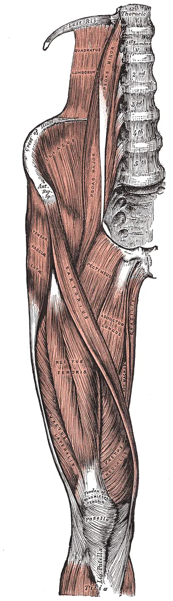
The iliacus is a flat, triangular muscle2. It fills the iliac fossa, a shallow hollow in the ilium (the wide upper part of the hip bone)2. It starts on the upper two-thirds of that hollow2. It also starts on the inner lip of the iliac crest, the curved top rim of the hip bone2.
The psoas major tendon takes in nearly all the fibres of the iliacus and attaches to the lesser trochanter2. The lesser trochanter is a small prominence on the inner side of the femur, just below its neck6. Some iliacus fibres carry on for roughly 2.5 cm along the femur’s shaft2.
In a dissection of 28 embalmed cadavers (bodies donated for study), the iliopsoas ended in one tendon in 128. It ended in two tendons in another 12, and in three tendons in 48.
A large bursa, a fluid-filled sac that cushions tissues where they rub, separates the tendon from the pubis and from the hip joint’s capsule2. It may open into the joint cavity2.
Its nerves
Sources differ on which nerves supply the psoas major1,2. One classic account names branches of lumbar nerves two and three1,2. A current reference names the first to third1,2. The iliacus is supplied by the femoral nerve1,2.
What the muscles do in movement
Acting from above, the psoas major flexes the thigh on the pelvis, and the iliacus assists2. With the femur fixed, the pull works the other way2. The psoas major bends the lumbar spine forward and to its own side, then tilts the pelvis forward with the iliacus2. When both sides keep working with the thighs fixed, the trunk and pelvis bend forward, as in sitting up from lying2.
Three studies recorded these deep muscles with fine wires placed in them11,12,13:
- In 7 healthy adults, the psoas and iliacus were generally active together, particularly when hip flexor torque (a turning force) was needed11.
- In 11 healthy subjects on a treadmill, iliacus and psoas activity rose markedly at speeds above 2.0 m/s12.
- In seven skilled soccer players doing maximal place kicks, the iliopsoas was active through the whole kick, even while the thigh was slowing down13.
In a study of eight people, the psoas stayed under 16% of its maximum while they lifted barbells of up to 100 kg3. The psoas was most active in maximal hip flexion efforts3. Each form of sit-up tested activated it more than the curl-up: 15 to 35% of maximum, against under 10%3.
The psoas and the lower back
Two modelling studies, which estimate forces with a model, not by measuring them, disagree about the psoas major and the lumbar spine14,15:
- One model used the dissection of three cadavers and X-rays of ten adult males14. At maximum contraction, it calculated severe compression forces and large shear (sliding) forces on the lumbar segments14.
- A three-dimensional model used seven dissected cadavers and MRI scans of 15 males15. It concluded that large forward shear forces arise at just one place, the lowest lumbar joint (L5-S1)15.
Breakdown
Where strains happen
NCAA injury surveillance of varsity teams from 25 sports recorded 770 hip flexor strains from 2009/10 to 2014/1516. That is 1.60 per 10,000 athlete-exposures, where one exposure is one athlete in one practice or game16. Of those strains, 59.4% happened without contact16.
A 2-year study followed 606 male professional footballers from 17 clubs17. Iliopsoas-related groin pain made up 12% of their time-loss groin injuries, those that cause missed training or matches17. Adductor-related groin pain made up 68%17. The hip flexor strain and athletic groin pain pages cover who is affected and the course over time.
Which muscle tears, and where
One study scanned male athletes with acute groin pain by MRI4. Of 156 athletes, 33 had a confirmed acute hip flexor muscle injury4. Their injuries were spread across five muscles4:
| Muscle | Injuries among the 33 athletes |
|---|---|
| Rectus femoris | 164 |
| Iliacus | 124 |
| Psoas major | 74 |
| Sartorius | 44 |
| Tensor fasciae latae | 14 |
Rectus femoris injuries came mainly during kicking (10 of 16) and sprinting (4)4. A change of direction injured the iliacus or psoas major, and no other muscle4.
Iliacus and psoas major injuries were mainly at the junction of muscle and tendon4. Of the 16 rectus femoris injuries, 10 (63%) involved the tendon4.
How the injury changes with age
One report covers 32 patients with iliopsoas injuries on MRI18. Those under 65 mostly had partial tendon tears and muscle strains, most often from an athletic injury18. In those 65 and older, the most common injury was a complete tendon tear, seen in 8 patients, each of them a woman18.
The snapping tendon
Among 87 elite ballet dancers who completed a questionnaire, 91% reported a snapping hip19. Of those, 58% had pain with the snap19. Ultrasound was used on 26 elite ballet dancers (50 hips) who could snap their hips at will19. In 59% of hips it showed the iliopsoas tendon snapping, and in a third it did not identify the cause19.
A moving ultrasound study looked at 18 snapping iliopsoas tendons in 14 patients20. In 14 hips, the tendon suddenly flipped around the iliacus muscle and struck the pubic bone20. This happened as the hip came back from a position bent, out to the side and turned outward20.
Recovery
What training studies found
Three studies trained the hip flexors directly5,21,22:
- Elastic bands, 6 weeks. A randomised controlled trial of 33 healthy adults used three 10-minute sessions a week5. Isometric hip flexion strength of the trained leg rose by 17%5.
- Resistance training, 8 weeks. In a controlled study of physically active, untrained adults, 24 people completed a hip flexion programme21. Their hip flexion strength rose by 12.2%, and their 40-yard dash time fell by 3.8%21. The 24 controls did not improve21.
- Resistance training added to running, 12 weeks. Eight well-trained male distance runners trained their hip flexors twice a week, with no control group22. The cross-sectional area of the psoas major, the size of a slice across it, went from 16.2 to 16.7 square centimetres22.
Muscle size in athletes
Two MRI studies related psoas size to speed23,24. In one, 56 male sprinters had a larger psoas major than 40 male non-sprinters, in absolute terms and relative to body mass23. Among the sprinters, the cross-sectional areas of the psoas major and gluteus maximus correlated with personal best 100 m time23. A correlation is how closely two measures rise and fall together.
In 66 collegiate American football players, psoas major volume on MRI explained 50% of the variance in on-field sprint speed24. That was more than any other lower-body muscle measured24.
A computer-model study of sprinting gives predicted figures, not measured ones25. In the model, a 10% rise in iliopsoas strength raised predicted maximum speed by 1.07%25.
After a strain
In the NCAA surveillance of 25 sports, 83.8% of hip flexor strains restricted participation for less than one week16. In the same data, 10.1% of hip flexor strains were recurrences16. The hip flexor strain page reports what studies found about return to sport.
Connections
The femoral nerve
The femoral nerve emerges from the outer border of the psoas major and runs between the psoas and the iliacus26. Below the inguinal ligament, the iliopsoas is the one muscle between the nerve and the front of the hip joint capsule27.
Bones and neighbouring muscles
The iliacus starts on the ilium, part of the pelvis, and the shared tendon ends on the femur2. The rectus femoris is covered on the quadriceps page and the pectineus on the hip adductors page6,7.
Exercises and stretches
Related exercise pages: split squat, running and side-lying hip abduction28,29,30. The related stretch page is the hip flexor stretch31.
| Page | What one study found |
|---|---|
| Split squat | In a simulation study, 11 participants did 10 split squat variations28. The rectus femoris was loaded most in the rear limb, at 13 to 27 N/kg28 |
| Running | In a computer-model study of nine subjects, runners went faster above 7 m/s by raising stride frequency29. The hip muscles, mainly the iliopsoas, gluteus maximus and hamstrings, did this by accelerating the hip and knee harder during swing29 |
| Side-lying hip abduction | In 20 healthy distance runners doing a loaded clamshell, the anterior hip flexors worked at 54% of maximum30. A clamshell is a side-lying exercise that opens the knees apart. That was more than the tensor fasciae latae (34%) and gluteus medius (33%), and not significantly different from the gluteus maximus (34%)30 |
| Hip flexor stretch | In 26 healthy men, iliacus stiffness was lower after a single 1-minute static hip extension stretch than before it31 |
Hip flexors is injured in
Hip flexors lies next to
Hip flexors crosses
Hip flexors starts on
Hip flexors attaches to
Hip flexors shares the rectus femoris with
Hip flexors shares the pectineus with
Hip flexors is stretched by
Hip flexors is loaded by
Sources
- Bordoni B, Varacallo MA (2026). Anatomy, Bony Pelvis and Lower Limb: Iliopsoas Muscle.. StatPearls. PMID 30285403
- Gray H; Lewis WH (editor) (1918). Anatomy of the Human Body. Lea & Febiger, Philadelphia (20th edition). Link
- Juker D, McGill S, Kropf P, Steffen T (1998). Quantitative intramuscular myoelectric activity of lumbar portions of psoas and the abdominal wall during a wide variety of tasks.. Medicine and Science in Sports and Exercise. doi:10.1097/00005768-199802000-00020 PMID 9502361
- Serner A, Weir A, Tol JL, Thorborg K, Roemer F, Guermazi A, Yamashiro E, Hölmich P (2018). Characteristics of acute groin injuries in the hip flexor muscles - a detailed MRI study in athletes.. Scandinavian journal of medicine & science in sports. doi:10.1111/sms.12939 PMID 28649793
- Thorborg K, Bandholm T, Zebis M, Andersen LL, Jensen J, Hölmich P (2016). Large strengthening effect of a hip-flexor training programme: a randomized controlled trial.. Knee surgery, sports traumatology, arthroscopy : official journal of the ESSKA. doi:10.1007/s00167-015-3583-y PMID 25796586
- Betts JG, Young KA, Wise JA, Johnson E, Poe B, Kruse DH, Korol O, Johnson JE, Womble M, DeSaix P (2022). Anatomy and Physiology 2e. OpenStax. Link
- Begum FA, Kayani B, Chang JS, Tansey RJ, Haddad FS (2020). The management of proximal rectus femoris avulsion injuries.. EFORT Open Reviews. doi:10.1302/2058-5241.5.200055 PMID 33312709
- Lin B, Bartlett J, Lloyd TD, Challoumas D, Brassett C, Khanduja V (2022). Multiple iliopsoas tendons: a cadaveric study and treatment implications for internal snapping hip syndrome.. Archives of orthopaedic and trauma surgery. doi:10.1007/s00402-021-04009-5 PMID 34347120
- Neumann DA, Garceau LR (2015). A proposed novel function of the psoas minor revealed through cadaver dissection.. Clinical anatomy (New York, N.Y.). doi:10.1002/ca.22467 PMID 25227908
- Patra A, Asghar A, Pushpa NB, Chaudhary P, Ravi KS, Kaur H, Przybycień W, Musiał A, Walocha JA (2024). Reappraisal of the Morphological and morphometric diversity of the psoas minor muscle with clinical and developmental insights: cadaveric analysis.. Folia morphologica. doi:10.5603/fm.99128 PMID 38619066
- Andersson E, Oddsson L, Grundström H, Thorstensson A (1995). The role of the psoas and iliacus muscles for stability and movement of the lumbar spine, pelvis and hip.. Scandinavian journal of medicine & science in sports. doi:10.1111/j.1600-0838.1995.tb00004.x PMID 7882121
- Andersson EA, Nilsson J, Thorstensson A (1997). Intramuscular EMG from the hip flexor muscles during human locomotion.. Acta physiologica Scandinavica. doi:10.1046/j.1365-201X.1997.00225.x PMID 9401589
- Dörge HC, Andersen TB, Sørensen H, Simonsen EB, Aagaard H, Dyhre-Poulsen P, Klausen K (1999). EMG activity of the iliopsoas muscle and leg kinetics during the soccer place kick.. Scandinavian journal of medicine & science in sports. doi:10.1111/j.1600-0838.1999.tb00233.x PMID 10407926
- Bogduk N, Pearcy M, Hadfield G (1992). Anatomy and biomechanics of psoas major.. Clinical biomechanics (Bristol, Avon). doi:10.1016/0268-0033(92)90024-X PMID 23915688
- Santaguida PL, McGill SM (1995). The psoas major muscle: a three-dimensional geometric study.. Journal of biomechanics. doi:10.1016/0021-9290(94)00064-b PMID 7730392
- Eckard TG, Padua DA, Dompier TP, Dalton SL, Thorborg K, Kerr ZY (2017). Epidemiology of Hip Flexor and Hip Adductor Strains in National Collegiate Athletic Association Athletes, 2009/2010-2014/2015.. The American journal of sports medicine. doi:10.1177/0363546517716179 PMID 28745561
- Mosler AB, Weir A, Eirale C, Farooq A, Thorborg K, Whiteley RJ, Hӧlmich P, Crossley KM (2018). Epidemiology of time loss groin injuries in a men's professional football league: a 2-year prospective study of 17 clubs and 606 players.. British journal of sports medicine. doi:10.1136/bjsports-2016-097277 PMID 28666981
- Bui KL, Ilaslan H, Recht M, Sundaram M (2008). Iliopsoas injury: an MRI study of patterns and prevalence correlated with clinical findings.. Skeletal radiology. doi:10.1007/s00256-007-0414-3 PMID 18026948
- Winston P, Awan R, Cassidy JD, Bleakney RK (2007). Clinical examination and ultrasound of self-reported snapping hip syndrome in elite ballet dancers.. The American journal of sports medicine. doi:10.1177/0363546506293703 PMID 17021311
- Deslandes M, Guillin R, Cardinal E, Hobden R, Bureau NJ (2008). The snapping iliopsoas tendon: new mechanisms using dynamic sonography.. AJR. American journal of roentgenology. doi:10.2214/AJR.07.2375 PMID 18287424
- Deane RS, Chow JW, Tillman MD, Fournier KA (2005). Effects of hip flexor training on sprint, shuttle run, and vertical jump performance.. Journal of strength and conditioning research. doi:10.1519/14974.1 PMID 16095411
- Yamanaka R, Wakasawa S, Yamashiro K, Kodama N, Sato D (2021). Effect of Resistance Training of Psoas Major in Combination With Regular Running Training on Performance in Long-Distance Runners.. International journal of sports physiology and performance. doi:10.1123/ijspp.2020-0206 PMID 33668016
- Tottori N, Suga T, Miyake Y, Tsuchikane R, Tanaka T, Terada M, Otsuka M, Nagano A, Fujita S, Isaka T (2021). Trunk and lower limb muscularity in sprinters: what are the specific muscles for superior sprint performance?. BMC research notes. doi:10.1186/s13104-021-05487-x PMID 33632290
- Martin JA, Joachim MR, Blemker SS, Opar DA, Mortensen B, Heiderscheit BC (2026). Lower-Body Muscle Volumes Can Explain Half of the Variance in Sprint Speed Between Collegiate American Football Players.. Scandinavian journal of medicine & science in sports. doi:10.1111/sms.70283 PMID 41999352
- Yang K, Lin YC, Pandy MG (2026). Running Speed is Maximized by Strengthening the Hip Flexors and Hip Adductors.. Annals of biomedical engineering. doi:10.1007/s10439-026-04184-9 PMID 42189432
- Valera-Calero JA (2026). A Didactic Ultrasound Manual for identifying lumbar Plexus Peripheral Nerves in the lower limb: Lower Abdominal Wall, Groin and Thigh.. Ultrasound international open. doi:10.1055/a-2920-0129 PMID 42591916
- Patton RS, Runner RP, Lazarus D, Bradbury TL (2018). Femoral neuropathy following direct anterior total hip arthroplasty: an anatomic review and case series†.. Journal of surgical case reports. doi:10.1093/jscr/rjy171 PMID 30206472
- Schellenberg F, Taylor WR, Lorenzetti S (2017). Towards evidence based strength training: a comparison of muscle forces during deadlifts, goodmornings and split squats.. BMC sports science, medicine & rehabilitation. doi:10.1186/s13102-017-0077-x PMID 28725437
- Dorn TW, Schache AG, Pandy MG (2012). Muscular strategy shift in human running: dependence of running speed on hip and ankle muscle performance.. The Journal of experimental biology. doi:10.1242/jeb.064527 PMID 22573774
- McBeth JM, Earl-Boehm JE, Cobb SC, Huddleston WE (2012). Hip muscle activity during 3 side-lying hip-strengthening exercises in distance runners.. Journal of Athletic Training. doi:10.4085/1062-6050-47.1.15 PMID 22488226
- Nojiri S, Yagi M, Mizukami Y, Ichihashi N (2019). Static stretching time required to reduce iliacus muscle stiffness.. Sports biomechanics. doi:10.1080/14763141.2019.1620321 PMID 31232167
Changes
- First published.