Quadratus lumborum
Also called QL
A four-sided muscle of the lower back, between the hip bone, the last rib and the lumbar spine. Where it attaches, what recordings show it doing, how its size differs in athletes, and what it works with.
- Region
- Spine
- Starts on
- the iliolumbar ligament and the nearby iliac crest, the top rim of the hip bone, along about 5 cm (Pelvis)1,2
- Ends on
- the lower border of the last (twelfth) rib, along about half of its length1,2; the tips of the side spurs (transverse processes) of the upper four lower back (lumbar) vertebrae, by four small tendons (Lumbar vertebrae)1,2
- Updated
- October 9, 2026
- Reviewed
- October 9, 2026 · how entries are checked
Function
Where it sits and what it attaches to
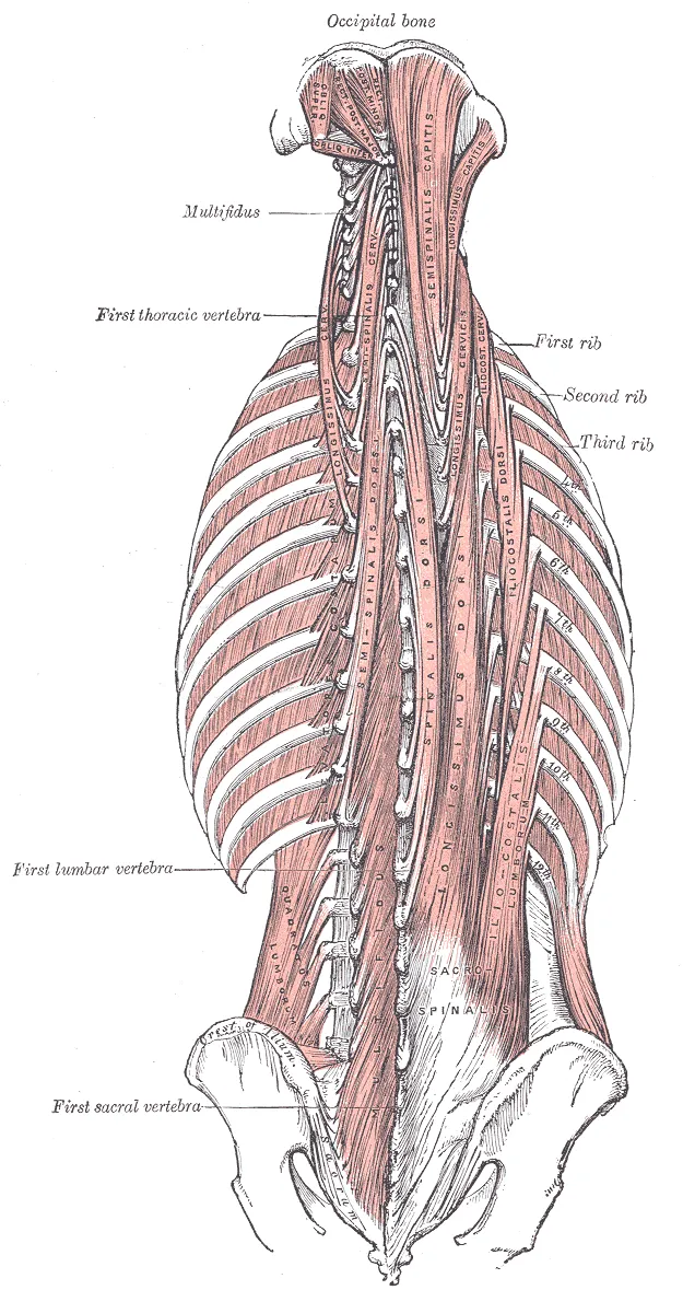
The quadratus lumborum sits in the lower back part of the trunk, to the side of the spine1. It forms part of the back wall of the abdomen, as do the lumbar vertebrae3. A vertebra is one of the bones stacked to form the spine, and lumbar means of the lower back. The muscle is irregularly four-sided, and broader at the bottom than at the top2.
It has three sets of attachments1,2:
| End | Where it attaches |
|---|---|
| Bottom | The iliolumbar ligament and the nearby iliac crest, along about 5 cm, by flat tendon fibres1,2 |
| Top | The lower border of the last (12th) rib, along about half of the rib’s length1,2 |
| Spine | The upper four lumbar vertebrae, at the tips of their transverse processes, by four small tendons1,2 |
The iliolumbar ligament is a band of tissue from the lowest lumbar vertebrae to the hip bone. A tendon is a tough cord of tissue that connects a muscle to a bone. A transverse process is a small spur of bone on each side of a vertebra. The number of attachments to the vertebrae varies, and so does how far the muscle attaches along the last rib2.
Three layers
One published description divides the muscle into three layers9:
- A front (anterior) layer, from the iliac crest to the 12th rib9.
- A middle layer, from the transverse processes of the first four lumbar vertebrae to the 12th rib9.
- A back (posterior) layer, from the iliac crest to those four transverse processes9.
What it is said to do
Sources describe three actions2,3:
- One textbook lists it as the muscle for lateral flexion, which means bending the spine to the side3.
- When the chest and spine are fixed, one quadratus lumborum acting alone may raise the pelvis toward its own side2.
- It draws down the last rib2. This helps fix the starting point of the diaphragm, the dome-shaped main breathing muscle, during breathing in2.
Other sources are less sure. An anatomy chapter says the muscle’s complex organisation makes its actions hard to identify precisely from how its fibres contract1. The authors of one cadaver model concluded that the muscle’s actual role in how the spine works has still to be determined4.
Their model, built from the dissection of six embalmed cadavers, worked out the forces the muscle could exert on the lumbar spine4. These were its compression force and its turning effects for bending backward and sideways4. Each was no more than 10 per cent of what two back muscles exert, the erector spinae and multifidus4. The erector spinae is the group of long muscles beside the spine that straighten the back. The multifidus is a deep muscle running along the spine.
Another group recorded the muscle’s electrical activity in four young adults10. They judged that these recordings, with the way the muscle is built, make it a better stabiliser of the spine than the psoas10. The psoas is a deep muscle that bends the hip.
What recordings of the muscle show
The studies below used fine-wire electrodes, very thin wire sensors placed into a muscle to record its activity10,11,12:
| Who was measured | Task | What was found |
|---|---|---|
| 7 healthy people | Bending the trunk toward the same side, lying on the side | The highest quadratus lumborum activity in the study11 |
| 12 healthy men | Lifting the pelvis sideways on the measured side | 19% of maximum, significantly higher than in forward, backward or opposite-side pelvic tilts12 |
| 4 young adults | Heavy barbell lifts, with weights of up to 70 kg | 74% of maximum on average10 |
Its nerve and blood supply
Two sources give different accounts of the nerve supply1,2:
- Gray’s Anatomy (1918) gives the twelfth thoracic nerve and the first and second lumbar nerves2. Thoracic means of the chest.
- A StatPearls anatomy chapter gives the twelfth thoracic nerve and the iliohypogastric and ilioinguinal nerves1. These last two run through the lower wall of the abdomen.
The twelfth thoracic, ilioinguinal and iliohypogastric nerves run between the muscle and the fascia that covers it2. Fascia is a sheet of tough connective tissue. The StatPearls chapter says the muscle’s blood comes from a lumbar branch of the iliolumbar artery and from the lumbar arteries1.
Breakdown
One reported strain in sport
A strain is an overstretched or torn muscle. A case report, a report on a single patient, described a quadratus lumborum strain in a 16-year-old soccer player9. Its authors called it the first report of a sports-related strain of this muscle9. The strain happened just after he kicked the ball, as the kicking leg landed9. The suspected cause was side bending of the trunk while the pelvis was forced to tilt backward9.
One side larger than the other
Three studies compared the left and right muscles in cricketers using MRI, a scan that shows soft tissue such as muscle in detail6,13,14. Cross-sectional area is the size of a slice cut straight across the muscle.
| Group | What was found |
|---|---|
| 21 male elite cricketers, from an eligible group of 26 with a mean age of 21.2 | Cross-sectional area was larger on the dominant-arm side13 |
| 51 adolescent fast bowlers and 18 swimmers | Volume asymmetry averaged 110.5% in the bowlers, significantly different from 96.6% in the swimmers14. A value above 100% meant a larger muscle on the bowling-arm side14 |
| 38 junior fast bowlers, mean age 14.9 | The mean side-to-side difference in cross-sectional area was 13%6 |
Asymmetry and lumbar injury in fast bowlers
One systematic review, of 16 studies of adolescent pace bowlers, found conflicting results on asymmetry as a risk factor for non-contact injury7. Another, of 16 studies of adult fast bowlers, found conflicting results for its association with injury8. Single studies show the disagreement, and one concerns the pars interarticularis14. This is a small segment of bone in a vertebra, between its upper and lower articular processes15:
- The 51 adolescent fast bowlers from the table above, followed with MRI. Greater asymmetry was significantly associated with one-sided pars interarticularis lesions that caused symptoms, at L4, the fourth lumbar vertebra14. These lesions developed on the side opposite the bowling arm14.
- 38 adolescent fast bowlers, scanned before a season. In this group, 21% developed lumbar bone stress injuries, meaning overuse damage to bone16. The muscle’s cross-sectional area was not significantly related to lumbar spine injury16.
- 23 elite adult fast bowlers, scanned at the start of a season. The bowlers who stayed free of lumbar spine injury had significantly larger asymmetries than those who went on to such an injury17.
- The 21 male elite cricketers from the table above. The side-to-side difference was greatest in the subgroup of fast bowlers with low back pain13.
One study modelled fast bowling postures with mathematical and finite element models, computer models that split a structure into many small pieces18. A muscle enlarged by 30% on one side went with significant rises in stress in the pars only when stress was fairly low18. When stress was already high, it changed that stress only slightly18. The authors took this to suggest that the asymmetry is not the cause of the stress fractures18. Related pages are lumbar vertebrae and spondylolysis.
What studies of low back pain measured
Studies of people with low back pain have measured the muscle’s size and its trigger points19,20:
- Size. A systematic review of 25 studies found conflicting evidence for a link between the muscle’s cross-sectional area and low back pain19. A 2025 meta-analysis, a study that pools earlier studies, combined 17 MRI studies21. It found no significant difference in the muscle’s functional cross-sectional area between people with and without low back pain21.
- Trigger points. A trigger point is a tender knot in a muscle. A systematic review of nine articles reported active ones in the quadratus lumborum of 30% to 55% of patients with low back pain20. Its authors cautioned that finding them by hand is subjective, which limits how far the results apply20.
Recovery
After the one reported strain
In the case report of the 16-year-old soccer player, the strain healed and he returned to competition 3 weeks later9.
After spaceflight
A case series (a report on a group, without a comparison group) followed 17 astronauts and cosmonauts on long-duration flights22. The muscle’s cross-sectional area and its attenuation on CT were significantly lower after flight22. A CT scan builds detailed pictures of the inside of the body from X-rays, and attenuation is how strongly a tissue blocks them. After recovery on Earth, area equalled or exceeded its preflight value22. Attenuation was still below its preflight value 2 to 4 years later22.
What training studies measured
Two reports measured the muscle’s size before and after exercise programmes23,24:
- 28 male soccer players aged 12 to 13. The cross-sectional areas of five trunk muscles, the quadratus lumborum among them, were significantly larger after six months of soccer training in both groups23. The 16 who added a stabilisation programme four times a week showed no significantly greater increase than the 12 who did not23. That programme included the side bridge23.
- People with chronic (long-lasting) low back pain. A systematic review and meta-analysis of 34 articles pooled exercise programmes24. They had significant positive effects on the thickness and cross-sectional area of the lumbar multifidus24. They had no significant effect on the cross-sectional area of the quadratus lumborum or the lumbar erector spinae24.
Connections
The fascia and its neighbours
A StatPearls anatomy chapter describes the muscle as an integral part of the thoracolumbar fascia1. This fascia is a sheet of tough connective tissue over the lower and middle back. Its middle layer lies between the quadratus lumborum and the paraspinal muscles, the back muscles that lie along the spine2,25. The aponeurosis, or flat sheet of tendon, of the transversus abdominis fuses with the covering on the back of the quadratus lumborum25. The transversus abdominis is the deepest of the three flat muscles on each side of the abdomen.
In front of the quadratus lumborum lie the kidney, the colon, the diaphragm and the psoas major and minor2. The psoas is covered on the hip flexors page.
Exercises that load it
A 2013 systematic review of core muscle activity in fitness exercises covered 17 studies and 252 healthy adults26. It found no studies of quadratus lumborum activity26. Related exercise pages: side plank and side-lying hip abduction10,27. A side bridge is a side plank, held propped on one forearm and the side of the feet.
| Exercise | What single studies found |
|---|---|
| Side plank | In four young adults holding a side support on one elbow and both feet, the muscle was more active (54%) than other muscles10 |
| Side plank | Thirteen healthy men did 14 bridge exercises with fine-wire electrodes in the muscle5. The front and back layers were each most active in a side bridge done with hip abduction5. The front layer reached 47.3% of maximum and the back layer 43.0%5 |
| Side-lying hip abduction | Twenty-four women with a weak gluteus medius, a buttock muscle, lifted with ankle loads of 3% or 5% of body weight27. Quadratus lumborum activity rose from 49% to 82% and 92% of maximum27 |
The related stretch page is child’s pose28. A 2025 review covered yoga programmes for the spine and chronic low back pain28. Its authors describe forward-bending poses, this one among them, as lengthening the erector spinae and quadratus lumborum28. This is their description, not a measurement28.
Quadratus lumborum was compared in a force model with
Quadratus lumborum has a back covering that fuses with the flat tendon of
Quadratus lumborum lies behind the psoas of
Quadratus lumborum has been measured in studies of
Quadratus lumborum attaches to
Quadratus lumborum is loaded by
Quadratus lumborum is described as lengthened by
Sources
- Bordoni B, Sina RE, Varacallo MA (2024). Anatomy, Abdomen and Pelvis, Quadratus Lumborum.. StatPearls. PMID 30571028
- Gray H; Lewis WH (editor) (1918). Anatomy of the Human Body. Lea & Febiger, Philadelphia (20th edition). Link
- Betts JG, Young KA, Wise JA, Johnson E, Poe B, Kruse DH, Korol O, Johnson JE, Womble M, DeSaix P (2022). Anatomy and Physiology 2e. OpenStax. Link
- Phillips S, Mercer S, Bogduk N (2008). Anatomy and biomechanics of quadratus lumborum.. Proceedings of the Institution of Mechanical Engineers, Part H: Journal of Engineering in Medicine. doi:10.1243/09544119JEIM266 PMID 18441751
- Oshikawa T, Adachi G, Akuzawa H, Okubo Y, Kaneoka K (2020). Change in Regional Activity of the Quadratus Lumborum During Bridge Exercises.. Journal of Sport Rehabilitation. doi:10.1123/jsr.2019-0225 PMID 32320945
- Kountouris A, Portus M, Cook J (2012). Quadratus lumborum asymmetry is not isolated to the dominant side in junior cricket fast bowlers.. British Journal of Sports Medicine. doi:10.1136/bjsm.2010.077453 PMID 21616940
- Forrest MRL, Hebert JJ, Scott BR, Brini S, Dempsey AR (2017). Risk Factors for Non-Contact Injury in Adolescent Cricket Pace Bowlers: A Systematic Review.. Sports Medicine. doi:10.1007/s40279-017-0778-z PMID 28879571
- Olivier B, Taljaard T, Burger E, Brukner P, Orchard J, Gray J, Botha N, Stewart A, Mckinon W (2016). Which Extrinsic and Intrinsic Factors are Associated with Non-Contact Injuries in Adult Cricket Fast Bowlers?. Sports Medicine. doi:10.1007/s40279-015-0383-y PMID 26365510
- Tanaka K, Funasaki H, Kubota D, Murayama Y, Saito M, Iguchi Y (2023). Quadratus lumborum muscle strain in a youth soccer player: a case report.. Radiology Case Reports. doi:10.1016/j.radcr.2022.10.084 PMID 36439922
- McGill S, Juker D, Kropf P (1996). Quantitative intramuscular myoelectric activity of quadratus lumborum during a wide variety of tasks.. Clinical Biomechanics. doi:10.1016/0268-0033(95)00056-9 PMID 11415616
- Andersson EA, Oddsson LI, Grundström H, Nilsson J, Thorstensson A (1996). EMG activities of the quadratus lumborum and erector spinae muscles during flexion-relaxation and other motor tasks.. Clinical Biomechanics. doi:10.1016/0268-0033(96)00033-2 PMID 11415651
- Oshikawa T, Adachi G, Akuzawa H, Okubo Y, Kaneoka K (2020). Activity of the Quadratus Lumborum and Trunk Muscles Relates to Pelvic Tilt Angle During Pelvic Tilt Exercises.. American Journal of Physical Medicine & Rehabilitation. doi:10.1097/PHM.0000000000001496 PMID 32541348
- Hides J, Stanton W, Freke M, Wilson S, McMahon S, Richardson C (2008). MRI study of the size, symmetry and function of the trunk muscles among elite cricketers with and without low back pain.. British Journal of Sports Medicine. doi:10.1136/bjsm.2007.044024 PMID 18065440
- Engstrom CM, Walker DG, Kippers V, Mehnert AJ (2007). Quadratus lumborum asymmetry and L4 pars injury in fast bowlers. Medicine & Science in Sports & Exercise. doi:10.1249/mss.0b013e3180408e25 PMID 17545879
- Madden V, Ayoub A, Thomas J, Thomas I (2026). Spondylolysis: A Narrative Review of Etiology, Diagnosis, and Management.. International journal of environmental research and public health. doi:10.3390/ijerph23020153 PMID 41752236
- Kountouris A, Portus M, Cook J (2012). Quadratus lumborum asymmetry and lumbar spine injury in cricket fast bowlers.. Journal of Science and Medicine in Sport. doi:10.1016/j.jsams.2012.03.012 PMID 22578717
- Kountouris A, Portus M, Cook J (2013). Cricket fast bowlers without low back pain have larger quadratus lumborum asymmetry than injured bowlers.. Clinical Journal of Sport Medicine. doi:10.1097/JSM.0b013e318280ac88 PMID 23377354
- de Visser H, Adam CJ, Crozier S, Pearcy MJ (2007). The role of quadratus lumborum asymmetry in the occurrence of lesions in the lumbar vertebrae of cricket fast bowlers.. Medical Engineering & Physics. doi:10.1016/j.medengphy.2006.09.010 PMID 17088094
- Ranger TA, Cicuttini FM, Jensen TS, Peiris WL, Hussain SM, Fairley J, Urquhart DM (2017). Are the size and composition of the paraspinal muscles associated with low back pain? A systematic review.. The Spine Journal. doi:10.1016/j.spinee.2017.07.002 PMID 28756299
- Monclús-Díez G, Díaz-Arribas MJ, Fernández-de-Las-Peñas C, Kosson D, Kołacz M, Kobylarz MD, Sánchez-Jorge S, Valera-Calero JA (2025). Prevalence of Myofascial Trigger Points in Patients with Radiating and Non-Radiating Low Back Pain: A Systematic Review.. Biomedicines. doi:10.3390/biomedicines13061453 PMID 40564172
- Li Y, Zhang X, Xu X, Xie S, Sun C, Dong J (2025). The Morphology and Composition of Paravertebral Muscles Evaluated by MRI Technology are Used to Predict Low Back Pain: A Systematic Review and Meta-analysis.. Academic Radiology. doi:10.1016/j.acra.2025.04.026 PMID 40348711
- Burkhart K, Allaire B, Bouxsein ML (2019). Negative Effects of Long-duration Spaceflight on Paraspinal Muscle Morphology.. Spine. doi:10.1097/BRS.0000000000002959 PMID 30624302
- Hoshikawa Y, Iida T, Muramatsu M, Ii N, Nakajima Y, Chumank K, Kanehisa H (2013). Effects of stabilization training on trunk muscularity and physical performances in youth soccer players.. Journal of Strength and Conditioning Research. doi:10.1519/JSC.0b013e31828bed36 PMID 23442276
- Karagiannopoulou V, Meirezonne H, De Greef I, Van Oosterwijck J, Matheve T, Danneels L, Willems TM (2025). The effects of exercise therapy on lumbar muscle structure in low back pain: A systematic review and meta-analysis.. Annals of Physical and Rehabilitation Medicine. doi:10.1016/j.rehab.2025.101988 PMID 40311262
- Willard FH, Vleeming A, Schuenke MD, Danneels L, Schleip R (2012). The thoracolumbar fascia: anatomy, function and clinical considerations.. Journal of Anatomy. doi:10.1111/j.1469-7580.2012.01511.x PMID 22630613
- Martuscello JM, Nuzzo JL, Ashley CD, Campbell BI, Orriola JJ, Mayer JM (2013). Systematic review of core muscle activity during physical fitness exercises.. Journal of Strength and Conditioning Research. doi:10.1519/JSC.0b013e318291b8da PMID 23542879
- Nam SJ, Oh DW (2021). Is it appropriate to use external loads during side-lying hip abduction exercise for weakened gluteus medius?. Journal of Back and Musculoskeletal Rehabilitation. doi:10.3233/BMR-200108 PMID 34092590
- Gandolfi MG, Zamparini F, Spinelli A, Saper RB, Prati C (2025). Yoga For Musculoskeletal Disorders: A Review of Prospective Clinical Studies.. Global advances in integrative medicine and health. doi:10.1177/27536130251388385 PMID 41282368
Changes
- First published.